
Systems and methods for assessment and monitoring of a mucosal disease in a subject's gastrointestinal tract

a system and mucosal disease technology, applied in the field of systems and methods for assessment and monitoring of mucosal disease in subjects' gastrointestinal tract, can solve the problems of not being widely available, unable to arrive at a comprehensive assessment of mucosal disease severity, and unable to assess the extent and location of mucosal disease by such methods

Active Publication Date: 2019-08-08
GIVEN IMAGING LTD
View PDF0 Cites 7 Cited by
  • Summary
  • Abstract
  • Description
  • Claims
  • Application Information

AI Technical Summary

Benefits of technology

The patent describes a method to monitor a mucosal disease in a person's gastrointestinal tract. The method involves obtaining multiple representations of the disease's manifestations in the tract, based on images captured during a procedure with a unique date. These representations are then displayed adjacent to each other, and tagged with their corresponding unique date. This allows users to monitor the condition of the disease over time and make informed decisions about treatment options.

Problems solved by technology

As such, these modalities do not directly show the extent nor the actual pathologies manifest in the mucosa, and are usually associated with undesirable radiation.
MRE, a radiation-free procedure, is an effective method for trans-mural assessment of mucosal diseases, but it can be expensive and as a result is not widely available.
Correlation between inflammatory markers and endoscopic findings exists, but while these methods can be easy and inexpensive, they cannot arrive at a comprehensive assessment of mucosal disease severity.
This is because, for example, mucosal disease extent and location cannot be assessed by such methods.
In addition, it has been shown that inflammatory markers have a high false negative (FN) rate, resulting in low sensitivity for mild disease.
On the one hand, only considering a few particular images during assessment can result in a limited and possible inaccurate or even detrimental assessment of the disease.
This can require the physician to spend an extensive amount of time (over 90 minutes) viewing a video and either taking notes, or remembering what is being viewed.
This of course can also lead to inaccurate assessment of the disease.
However, to date, there is not a wide agreement regarding a gold standard that should be used to monitor mucosal involvement throughout the GIT, nor is there agreement regarding indices for mucosal healing, disease activity and / or response to treatment.

Method used


Image

  • Systems and methods for assessment and monitoring of a mucosal disease in a subject's gastrointestinal tract
  • Systems and methods for assessment and monitoring of a mucosal disease in a subject's gastrointestinal tract
  • Systems and methods for assessment and monitoring of a mucosal disease in a subject's gastrointestinal tract

Examples


Embodiment Construction

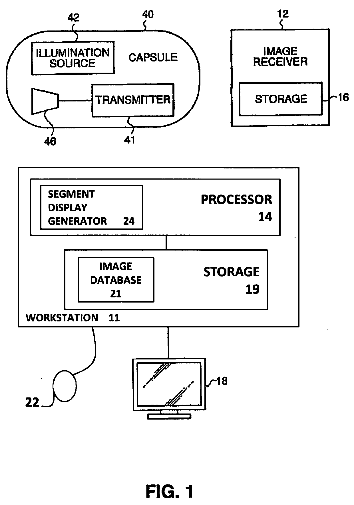
[0057]According to some embodiments, assessment of mucosal manifestations of a mucosal disease in a gastrointestinal track of a subject (e.g., patient) can be performed.

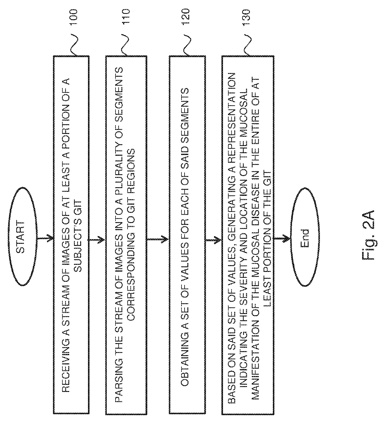
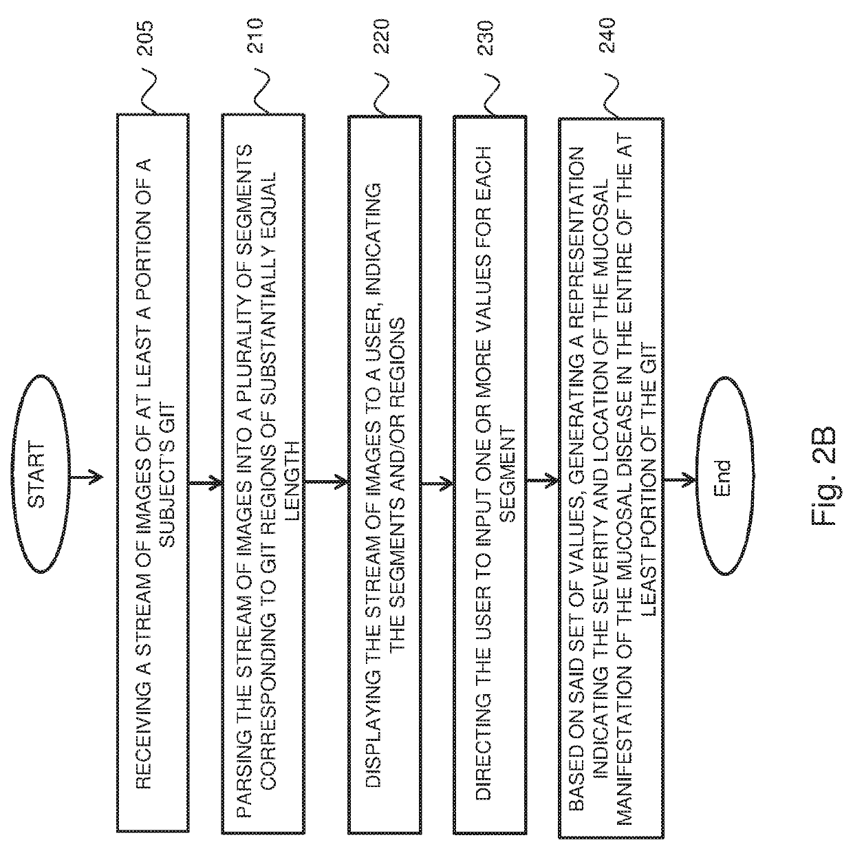
[0058]A stream of images of the subject's GIT can be received. The stream of images can be captured by procedures which image or at least indicate characteristics referring to one or more mucosal manifestations of disease within the GIT, such as capsule endoscopy, colonoscopy or gastroscopy. The stream of images can be parsed into a plurality of segments, each segment can show a region of the subject's GIT. In some embodiments, the regions can be of substantially equal length.

[0059]A set of values that refer to and / or are indicative of, at least, the pathological involvement and disease severity of the segment with respect to the mucosal manifestation of the disease are assigned to each segment and its corresponding GIT region. The set of values can include, for example, a most typical or common mucosal manifestation...


PUM


Abstract

A computerized-method for a mucosal assessment of a mucosal disease in a Gastrointestinal Tract (GIT) of a subject, including receiving a stream of images of at least a portion of the GIT, parsing the stream into a plurality of segments, wherein each segment corresponds to a region of the at least portion of the GIT, obtaining a set of values for each segment, wherein the set of values refers to the pathological involvement of the segment in the mucosal disease and to severity of mucosal manifestation of the mucosal disease in the segment, and based on said set of values for each segment, generating a representation indicating the location and severity of the mucosal manifestation of the mucosal disease in the entirety of the at least portion of the subject's GIT, thereby allowing to assess the condition of the mucosal disease in the at least portion of the GIT.

Description

FIELD OF THE INVENTION[0001]The present invention relates to a method and system for standardized assessment and / or monitoring of a mucosal disease in the Gastrointestinal Tract (“GIT”) of a subject. More specifically, but not limited to, the present invention relates to systems and methods for assessing and / or monitoring of a mucosal disease in a standardized manner by characterizing, scoring, summarizing and / or displaying images of regions of a subject's GIT based on images captured during one or more procedures.BACKGROUND OF THE INVENTION[0002]The GIT, referring to the alimentary tract extending from the mouth until the anus, is one of the largest organs in the body, serving a number of functions centered on consumption, digestion and absorption of nutritional products and water; and excretion of waste products. This alongside the important immunological defense against the concomitant exposure to foreign substances, making it the largest immune organ in the body. The GIT is divi...

Claims


Application Information

Patent Timeline
IPC IPC(8): G06T7/00A61B1/04A61B1/00A61B5/00G06T7/11G16H30/40G16H10/60
CPCG06T7/0012A61B1/041A61B1/00009A61B5/42G06T7/11G16H30/40G16H10/60G06T2207/30092G06T2207/10068G06T2207/20021G06T2207/10016A61B5/6861A61B5/0031A61B5/0066A61B5/0073A61B5/07A61B5/073A61B5/4836G06T2207/30028A61B1/000094
Inventor DOLNIK, INESSAELKAYAM, ORITSTEIN, ASSAFLAPIDUS, ALON
Owner GIVEN IMAGING LTD
Learn More