Multiplex detection of intracellular or surface molecular targets in single cells
a multi-functional, single cell technology, applied in the direction of microbiological testing/measurement, biochemistry apparatus and processes, etc., can solve the problem that human cancers are highly heterogeneous
- Summary
- Abstract
- Description
- Claims
- Application Information
AI Technical Summary
Benefits of technology
Problems solved by technology
Method used
Image
Examples
example 1
and Methods
[0328]I. Assay procedure (using miRNA detection as an example)[0329]1. Spin down harvested cells and wash them with DPBS twice[0330]2. Fix cells with paraformaldehyde solution (4% in PBS) inside the tube. Incubate cells for 10 min at 37° C.[0331]3. After 10 min, put the cells on ice for 1 min and then spin them down to remove paraformaldehyde solution[0332]4. Add 90% methanol into the cells and leave them on ice for 30 min.[0333]5. After half an hour, spin down the cells and remove methanol solution.[0334]6. Add blocking buffer (3% BSA+2% NaCl in TET) inside the tube and incubate cells at 37° C. for 1 hour.[0335]7. After an hour, the cells were spin down and blocking buffer was removed.[0336]8. Solution A (T4 DNA ligase inside NEB2A and TET) should be added to the cells[0337]9. Add 1 uL PC linker conjugated micro RNA probe solution (1 ul*100 uM=100 nmole) to 1 mL cell suspension and incubated it at 37° C. for 2 hours.[0338]At the same time, the glass slide with DNA probe ...
example 2
Depiction of Multiplexed Identification of Intracellular Targets from an Individual Cell
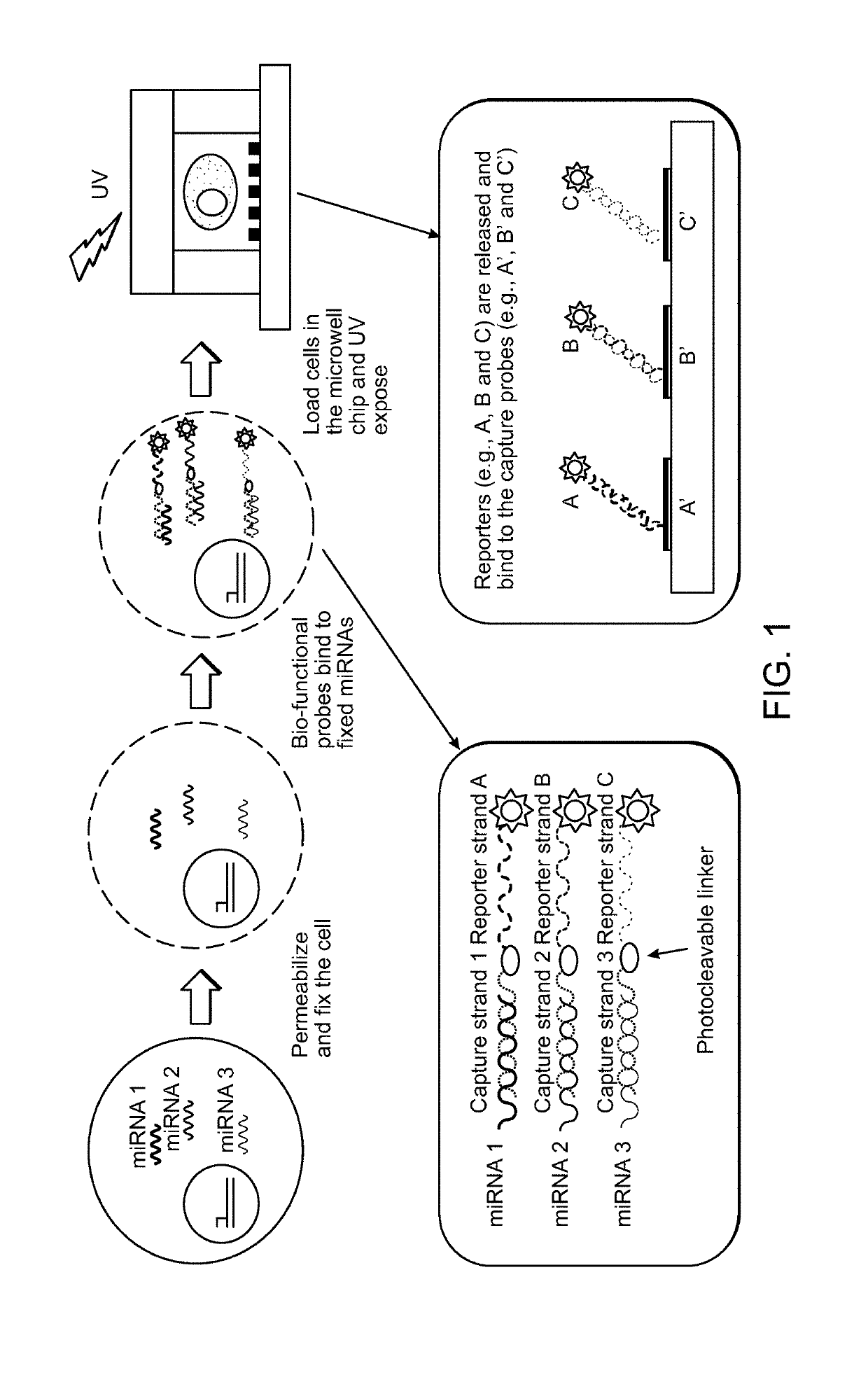
[0348]FIG. 1 depicts a schematic depiction of some embodiments of the multiplexed detecting of intracellular targets from an individual cell. In this non-limiting embodiment, miRNA capture strands serve as nucleic acid bait of the reporter-probe conjugates (RPCs).
[0349]In this embodiment, the method of multiplexed detection of intracellular targets in an individual cell comprises (a) producing a suspension of fixed, permeabilized cells, (b) incubating cells of (a) with at least two reporter-probe conjugates (RPC) each of which comprises, in the following order: (1) a nucleic acid bait (FIG. 1, any of the miRNA 1-3) that hybridizes to an intracellular target or a cell surface target; (2) a cleavable linker; and (3) a nucleic acid reporter molecule of known sequence (FIG. 1 A, B or C), under conditions under which nucleic acid bait component of the RPCs bind to their intracellular targets, thereby ...
example 3
Depiction of an Alternative Embodiment of Multiplexed Identification of Intracellular Targets from an Individual Cell
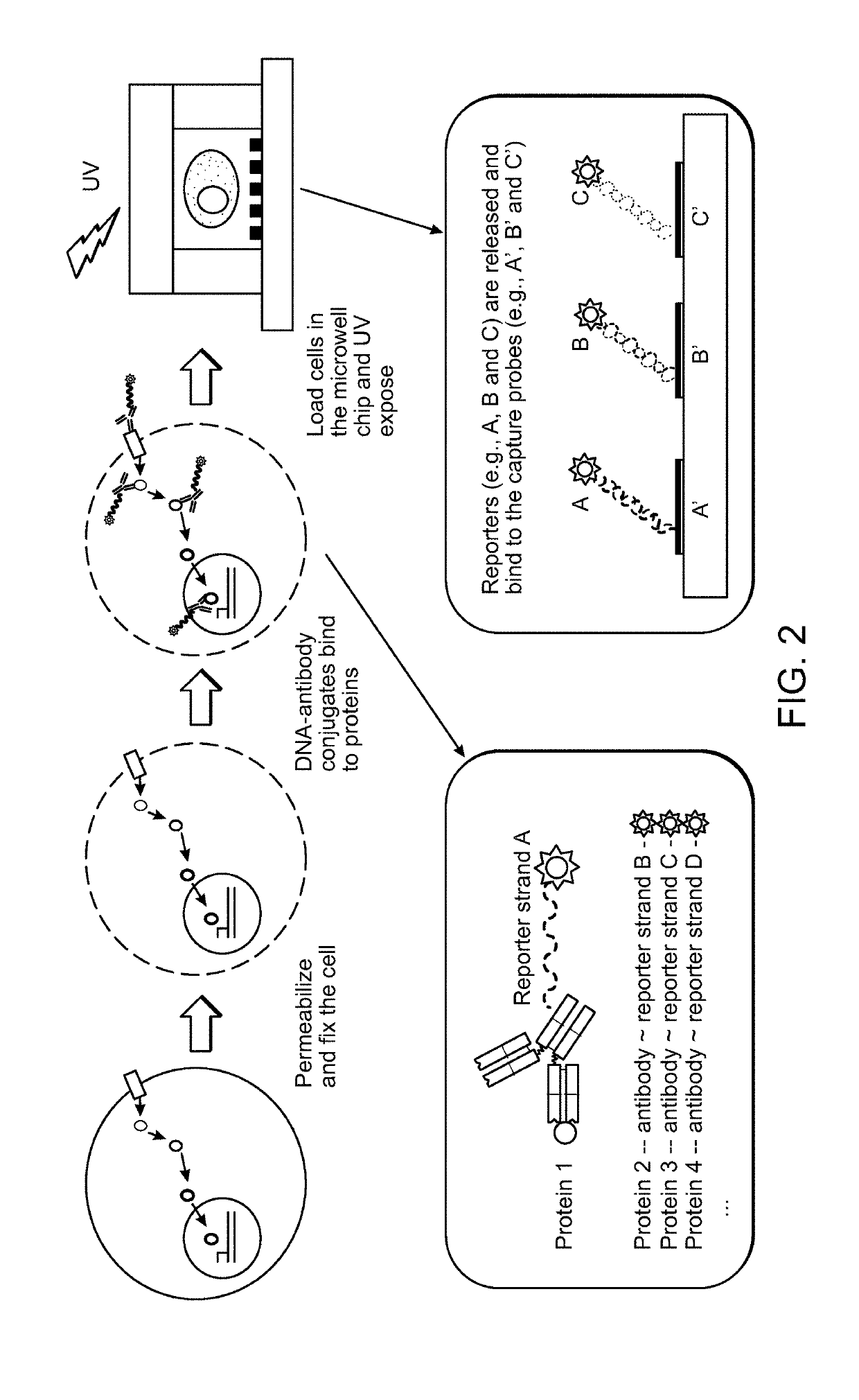
[0351]FIG. 2 depicts another non-limiting embodiment of the instant disclosure. This embodiment follows the same general method as the embodiment described in Example 2, however the capture portion of the RPC comprises an antibody that binds to an intracellular target rather than a nucleic acid bait sequence.
PUM
| Property | Measurement | Unit |
|---|---|---|
| Volume | aaaaa | aaaaa |
| Volume | aaaaa | aaaaa |
| Fluorescence | aaaaa | aaaaa |
Abstract
Description
Claims
Application Information
 - R&D
- Intellectual Property
- Life Sciences
- Materials
- Tech Scout
- Unparalleled Data Quality
- Higher Quality Content
- 60% Fewer Hallucinations
Browse by: Latest US Patents, China's latest patents, Technical Efficacy Thesaurus, Application Domain, Technology Topic, Popular Technical Reports.
© 2025 PatSnap. All rights reserved.Legal|Privacy policy|Modern Slavery Act Transparency Statement|Sitemap|About US| Contact US: help@patsnap.com


