Optical detection of biological components

a biological component and optical detection technology, applied in the direction of material excitation analysis, instruments, measurement devices, etc., can solve the problems of complex screening, difficult operation, and lack of mobility, and achieve the effects of convenient processing, low magnification, and convenient imag

Pending Publication Date: 2020-01-02
UNIV OF MASSACHUSETTS
View PDF1 Cites 0 Cited by
  • Summary
  • Abstract
  • Description
  • Claims
  • Application Information

AI Technical Summary

Benefits of technology

[0012]There are several advantages associated with the substrates, systems, and methods of the present disclosure, some of which are unexpected. According to some embodiments, the boronic acid component or the nucleic acid component of the substrate provides an attractive and at least somewhat selective binding target for bacteria. Nucleic acids can be particularly well suited for selective binding to target bacteria. According to some embodiments, the presence of bacteria bound to the boronic acid component or nucleic acid of the substrate can be visually confirmed under relatively low magnification power. According to some embodiments, the bacteria can be imaged conveniently with a relatively low magnification power lens attached to a camera of a smartphone, tablet, or computer. According to some embodiments, the image can be conveniently processed using a program or application associated with the smartphone, tablet, or computer. According to some embodiments, the presence or absence of bacteria on a food sample such as meat or a vegetable can be visually confirmed by contacting the food sample, or a portion thereof, with the substrate and imaging the substrate. According to some embodiments, the ability to conveniently confirm the presence or absence of bacteria in a food sample can allow for onsite screening of food samples for safety prior to consumption. According to some embodiments, the presence or absence of bacteria in an environmental sample, such as a pond or a soil sample, can be visually confirmed by contacting the environmental sample, or a portion thereof, with the substrate and imaging the substrate. According to some embodiments, the ability to conveniently confirm the presence or absence of bacteria can be applied to any aqueous sample. According to some embodiments, the systems and methods described herein can be incorporated into a portable system than can be used for educational purposes or for onsite screening. According to some embodiments, where the boronic acid component of the substrate comprises 3-mercaptophenylboronic acid the presence or absence of bacteria is readily confirmed using a lower magnification than a corresponding substrate that is free of 3-mercaptophenylboronic acid. According to some embodiments, images of the substrate can be pixilated and used to determine the concentration of the bacteria in the sample. According to some embodiments, the substrates can be used in conjunction with a surface enhanced Raman spectroscopy method to confirm the presence of the bacteria.

Problems solved by technology

The presence of bacteria in a food source or in an environment can be hazardous to health.
However, screening for bacteria can be complicated and require intricate systems that are difficult to operate and lack mobility to perform rapid on-site testing.

Method used


Image

  • Optical detection of biological components
  • Optical detection of biological components
  • Optical detection of biological components

Examples


example 1 preparation

of the Substrate

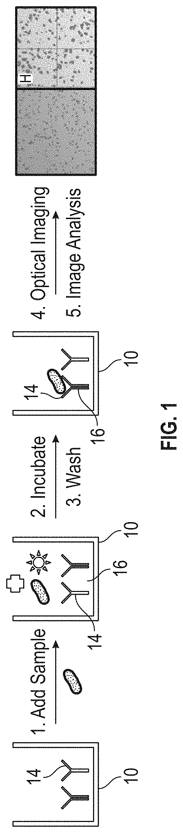
[0077]A gold coated slide (Thermo Scientific Madison, Wis.) was washed with deionized water and cut into rectangular pieces (approximately 2.5 by 3.5 mm) using a glass knife and cutter to form substrate precursors. The substrate precursors were then washed with ethanol (Pharmco-aaper Chicago, Ill.) for 30 seconds and placed into 4 mL of 1 mM 3-mercaptophenylboronic acid (3-MPBA)-ethanol (AstaTech Bristol, Pa.) solution in a sterile 5 mL test tube. The test tube was put onto a shaker (speed=20 RPM) for approximately 17 hours. After 24 hours, the substrate was formed and was washed with ethanol for 20 seconds and put into a 96-well plate. 150 μL of ethanol was pipetted into each well to stabilize the substrate until use (approximately 1 hour).

example 2 bacterial detection

[0078]Water, buffering chemicals (50 mM ammonia bicarbonate), and the biological sample, were added into the vial including the substrate. The mixture was incubated for 30 minutes. The substrate was removed and rinsed with water. A smartphone including a magnification lens was positioned over the substrate and a photograph was taken.

example 3

Example 3.1 Using a High Precision Lab Microscopy

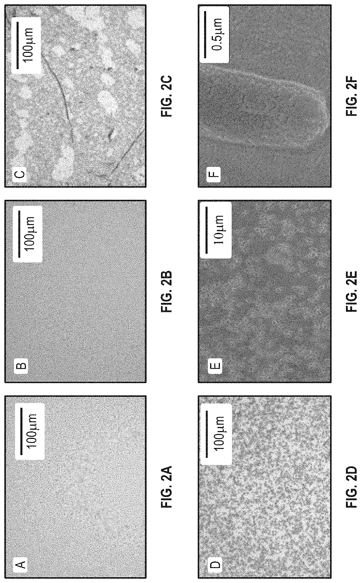
[0079]FIG. 2 shows that after bacteria cells bound to the 3-MPBA coated substrates, the bacterial cells were clearly visible under the 20× objective lens (FIG. 2D). Those black spots were confirmed as individual bacterial cells using scanning electron microscope (FIGS. 2E and 2F). When the substrates are free of coating with 3-MPBA, there were no clear black spots.

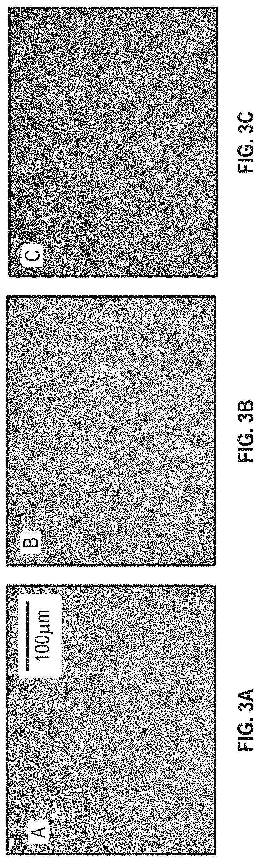
[0080]Ammonia bicarbonate was found to be helpful in this Example. As shown in FIG. 4A, bacteria suspended in water were not captured effectively compared to sodium hydroxide (FIG. 4B) and ammonia bicarbonate (both of the pH was 8.4) (FIG. 4C). This is because the boronic acid-diol reaction is more favorable in the alkaline condition. There also appears to be a noticeabble difference between sodium hydroxide and ammonia bicarbonate at the same pH. This demonstrates that ammonia bicarbonate has the ability to enhance the boronic acid-diol reaction more than just providing the...


PUM

PropertyMeasurementUnit
pHaaaaaaaaaa
distanceaaaaaaaaaa
distanceaaaaaaaaaa

Abstract

The present disclosure provides a substrate. The substrate includes a metallic layer. The substrate further includes a boronic acid component, a nucleic acid component, or both at least partially coating the metallic layer.

Description

CROSS-REFERENCE TO RELATED APPLICATION[0001]This application claims the benefit of priority to U.S. Provisional Patent Application Ser. No. 62 / 653,322 entitled “OPTICAL DETECTION OF BIOLOGICAL COMPONENTS,” filed Apr. 5, 2018, the disclosure of which is incorporated herein in its entirety by reference.STATEMENT OF GOVERNMENT SUPPORT[0002]This invention was made with Government support under 2015-67021-22993 awarded by the United States Department of Agriculture, National Institute of Food and Agriculture (USDA / NIFA). This invention was also made with Government support under 2016-31100-06025 awarded by the United States Department of Agriculture, National Institute of Food and Agriculture (USDA / NIFA) Hatch Capacity Funding. This invention was also made with Government support under NI17HFPXXXXXG052 awarded by the United States Department of Agriculture, National Institute of Food and Agriculture (USDA / NIFA) Hatch Capacity Funding. The U.S. Government has certain rights in this invent...

Claims


Application Information

Patent Timeline
Patent Type & Authority Applications(United States)
IPC IPC(8): G01N33/553G01N33/569G01N21/65
CPCG01N21/658G01N33/56916G01N33/553G01N33/56911C12Q1/22G01N33/521
Inventor HE, LILIPEARSON, BROOKE
Owner UNIV OF MASSACHUSETTS
Why Eureka
Try Eureka