Suppression of Microglial Activation with Innate Lymphoid Cells
- Summary
- Abstract
- Description
- Claims
- Application Information
AI Technical Summary
Benefits of technology
Problems solved by technology
Method used
Image
Examples
example 1
Enriched in Meninges
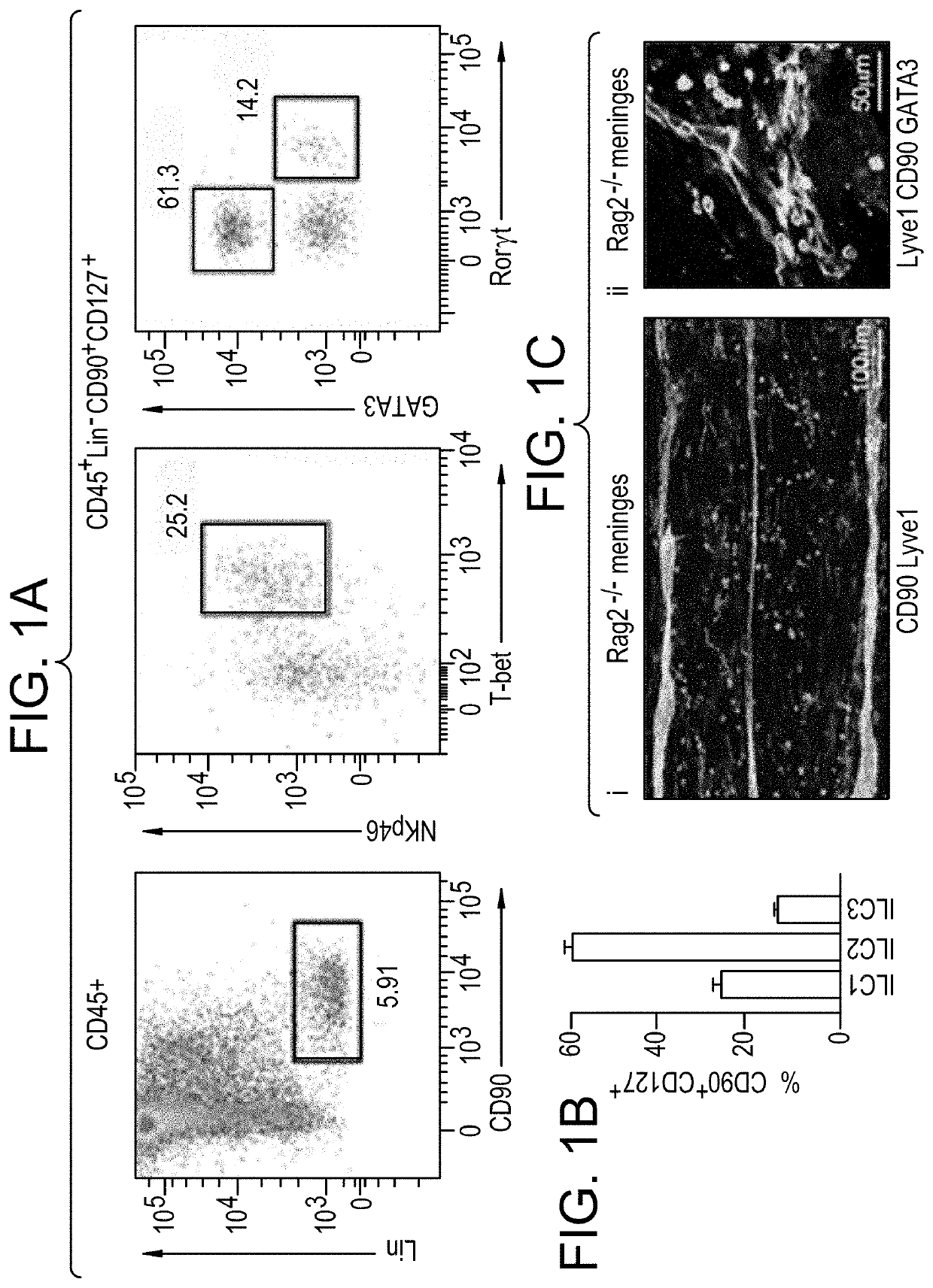
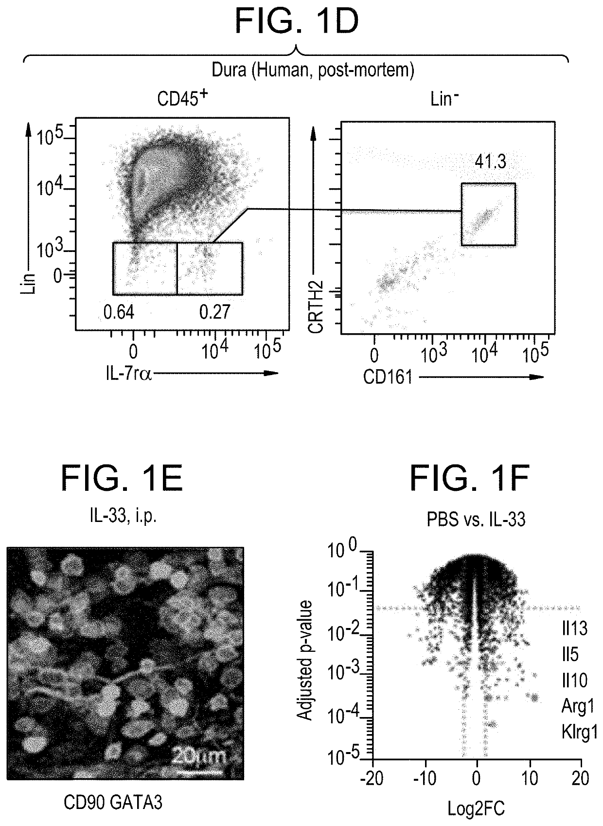
[0241]Transcription factor labeling of CD45+Lin−FCϵr1α−DX5−CD90+IL7rα+cells (FIG. 1A) was used to resolve ILC1 (Tbet+) ILC2 (Gata3Hi) and ILC3 (RORγt+Gata3− / lo) subsets. First, meningeal innate lymphoid cells (mILCs) were profiled to determine the most abundant type. Brains of nave mice were carefully dissected and placed immediately into 4% paraformaldehyde for fixation. After 48 hours (hr) of fixation at 4° C., brains were incubated for 24 hrs in 30% sucrose in H2O and subsequently stored at −80° C. Skullcaps were placed in 20 ml fluorescence activated cell sorting (FACS) buffer (DMEM / F-12 +2% FBS) in petri dishes and meninges peeled carefully away using forceps. Meninges were placed in a 70 μm cell filter and gently dissociated using the reverse side of a syringe plunger with (minimum 100) circular strokes. Cells were then washed through the filter three times to further release cells from dissociated tissue. Tubes were spun using a centrifuge for 7 minutes (m...
example 2
IL-25 Activates Meningeal ILC2
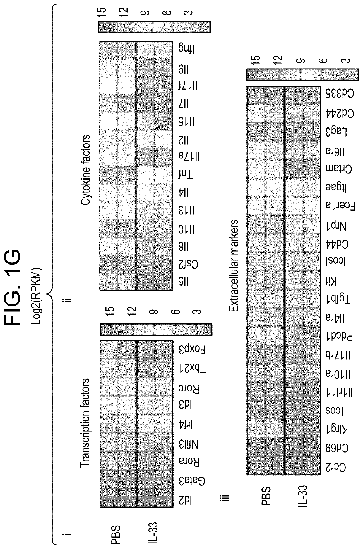
[0244]To probe relative functional response of IL-25 and IL-33, Rag2− / − mice were treated with each cytokine by intraperitoneal (i.p.) injection; mice were injected with 0.033 mg / kg, daily, for 3 days 24 hours apart. Brains and meninges were removed as described in Example 1. Cells were isolated, labeled with antibodies to Lineage markers, FCϵrlα, DX5, CD45, CD90, IL7rα, KLRG1, ST2, GATA3, KI-67, and analyzed with FACS. Increased proliferation as measured by mILC2 percentage of CD45+, counts, and K1-67 labeling, was seen with injection of either factor, but most robustly with IL-33.
example 3
cient Mice Exhibit Microglial Activation
[0245]Microglia, the principal immune presence within CNS, are engaged in constant surveillance and respond readily to protect delicate surrounding neural tissues. To examine the role of mILC2-derived factors in CNS immunomodulation, microglia were isolated from brains of ILC2-deficient Rag2− / − γ− / − mice and compared to those from control Rag2− / − mice using FACS.
[0246]Immunolabeling with Iba-1 indeed suggested visible disparities in microglial density (FIG. 3A) with brains from ILC-deficient mice showing increased numbers of microglia per unit area (FIG. 3B). Subsequent analysis of acutely-isolated brain cell suspensions by flow cytometry revealed elevated side scatter (a general indication of cell granularity thus implying intracellular activity) (FIG. 3C), increased expression of CD45 (FIG. 3D) and other surface activation markers, e.g., FcRII / III and MARCO (FIG. 3E) by microglia from ILC-deficient mice. These observations indeed suggested ...
PUM
| Property | Measurement | Unit |
|---|---|---|
| Permeability | aaaaa | aaaaa |
Abstract
Description
Claims
Application Information
 - R&D
- Intellectual Property
- Life Sciences
- Materials
- Tech Scout
- Unparalleled Data Quality
- Higher Quality Content
- 60% Fewer Hallucinations
Browse by: Latest US Patents, China's latest patents, Technical Efficacy Thesaurus, Application Domain, Technology Topic, Popular Technical Reports.
© 2025 PatSnap. All rights reserved.Legal|Privacy policy|Modern Slavery Act Transparency Statement|Sitemap|About US| Contact US: help@patsnap.com


