Tissue cutting systems, devices and methods

a technology of tissue cutting and cutting blades, applied in the field of tissue cutting systems, can solve the problems of progressive heart failure, affecting the patient's recovery, and reducing the pumping efficiency of the heart,

Pending Publication Date: 2020-04-23
EVALVE
View PDF3 Cites 14 Cited by
  • Summary
  • Abstract
  • Description
  • Claims
  • Application Information

AI Technical Summary

Benefits of technology

[0011]The present disclosure describes methods, devices and systems that may be employed to detach a previously sutured “bowtie” or implanted device that clips together the anterior and posterior leaflets of a valve from one or all of the leaflets. The technology described and claimed herein could also be adapted to selectively target

Problems solved by technology

Regurgitation of the valve can significantly decrease the pumping efficiency of the heart, placing the patient at risk of severe, progressive heart failure.
Mitral valve regurgitation can result from many different mechanical defects in the mitral valve or the left ventricular wall.
The valve leaflets, the valve chordae which connect the leaflets to the papillary muscles, the papillary muscles themselves or the left ventricular wall may be damaged or otherwise dysfunctional.
Commonly, the valve annulus may be damaged, dilated, or weakened, limiting the ability of the mitral valve to close adequately against the high pressures of the left ventricle.
The need to both open the chest and place the patien

Method used


Image

  • Tissue cutting systems, devices and methods
  • Tissue cutting systems, devices and methods
  • Tissue cutting systems, devices and methods

Examples


Embodiment Construction

[0040]As mentioned earlier, sometimes, after installation of an edge-to-edge repair device, such as a suture “bow-tie” or fixation device, in the heart, it needs to be removed or at least detached from one or both of anterior and posterior leaflets. Ordinarily, this has been done during a high-risk invasive procedure such as open-heart surgery. The presently described system 100, and associated devices and methods, however, allow a physician or clinician to address heart problems in a minimally invasive procedure.

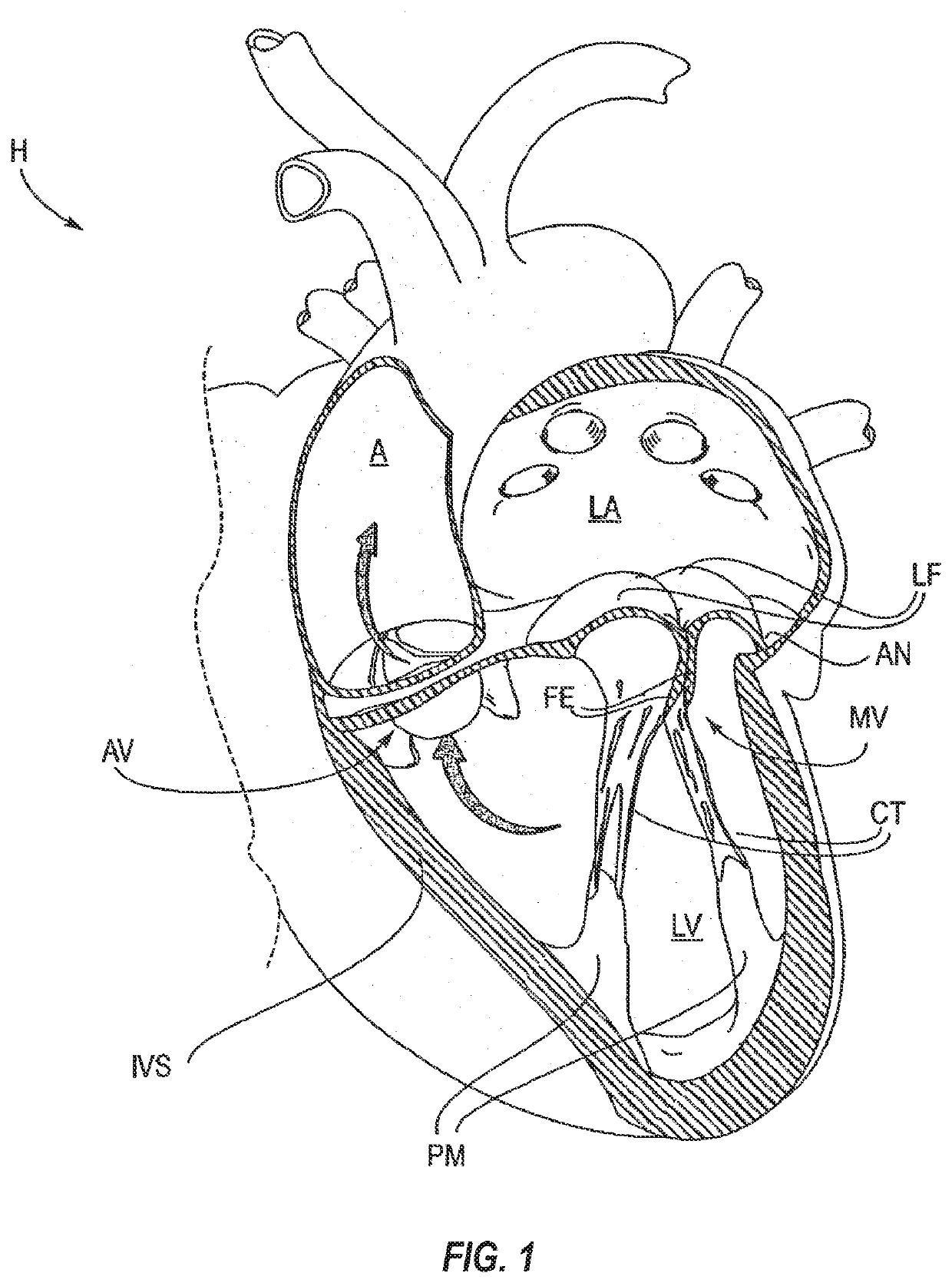
[0041]For ease of explanation, FIG. 1 illustrates the left ventricle (LV) of a normal heart H in systole. The left ventricle (LV) is contracting and blood flows outwardly through the tricuspid (aortic) valve (AV) in the direction of the arrows. Back flow of blood or “regurgitation” through the mitral valve (MV) is prevented since the mitral valve is configured as a “check valve” which prevents back flow when pressure in the left ventricle is higher than that in the left atr...


PUM


Abstract

Methods, devices and systems for disabling and/or removing a mitral valve edge-to-edge repair device via minimally invasive, endovascular procedures. Additional procedures on a heart may sometimes become necessary after the installation of a mitral valve edge-to-edge repair device. To prepare for such additional procedures, the edge-to-edge repair device may be removed or disabled in minimally invasive ways (e.g., through an endovascular procedure), without requiring open access to the heart.

Description

CROSS-REFERENCE TO RELATED APPLICATIONS[0001]This application claims the benefit of priority based on U.S. Provisional Patent Application Ser. No. 62 / 748,947, filed Oct. 22, 2018 and entitled “Leaflet Cutting Devices and Methods.”BACKGROUND[0002]The present invention relates generally to medical methods, devices, and systems. In particular, the present invention relates to methods, devices, and systems for the endovascular, percutaneous or minimally invasive surgical treatment of bodily tissues, such as tissue approximation or valve repair. More particularly, the present invention relates to cutting of leaflet tissue in preparation of subsequent implanting of a medical implant through minimally invasive procedures.[0003]Surgical repair of bodily tissues often involves tissue approximation and fastening of such tissues in the approximated arrangement. When repairing valves, tissue approximation includes coapting the leaflets of the valves in a therapeutic arrangement which may then b...

Claims


Application Information

Patent Timeline
IPC IPC(8): A61F2/24A61B17/32
CPCA61B17/32A61B2017/00783A61B2017/00243A61B2018/00601A61B2017/00867A61F2/2466A61F2/2445A61B17/1285A61B18/1492A61B2017/003A61B2017/00358A61B2017/00876A61B2018/00369A61B2090/3966A61F2/246A61F2/2463A61B2018/144A61B2018/1407A61B2018/1475
Inventor DALE, THEODORE PAULEIDENSCHINK, TRACEE ELIZABETH JOHNSON
Owner EVALVE
Why Eureka
Try Eureka