
Treatment of bone growth disorders

a technology of bone growth disorder and activator, which is applied in the field of activator, can solve the problems of disease group a major challenge for the health care system, bone growth disorder, and bone growth disturbance, and achieve the effect of increasing the production of phosphatidylinositol-3-phosphate, and being easy to measur

Pending Publication Date: 2020-07-23
FOND AZIONE TELETHON
View PDF0 Cites 1 Cited by
  • Summary
  • Abstract
  • Description
  • Claims
  • Application Information

AI Technical Summary

Benefits of technology

The present invention provides an activator of beclin 1-Vps 34 complex for use in the treatment and prevention of bone growth disorders. The activator can increase phosphatidylinositol 3-phosphates (PI3P) production in a cell and can be selected from a polypeptide, a polynucleotide coding for the polypeptide, a vector comprising the polynucleotide, a host cell expressing the polypeptide or the vector, a small molecule selected from a mTORC1 inhibitor or a BH3 mimetic, and a functional fragment of beclin 1 consisting of residues 270-278. The activator can be used in the treatment of achondroplasia, hypochondroplasia, spondyloepiphyseal dysplasia, a lysosomal storage disorder, or other bone growth disorders.

Problems solved by technology

Defects in the development and maintenance of the growth plates lead to disorders of the bone growth.
These mutations cause the FGFR3 protein to be overly active, which interferes with skeletal development and leads to the disturbances in bone growth seen with this disorder.
Although individually rare, the lysosomal storage disorders (LSDs) as a group have a frequency of about 1:8000 live births, making this disease group a major challenge for the health care system.
However, the frequency of the disease may be underestimated as the most frequent presentation is the antenatal form, which remains underdiagnosed.
Currently, MPS VII lacks an efficient treatment.
However, effects of these interventions on skeletal disease manifestations are less well established and outcomes are highly dependent on disease burden at treatment initiation.
Furthermore, the efficacy of these therapeutic strategies has several major limitations, such as the difficulty of reaching particular tissues such as the skeleton.
Indeed, gene therapy approaches in different MPS animal models showed very little efficacy on bone defects (Ferla R et al., 2014, Stevenson D A and Steiner R D, 2013).
However, they do not provide any evidence or suggestion that induction of autophagy and in particular that activation of the Beclin-1 / Vps34 complex could be effective in the treatment of bone disorders.

Method used


Image

  • Treatment of bone growth disorders
  • Treatment of bone growth disorders
  • Treatment of bone growth disorders

Examples


example 1

n of Autophagy Prevents the Skeletal Defects Associated with LSDs

[0263]Tat-Beclin 1 peptide is capable of inducing autophagy in a cell by activating Beclin 1-Vps34 complex (see FIG. 4A1).

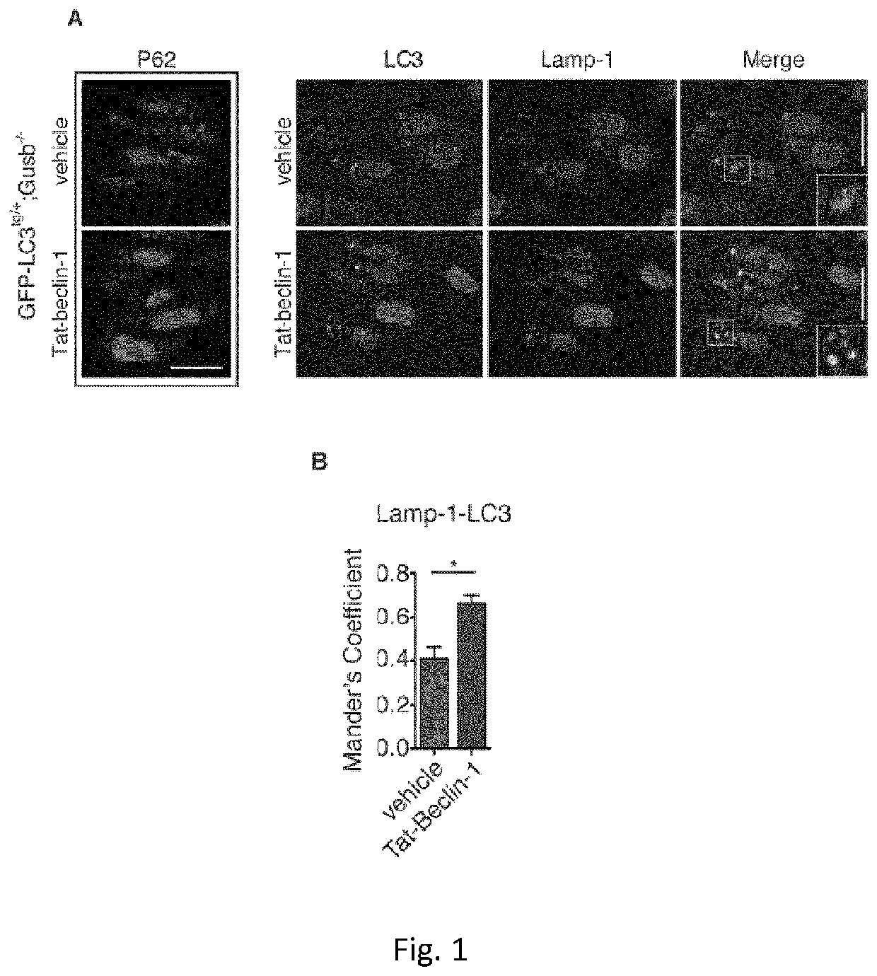
[0264]Daily injection of Tat-Beclin1 peptide promoted Av-Lys fusion and p62 / SQSTM1 degradation in the growth plate of MPS VII (Gusb− / −) mice expressing the fluorescent autophagy reporter GFP-LC364 (Gush− / −; GFP-LC3tg / + mice) (FIG. 1a,b).

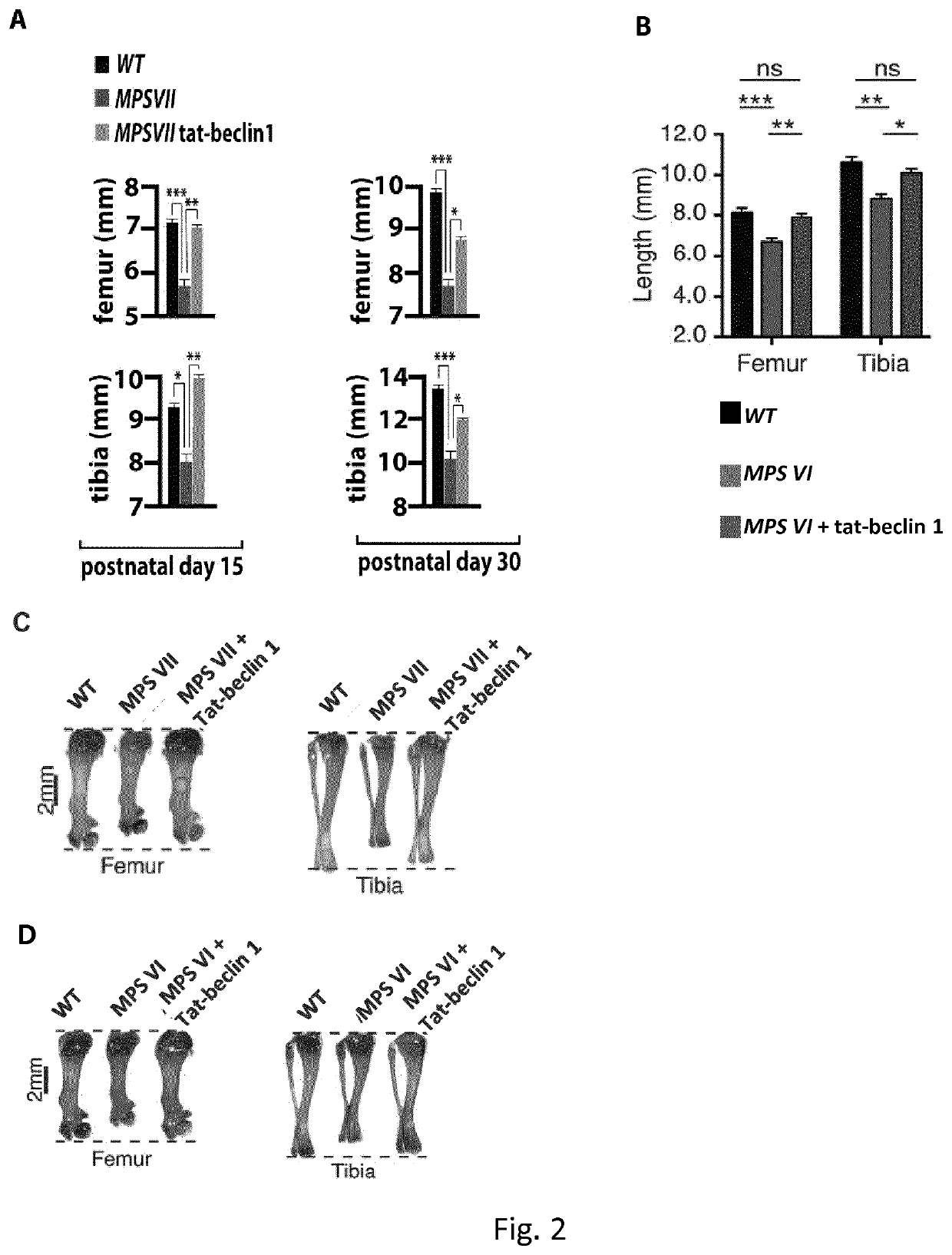
[0265]Newborn MPS VII and MPS VI mice were intraperitoneally injected daily with retro-inverso Tat-Beclin 1 peptide (Beclin 1Activator II, retro-inverso Tat-Beclin 1, Millipore) at 2 mg / kg resuspended in PBS, according to a preferred embodiment of the invention. Control mice were injected with vehicle only. Mice were sacrificed after 15 (P15) and 30 (P30) days.

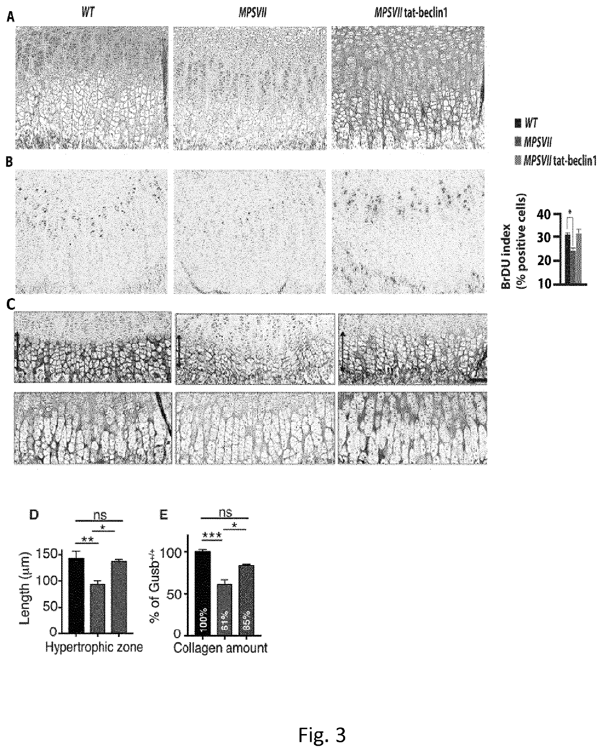
[0266]Starting at postnatal day 15 (P15) MPSVII mice show significant reduced femur and tibia lengths compared to wild type mice (FIG. 2a,c). A similar phenotype was also observed in P15 MPSVI mice (FIG. 2b, d)...

example 3

Flux Increases During Early Post-Natal Bone Development

[0273]The femoral growth plates of mice that ubiquitously express the autophagosome marker MAP1LC3 tagged with green fluorescent protein (GFP) (GFP-LC3tg / +) (Mizushima N et al, Mol Biol Cell 2004) were analyzed. Very few autophagic vesicles (AVs) were detected in the growth plates of newborn mice (P0) (FIG. 5a, quantification in 5b). Sections obtained from older mice (P2 to P8) showed a progressive age-dependent increase in the number of AVs (FIG. 5a, quantification in 5b). This observation was confirmed by TEM analysis (FIG. 9a) and biochemically by quantifying the conversion of the non-lipidated form of LC3 (LC3I) to the autophagosome-associated lipidated form (LC3II) (Kabeya Y et al., 2005) in femoral growth plates of wild type mice at different time points (FIG. 5c). In vivo inhibition of lysosomal function by Leupeptin administration further increased the levels of LC3II in the growth plate of P6 but not P2 mice, indicating...

example 4

Regulates Skeletogenesis and the Composition of Growth Plate ECM

[0274]The essential autophagy gene 7 (Atg7) was deleted in chondrocytes by crossing a mouse line carrying the Atg7 floxed allele (Atg7f / f) (Komatsu, M. et al., J. Cell Biol. 2005) with two different Cre mouse lines: 1) the Prx1-Cre line, in which the Cre protein is expressed in the mesenchymal cells of the limbs during embryogenesis (Logan M et al., Genes 2002) and 2) the Col2a1-Cre line, in which the expression of the Cre protein is mainly restricted to mature chondrocytes before and after birth (Ovchinnikov DA, Genes 2002).

[0275]The selective lack of Atg7 protein and the inhibition of functional autophagy in the femoral growth plates of Atg7f / f; Prx1-Cre and Atg7f / f; Col2a1-Cre mice was verified (FIG. 9c-f).

[0276]Atg7f / f; Prx1-Cre and Atg7f / f; Col2a1-Cre mice were born at the expected Mendelian ratio, with bones of normal shapes and sizes, suggesting that chondrocyte autophagy is dispensable during embryonic skeletal ...


PUM

PropertyMeasurementUnit
Disorderaaaaaaaaaa

Abstract

The present invention relates to an activator of beclin 1-Vps 34 complex for use in the treatment and / or prevention of a bone growth disorder. The activator may be a polypeptide, a polynucleotide, a vector, a host cell or a small molecule. In particular the activator may be a Beclin 1 peptide or a fragment or a derivative thereof, a mTORC1 inhibitor or a BH3 mimetic. The present invention also relates to pharmaceutical composition comprising said activator.

Description

FIELD OF THE INVENTION[0001]The present invention relates to an activator of beclin 1-Vps 34 complex for use in the treatment and / or prevention of a bone growth disorder. The activator may be a polypeptide, a polynucleotide, a vector, a host cell or a small molecule. In particular the activator may be a Beclin 1 peptide or a fragment or a derivative thereof, a mTORC1 inhibitor or a BH3 mimetic. The present invention also relates to pharmaceutical composition comprising said activator.BACKGROUND[0002]Bones in different parts of the skeleton develop through two distinct processes, intramembranous ossification and endochondral ossification. Intramembranous ossification occurs in the flat bones of the skull and involves direct differentiation of embryonic mesenchymal cells into the bone-forming osteoblasts. Endochondral ossification is responsible for the initial bone development from cartilage, in utero and infants; furthermore it is an essential process during formation of long bones,...

Claims


Application Information

Patent Timeline
IPC IPC(8): A61K38/17A61P19/08A61K45/06A61K31/4745
CPCA61K48/00A61K45/06A61K38/1761A61P19/08A61K31/4745A61K31/436C07K14/82C07K14/4747C07K2319/10A61K2300/00
Inventor SETTEMBRE, CARMINECINQUE, LAURABARTOLOMEO, ROSAAURICCHIO, ALBERTOTRAPANI, IVANATORIELLO, ELISABETTA
Owner FOND AZIONE TELETHON
Learn More