
Image based ultrasound probe calibration

Pending Publication Date: 2021-02-18
BRAINLAB
View PDF4 Cites 1 Cited by
  • Summary
  • Abstract
  • Description
  • Claims
  • Application Information

AI Technical Summary

Benefits of technology

The patent describes a method for analyzing ultrasound images using a parametrical approach to capture the texture of the image. This approach is able to overcome the difficulty of dealing with speckle noise and low trend intensity inhomogeneity commonly present in ultrasound images. Additionally, the method allows for better estimations of similarities with less data and results in more localized analysis on the ultrasound image.

Problems solved by technology

The usability of phantoms for calibration is however limited.
Sterility requirements in the operation theatre require cumbersome draping.
The need for tracking involves line-of-sight problems between tracking camera and phantom's markers.

Method used


Image

  • Image based ultrasound probe calibration
  • Image based ultrasound probe calibration
  • Image based ultrasound probe calibration

Examples


Embodiment Construction

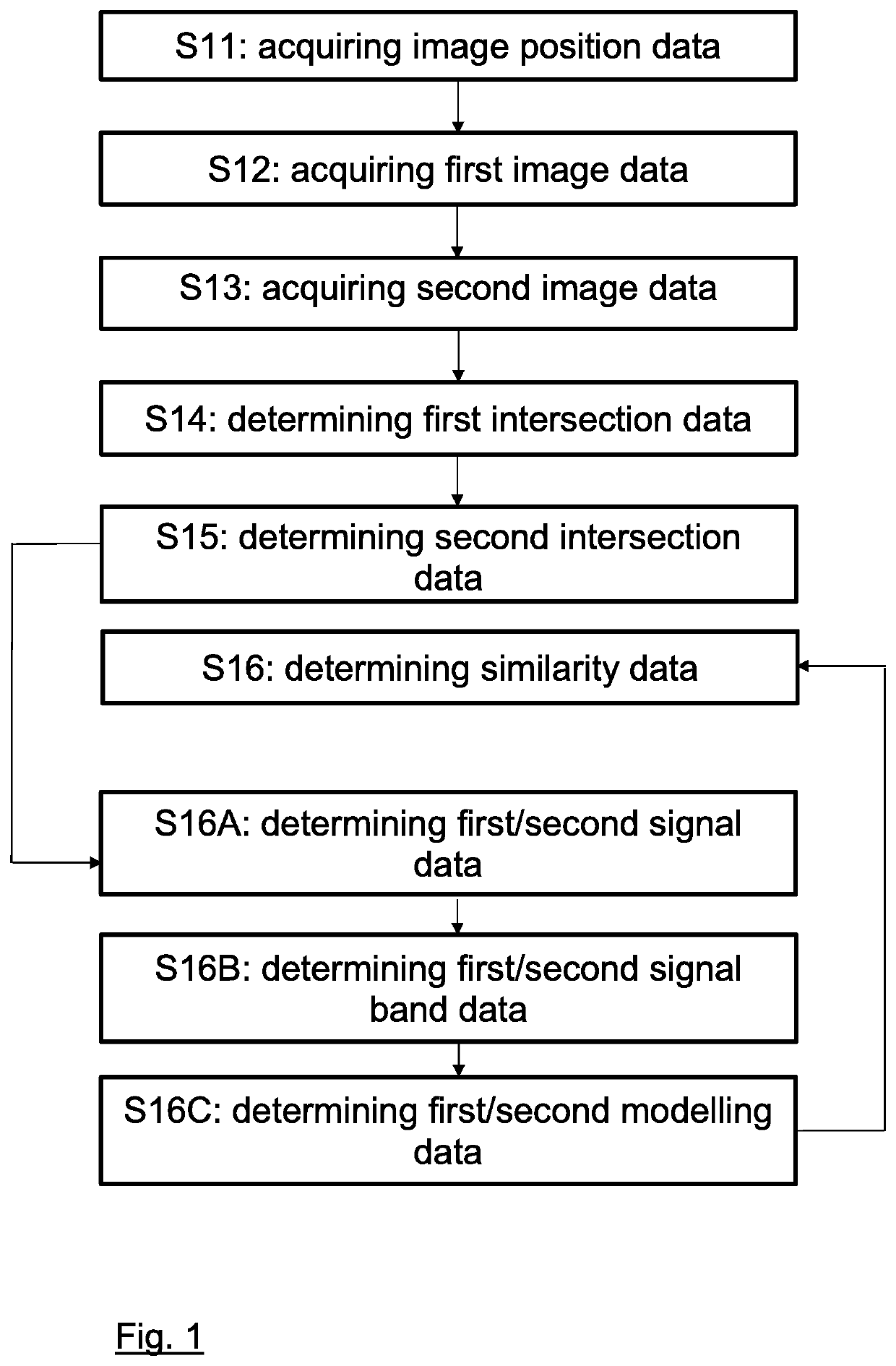
[0112]FIG. 1 illustrates the basic steps of the method according to the first aspect and to the second aspect.

[0113]In steps S11 to S13, at least two intersecting ultrasound-images are acquired, wherein the spatial position of the ultrasound image plane is initially predefined.

[0114]Then, the content of each ultrasound image within the image intersection is determined in steps S14 and S15, and compared with each other in step S16. The grade of similarity between the image content indicates how well the ultrasound probe is calibrated.

[0115]In this specific example, the method according to the second aspect is used to compare the image content by performing steps S16A to S160.

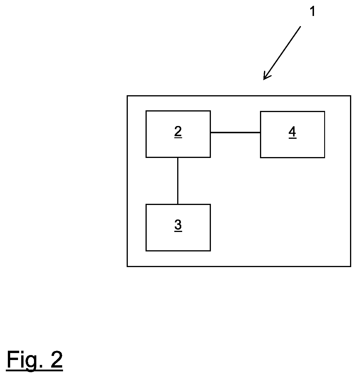
[0116]FIG. 2 is a schematic illustration of the medical system 1 according to the sixth aspect. The system is in its entirety identified by reference sign 1 and comprises a computer 2, an electronic data storage device (such as a hard disc) 3 for storing at least the patient data and a medical device 4 (such as a...


PUM


Abstract

The disclosed invention encompasses an image-based approach of calibrating an ultrasound-probe, wherein at least two ultrasound-images which cross each other are acquired with a tracked ultrasound probe, and wherein the intersection areas of these images, which have been calculated on the basis of the tracked spatial position of the ultrasound probe are checked for similar image content. The grade of similarity gives an indication as to how well the ultrasound probe is calibrated.

Description

FIELD OF THE INVENTION[0001]The present invention relates to a computer-implemented method of calibrating an ultrasound probe, a corresponding computer program, a non-transitory program storage medium storing such a program and a computer for executing the program, as well as a medical system comprising an electronic data storage device and the aforementioned computer.TECHNICAL BACKGROUND[0002]An ultrasound probe placed at the human body provides insight at a spatially defined image plane. The location of that plane with respect to the probe's housing depends on the probe's internal construction. For use with navigation systems the exact spatial transformation between the image plane and the probe's housing has to be established by a calibration beforehand. Today's calibration procedures utilize a dedicated calibration phantom with well-defined inside structure and a tracked ultrasound probe. The usability of phantoms for calibration is however limited. The phantom's features have t...

Claims


Application Information

Patent Timeline
IPC IPC(8): A61B8/00A61B90/00
CPCA61B8/58A61B8/4245A61B2090/3925A61B90/39A61B8/463A61B8/145A61B8/587
Inventor GÖTTE, HUBERTILLANES MANRÍQUEZ, ALFREDO GUILLERMOFRIEBE, MICHAELBALAKRISHNAN, SATISHPOUDEL, PRABAL
Owner BRAINLAB
Features
  • Generate Ideas
  • Intellectual Property
  • Life Sciences
  • Materials
  • Tech Scout
  • Unparalleled Data Quality
  • Higher Quality Content
  • 60% Fewer Hallucinations
Learn More