Human pluripotent stem cell derived neurodegenerative disease models on a microfluidic chip

a microfluidic chip and neurodegenerative disease technology, applied in the field of cell culture, can solve the problem of no models that include human vascular components

Pending Publication Date: 2021-05-06
CEDARS SINAI MEDICAL CENT
View PDF0 Cites 4 Cited by
  • Summary
  • Abstract
  • Description
  • Claims
  • Application Information

AI Technical Summary

Benefits of technology

[0013]Described herein is a method, including: contacting a quantity of blood cells with one or more vectors encoding a reprogramming factor, and delivering a quantity of reprogramming factors into the blood cells, culturing the blood cells in a reprogramming media, wherein the quantity of blood cells are obtained from a human subject afflicted with a neurodegenerative disease, and further wherein delivering the reprogramming factors, and culturing in a reprogramming media generates blood cell derived induced pluripotent stem cells (iPSCs). In other embodiments, the neurodegenerative disease is Parkinson's disease (PD). In other embodiments, the neurodegenerative disease is amyotrophiclateral sclerosis (ALS). In other embodiments, the iPSCs are further cultured in fluidic communication with one or more of astrocytes, microglia, and vascular cells. In other embodiments, the one or more vectors are oriP / EBNA1 vectors. In other embodiments, the method includes differentiating the iPSCs into neuron. In other embodiments, the method includes d differentiating the iPSCs into vascular cells. In other embodiments, the method includes differentiating the iPSCs into astrocytes. In other embodiments, the method includes differentiating the iPSCs into microglia.
[0014]Described herein is a quantity of neurodegenerative disease derived induced pluripotent stem cells (iPSCs) made by a method including contacting a quantity of blood cells with one or more vectors encoding a reprogramming factor, and delivering a quantity of reprogramming factors into the blood cells, culturing the blood cells in a reprogramming media, wherein the quantity of blood cells are obtained from a human subject afflicted with a neurodegenerative disease, and further wherein delivering the reprogramming factors, and culturing in a reprogramming media generates blood cell derived iPSCs. In other embodiments, the neurodegenerative disease is Parkinson's disease (PD). In other embodiments, the neurodegenerative disease is amyotrophiclateral sclerosis (ALS)
[0015]Also described herein is a method of compound screening, including contacting a quantity of cells with one or more test compounds measuring one or more parameters, and selecting one or more test compounds based on the measured one or more parameters, wherein cells are differentiated from neurodegenerative disease derived induced pluripotent stem cells (iPSCs). In other embodiments, the differentiated cells are neurons. In other embodiments, the differentiated cells vascular cells. In other embodiments, the differentiated cells are astrocytes. In other embodiments, the differentiated cells are microglia. In other embodiments, the one or more parameters include permeability of the test compound across a quantity of vascular cells. In other embodiments, the iPSCs are made by a method including contacting a quantity of blood cells with one or more oriP / EBNA1 vectors encoding a reprogramming factor and delivering a quantity of reprogramming factors into the blood cells culturing the blood cells in a reprogramming media, wherein the quantity of blood cells are obtained from a human subject afflicted with a neurodegenerative disease, and further wherein delivering the reprogramming factors, and culturing in a reprogramming media generates blood cell derived iPSCs.

Problems solved by technology

While PD models using iPSCs have been attempted, none are from young onset sporadic patients.
In addition, there are no models that include human vascular component, a critical feature of therapeutic delivery, or incorporate the cast of support cells (e.g., microglia, astrocytes), believed to play roles in disease progression,

Method used


Image

  • Human pluripotent stem cell derived neurodegenerative disease models on a microfluidic chip
  • Human pluripotent stem cell derived neurodegenerative disease models on a microfluidic chip
  • Human pluripotent stem cell derived neurodegenerative disease models on a microfluidic chip

Examples


example 1

Preliminary Data

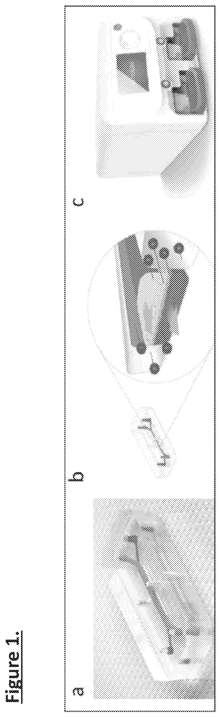
[0092]Organ-chips allow for MPS models of several organ and tissue systems, including the lung alveolus and small airway, intestine, liver, kidney, and blood-brain barrier and other organs. Organ-chips can be scaled-up while operating in parallel, and can be paired specially developed instruments to eliminate the need for the user to connect and disconnect tubing, reduce fluid dead-volumes to improve sampling, and remove undesired bubbles. These advantages increase experimental throughput, and reducing variability (FIG. 1).

[0093]Biological material on organ-chips include induced pluripotent stem cells (iPSCs), iPSC-derived cells and tissue. With a simple blood sample taken in the clinic, one can derive iPSC lines that carry the donor's genetic makeup. The patient's peripheral blood mononuclear cells (PBMCs) are directly reprogrammed, without the need for an expansion step, using non-integrating techniques that allow for transient ectopic expression of reprogramming f...

example 2

[0097]Development of Reliable APS Models of Control iPSC-Derived MNs and DANs

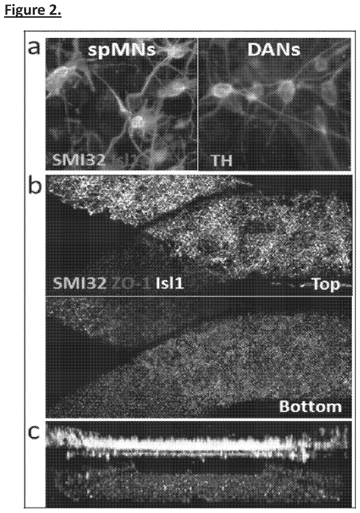
[0098]Prior studies by the Inventors seeding neural and endothelial cells with MPS devices described above has shown that both spMNs and DANs can differentiate in co-culture with BMECs seeded into the opposite channel. Without being bound by any particular theory, it is believed that addition of astrocytes and microglia will strengthen the model further. For example, Microglia are also a cell type that has been implicated in neurodegenerative disease and is known to lose function in traditional forms of culture. The MPS could therefore allow this cell type display novel biological and disease related physiology for the first time in vitro.

[0099]Initial studies have included seeding organ-chip with iPSC-derived astrocytes in the neural side and BMECs in the blood side 1-week prior to seeding neurons. Preliminary results indicate that the astrocyte layer increases stability of both the neural and blood compar...

example 3

Use Astrocytes and Microglia to Enhance Potential Biomarkers Through Maturation of spMN and DANs

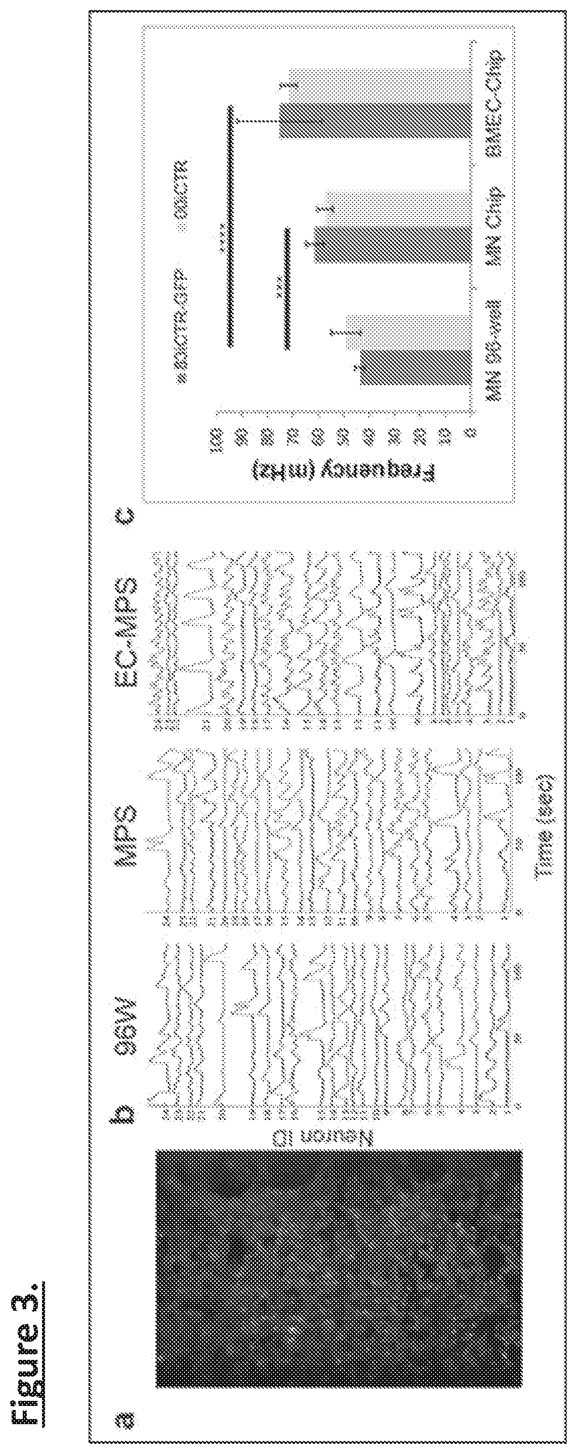
[0101]Either spMNs or DANs are cultured in chips under 4 different conditions: (i) neurons alone in the top channel (ii) neurons in top channel with BMEC in the bottom channel (as in FIG. 2). (iii) neurons seeded into chips pre-coated with astrocytes in top channel and BMEC on bottom channel and (iv) neurons and microglia (FIG. 5) seeded into chips pre-coated with astrocytes in the top channel and BMECs in the bottom channel. One can produce microglia using a cytoplasmic florescent reporter line under a constitutive promoter so that one can determine their persistence in the cultures. This overcomes difficulties characterizing these cells with specific markers. Both channels can have 10 μl flow rates, which appear to be optimal for neural and endothelial cell survival.

[0102]Cultures are analyzed at either 2 weeks or 4 weeks post-seeding to establish how the co-cultures mature over time in...


PUM

PropertyMeasurementUnit
concentrationsaaaaaaaaaa
flow ratesaaaaaaaaaa
time-to-therapeutic effectaaaaaaaaaa

Abstract

Described herein is a microphysiological system for models of disease. Specifically, induced pluripotent stem cells (iPSCs) and iPSC-derived cells, including those obtained from disease patients, are seeded onto microfluidic “chip” devices to study cellular development and disease pathogenesis. Herein, neurodegenerative disease modeling, including Parkinson's Disease (PD) is shown to reproduce key PD pathology in a vascularized human model that contains neurons relating to PD pathology. Such compositions and methods are used for research for PD biomarkers, patient screening for PD risk assessment, and therapeutic discovery and testing. A panel of biomarkers are generated through analysis of living PD-chips by neural activity, whole transcriptomic, proteomic, and metabolomic analysis, and functional enzyme tests of media and tissue. Introducing therapeutics through a vasculature channel, coupled with blood brain barrier penetration studies can be assessed for efficacy in the human neural cells present in the PD-Chip.

Description

STATEMENT REGARDING FEDERALLY-SPONSORED RESEARCH[0001]This invention was made with government support under NS105703 awarded by the National Institutes of Health. The government has certain rights in the invention.FIELD OF THE INVENTION[0002]The present invention relates to the field of culturing cells, and in particular, cells for disease modeling in a fluidic device, including but not limited to a microfluidic device or chip.BACKGROUND[0003]The physiological, molecular and cellular changes that underlie amyotrophic lateral scleroris (ALS) and Parkinson's disease (PD) are complex. Post-mortem brain tissues clearly have losses in motor and dopamine neurons that underlie the diseases, but the path to neuronal death remains open to speculation—with several existing theories. However, these mutant specific phenotypes have not yet explained adult onset diseases of a sporadic origin, which do not display overt cell death in current culture systems. While genetic forms of ALS and PD are i...

Claims


Application Information

Patent Timeline
Patent Type & Authority Applications(United States)
IPC IPC(8): C12N5/079C12N5/0793C12M3/06C12M1/12C12M1/34C12N5/074G01N33/50C12N5/071
CPCC12N5/0622C12N5/0619C12M23/16C12M25/02C12M41/46C12N5/0696C12N2506/45C12N5/0697G01N33/5088C12N2502/081C12N2502/086C12N2502/45G01N33/5058C12N5/069C12N2506/11C12N2502/28C12N2502/11C12M35/08B01L3/502761B01L2300/0877B01L2200/0668
Inventor SANCES, SAMUELLAPERLE, ALEXANDERYUCER, NURSVENDSEN, CLIVE N.
Owner CEDARS SINAI MEDICAL CENT
Why Eureka
Try Eureka