

Vascular endothelial growth factor/Anti-fibronectin antibody fusion proteins
a growth factor and vascular endothelial technology, applied in the field of vascular endothelial growth factor/antifibronectin antibody fusion proteins, can solve the problems of immunoconjugated with therapeutic effects, such as anti-inflammatory activity, and remains a challenge, so as to facilitate the resolution of oedema and inflammation, improve lymphatic function, and improve lymphatic clearance
- Summary
- Abstract
- Description
- Claims
- Application Information
AI Technical Summary
Benefits of technology
Problems solved by technology
Method used
Image

Examples
example 1
n of the F8-VEGF-C, F8-VEGF-C156Ser, and KSF-VEGF-C Fusion Proteins
Materials and Methods
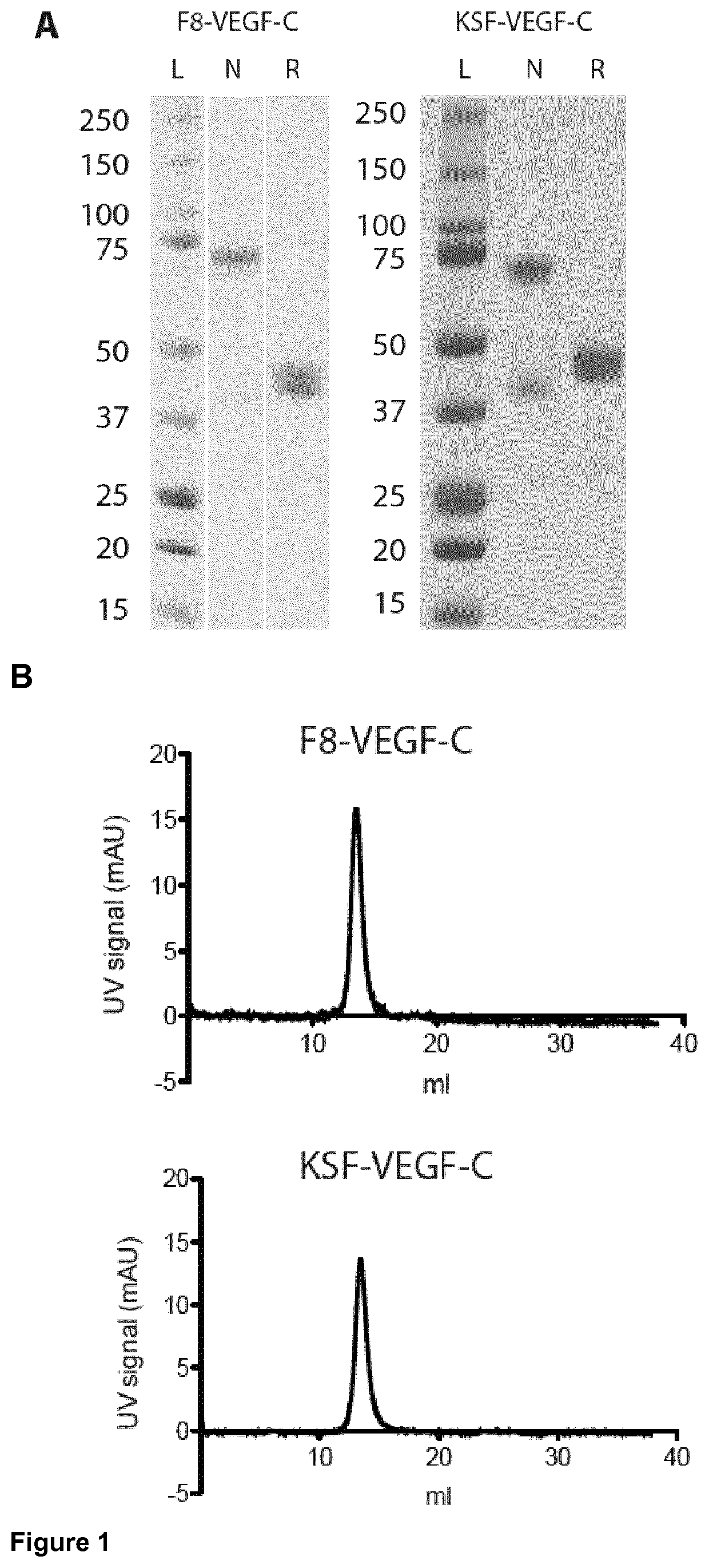
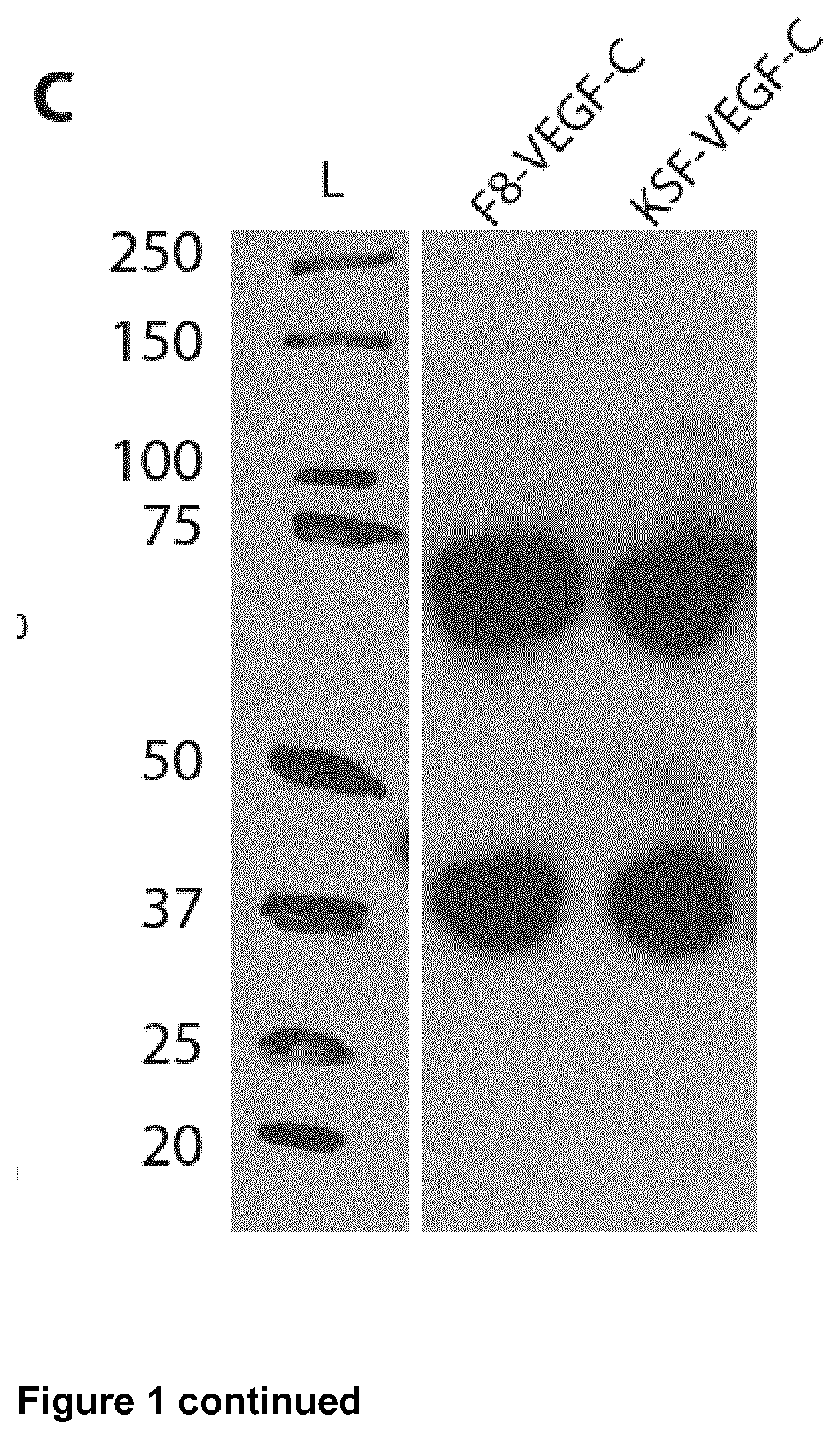
Cloning of Fusion Protein F8-VEGF-C
[0146]The genes encoding F8 and ΔNΔCVEGF-C (Joukov V, Kumar V, Sorsa T, Arighi E, Weich H, Saksela O, and Alitalo K. A recombinant mutant vascular endothelial growth factor-C that has lost vascular endothelial growth factor receptor-2 binding, activation, and vascular permeability activities. J Biol Chem. 1998; 273(12):6599-6602) were amplified and PCR assembled. The product was digested using HindIII / NotI and cloned into an identically digested pcDNA3.1(+) plasmid backbone (Invitrogen).
Cloning of Fusion Protein F8-VEGF-C156Ser
[0147]The genes encoding F8 and ΔNΔCVEGF-C156Ser (Joukov V, Kumar V, Sorsa T, Arighi E, Weich H, Saksela 0, and Alitalo K. A recombinant mutant vascular endothelial growth factor-C that has lost vascular endothelial growth factor receptor-2 binding, activation, and vascular permeability activities. J Biol Chem. 1998; 273(12):6599-6602) wer...
example 2
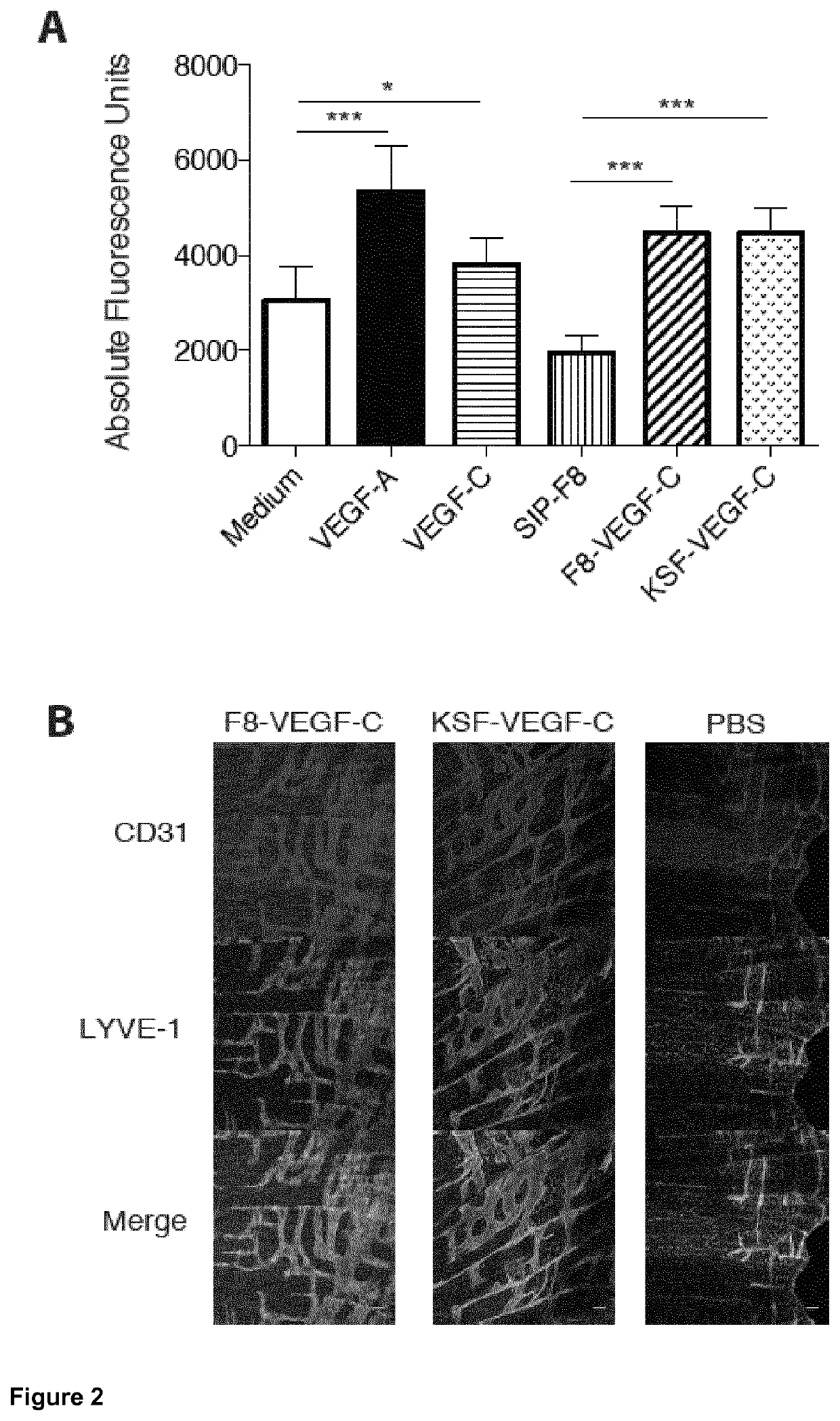
l Activity of F8-VEGF-C and KSF-VEGF-C In Vitro and In Vivo Materials and Methods
[0159]For proliferation assays, 2500 human lymphatic endothelial cells (LECs) were seeded into collagen-coated 96-well plates in EBM medium (Lonza) containing 20% FBS (Gibco), 25 μg / ml cAMP (Sigma) and 10 μg / ml hydrocortisone (Sigma). After an overnight starvation in EBM containing 0.5% FBS, the cells were incubated with VEGF-A (20 ng / ml, Cell Sciences), VEGF-C (200 ng / ml, kind gift of Dr. Kurt Ballmer-Hofer, Villingen, Switzerland), F8-SIP (400 ng / ml), F8-VEGF-C (600 ng / ml) or F8-VEGF-C156Ser (600 ng / ml). After 72 hours, 100 μg / ml 4-methylumbelliferyl heptanoate (MUH, Sigma) were added and the fluorescence intensity, corresponding to the number of viable cells was measured on a SpectraMax reader (Molecular Devices) at 355 nm excitation and 460 nm emission (Detmar et al. “Cytokine regulation of proliferation and ICAM-1 expression of human dermal microvascular endothelial cells in vitro”, J Invest Dermat...
example 3
F8-VEGF-C on Cell Populations
Materials and Methods
[0168]Ears were harvested from K14-VEGF-A transgenic mice on day 15 or 16 after challenge as described in Example 4 below. Following mechanical disruption of the tissue, samples were digested under rotation in DMEM (Gibco) containing 4 mg / ml of collagenase IV (Gibco) at 37° C. for 45 min. Samples were filtered through 40 μm strainers (Falcon), centrifuged and resuspended in FACS buffer containing 2% FBS (Gibco) and 2 mM EDTA (Fluka). Samples were incubated with anti-mouse CD16 / 32 antibody (93, Biolegend) on ice for 20 min and subsequently stained on ice for 30 min using Pacific Blue anti-mouse CD45 (30-F11, Biolegend), APC anti-mouse CD4 (GK1.5, Biolegend), FITC anti-mouse CD8 (53-6.7, Biolegend), PerCP-eFluor 710 anti-mouse gamma delta TCR (GL-3, eBioscience), or Brilliant Violet anti-mouse CD25 (PC61, Biolegend) antibodies and live / dead fixable aqua dye (Life Technologies). Staining for Foxp3 was performed using the anti-mouse / rat ...
PUM
| Property | Measurement | Unit |
|---|---|---|
| pH | aaaaa | aaaaa |
| pH | aaaaa | aaaaa |
| nucleic acid | aaaaa | aaaaa |
Abstract
Description
Claims
Application Information