
An application of exosome tbrii protein as a marker in the preparation of breast cancer detection kit

Active Publication Date: 2021-07-22
ZHEJIANG UNIV
View PDF0 Cites 0 Cited by
  • Summary
  • Abstract
  • Description
  • Claims
  • Application Information

AI Technical Summary

Benefits of technology

The present invention provides a kit for detecting breast cancer by detecting peripheral blood. This kit utilizes the specific binding of an antibody to a protein called TβRII on exosomes, which are small vesicles in the blood. The kit can qualitatively or quantitatively detect the presence of TβRII protein by detecting the label on the antibody. The kit has several advantages, such as minimal-invasive and real-time detecting, and can be used as a liquid biopsy for tumor diagnosis and metastasis. It is convenient, fast, and suitable for a large number of people and can help in breast cancer prevention and detection.

Problems solved by technology

During the later stage of breast cancer, TGF-β pathway is abnormally activated, leading to tumor metastasis.

Method used


Image

  • An application of exosome tbrii protein as a marker in the preparation of breast cancer detection kit
  • An application of exosome tbrii protein as a marker in the preparation of breast cancer detection kit
  • An application of exosome tbrii protein as a marker in the preparation of breast cancer detection kit

Examples


embodiment 1

[0039]1. Exaction and identification of exosome derived from human breast cancer cell line MDA-MB-231. The extraction method is as follows:

[0040]MDA-MB-231 cell culture supernatant was prepared, centrifuged at 4000 rpm / min for 10 min; the supernatant was collected and centrifuged at 100,000 g for 1 hour; the pellet was collected, and the pellet was resuspended in 28 ml of PBS buffer and centrifuged at 100,000 g for 1 hour. For exosomes. The exogenous body weight was suspended in 0.2 ml of PBS buffer and stored in −80° C. for use.

[0041]2. exosomes lysate lysis (composition: 2.5% SDS and 8 M urea, the balance is water) exosomes 30 minutes, determination of exosome concentration by BCA protein concentration; determination of isolated products by immunoblotting Tumor exosomes.

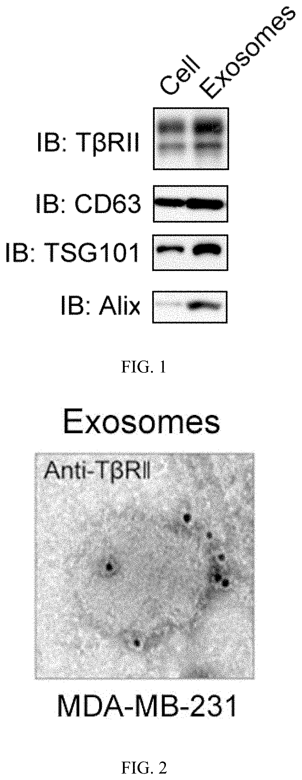
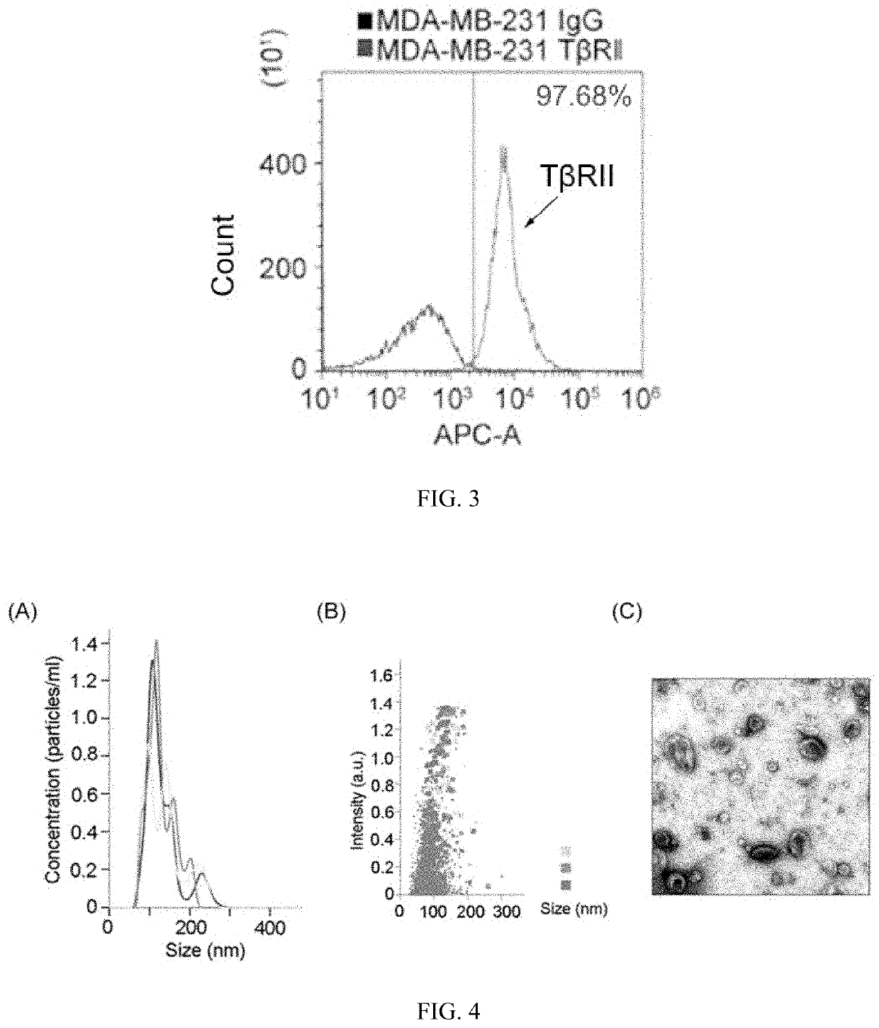
[0042]As shown in FIG. 1, the isolated exosomes of step 1 contained a high protein content of TβRII in addition to the known markers TSG101, ALIX, and CD63.

[0043]3. Detection of exosomal TβRII by flow cytometry:

[00...

embodiment 2

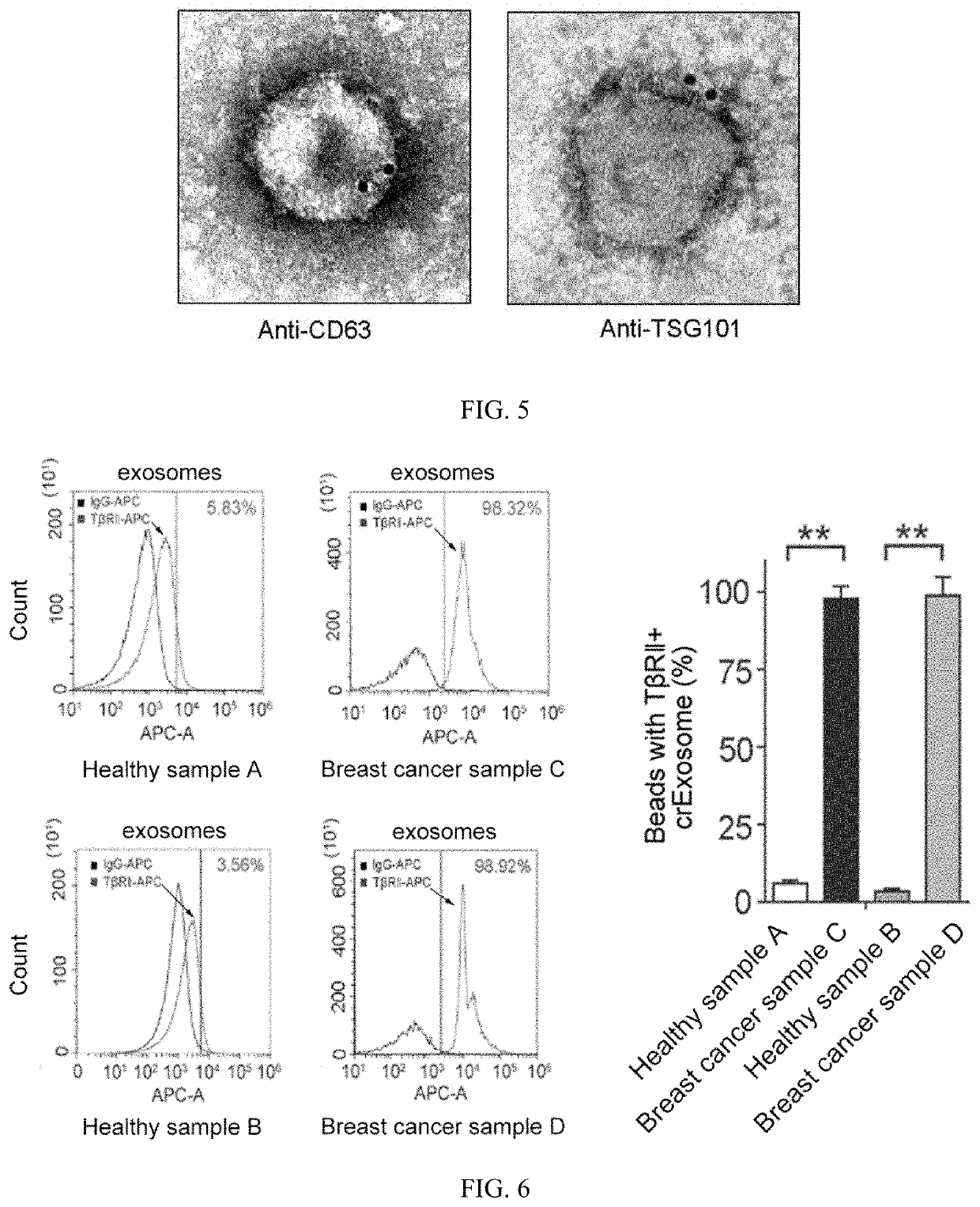
[0048]Using the technique of Example 1, the peripheral blood exosomes of 36 breast cancer patients (20 normal samples were also tested as negative controls) were isolated and obtained and identified, and the expression of TβRII was detected.

[0049]1. Using ultracentrifugation to obtain exosomes in the peripheral blood of the population to be tested:

[0050]1 ml of venous blood from each sample was collected, 0.5 ml of 2.5% sodium citrate anticoagulant was added, mixed well, centrifuged at 4000 rpm / min for 10 min, and the upper layer of plasma was taken.

[0051]PBS was added to dilute the upper layer plasma to 28 ml, poured into an ultracentrifuge tube, and centrifuged at 100,000 g for 1 hour. The supernatant was discarded with a pipette or pipette, 1 ml of liquid was left at the bottom of the tube, PBS was added to 28 ml, the pellet was resuspended, and centrifuged at 100,000 g for 1 hour. The supernatant was discarded with a pipette or pipette, and the ultracentrifuge tube was put upsid...


PUM


Abstract

The present invention discloses an application of the exosome TβRII protein as a marker in preparing a breast cancer diagnostic kit, and belongs to the technology field of breast cancer detection kits. The present invention finds that the content of TβRII positive exosomes in the serum of breast cancer patients is significantly higher than these of healthy people, and after the surgery, the content of TβRII exosomes in the serum of breast cancer patients reduces significantly, which suggests the value of the TβRII positive exosomes during the diagnosis and prognosis of breast cancer. The present invention provides a breast cancer diagnosis kit based on detecting exosome TβRII protein in peripheral blood, which is simple in operation, and only needs to extract a small amount of peripheral blood of the test population, thereby detecting whether the test population has breast cancer and evaluating the tumor of the patient. The degree of malignancy and the state of metastasis.

Description

TECHNOLOGY FIELD[0001]The present invention is related to the field of breast cancer detection kit and particularly related to an application of exosome TβRII protein as a marker in the preparation of breast cancer diagnostic kit.BACKGROUND TECHNOLOGY[0002]Breast cancer is the most common malignancy in women. It ranks second among the most common cancers worldwide. A prominent feature of breast cancer is its susceptibility to metastasis. The most common metastatic organ is bone, and 80% of patients who die because of bone metastases. Tumor metastasis is a very important cause of death in cancer patients, with up to 90% of solid tumor patients dying from tumor metastasis.[0003]At present, there are two commonly used methods for detecting breast cancer metastasis. One is immunohistochemical technique, wherein pathological sections are prepared by collecting pathological tissues, and tumor metastasis is detected by detecting related proteins. The other method is Western blot, which has...

Claims


Application Information

Patent Timeline
IPC IPC(8): G01N33/574G01N33/68
CPCG01N33/57415G01N33/6803G01N33/57488G01N33/574G01N2800/365G01N2333/495
Inventor ZHANG, LONGXIE, FENG
Owner ZHEJIANG UNIV
Learn More