Use of Anti-fam19a5 antibodies

a technology of anti-fam19a5 and antibodies, applied in the field of retinopathy treatment, can solve the problems of not cure the disease, undesirable side effects, significant discomfort, etc., and achieve the effects of reducing and/or inhibiting neovascularization, and reducing cd31 and/or vegf expression

Pending Publication Date: 2021-11-11
NEURACLE SCI CO LTD
View PDF0 Cites 1 Cited by
  • Summary
  • Abstract
  • Description
  • Claims
  • Application Information

AI Technical Summary

Benefits of technology

[0017]In some aspects, the FAM19A5 antagonist reduces and / or inhibits neovascularization within the retina of the subject, as evidenced by decreased CD31 and / or VEGF expression. In certain aspects, the FAM19A5 antagonist is capable of reducing and / or inhibiting neovascularization within the retina of a mouse having laser induced choroidal neovascularization, as evidenced by decreased CD31 and / or VEGF expression. In further aspects, wherein the FAM19A5 antagonist reduces expression of platelet-derived growth factor (PDGF) within a nerve fiber layer and a ganglion cell layer of the retina of the subject compared to a reference (e.g., corresponding value in the subject prior to FAM19A5 antagonist administration or in the subject without the FAM19A5 antagonist administration). In other aspects, the FAM19A5 antagonist improve the retinal potential of the subject compared to a reference (e.g., corresponding value in the subject prior to FAM19A5 antagonist administration or in the subject without the FAM19A5 antagonist administration). In some aspects, the improvement in retinal potential comprises an increase in B-wave. In certain aspects, the FAM19A5 antagonist is capable of reducing and / or inhibiting the accumulation or aggregation of drusen within the retina of the subject.

Problems solved by technology

While some treatment options are available and can help manage the symptoms associated with DR, they do not cure the disease and can also have undesirable side effects.
For example, laser therapy can cause significant discomfort during treatment, permanent retinal scarring, and decreased peripheral, color, and night vision.
AMD is thought to be caused by hardening of the arteries that nourish the retina, which deprives the retinal tissue of oxygen and thus, leading to central vision deterioration.
However, there is no known cure for AMD and many of the treatment options may not be viable for all AMD patients.
For instance, many patients who develop choroidal neovascularization (CNV) (i.e., wet AMD) are not good candidates for laser therapy because the CNV is too large for laser treatment, or the location cannot be determined accurately.
Moreover, even with treatment, many AMD patients experience progressive loss of central vision.

Method used


Image

  • Use of Anti-fam19a5 antibodies
  • Use of Anti-fam19a5 antibodies
  • Use of Anti-fam19a5 antibodies

Examples


example 1

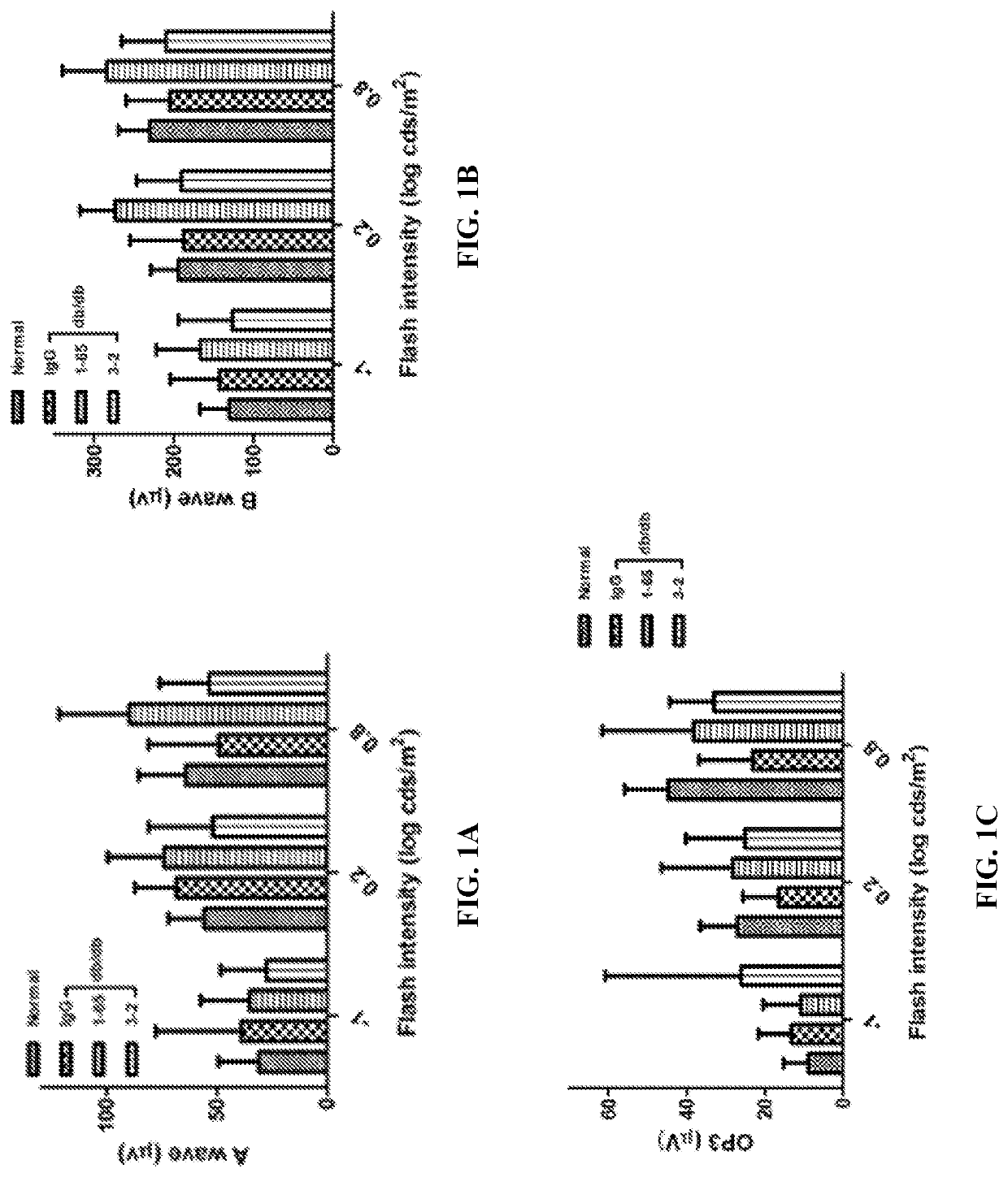
Evaluation of the Retinal Potential Difference After In Vivo Administration of Anti-FAM19A5 Antibody in a Mouse Model of Diabetic Retinopathy

[0348]To begin assessing the effect of anti-FAM19A5 antibody treatment on diabetic retinopathy, type 2 diabetic db / db mice (B6.BKS(D)-Leprdb / j) (25-week old) were used. Briefly, the mice were stabilized for one week, and then, either human IgG control antibody or an anti-FAM19A5 antibody (1-65 or 3-2 clones) were intravitreally administered to the mice (total of 7 doses at 4 μg / eye every 2 weeks) using a micro pump system. Normal (i.e., non-diabetic wild-type) mice were used as controls. Then, the retinal potential was assessed using electroretinography (ERG).

[0349]ERG is a diagnostic test that measures the electrical activity generated by various cells in the retina (i.e., retinal potential), including the photoreceptors (rods and cones), inner retinal cells (bipolar and amacrine cells), and the ganglion cells. Generally, upon a bright stimulu...

example 2

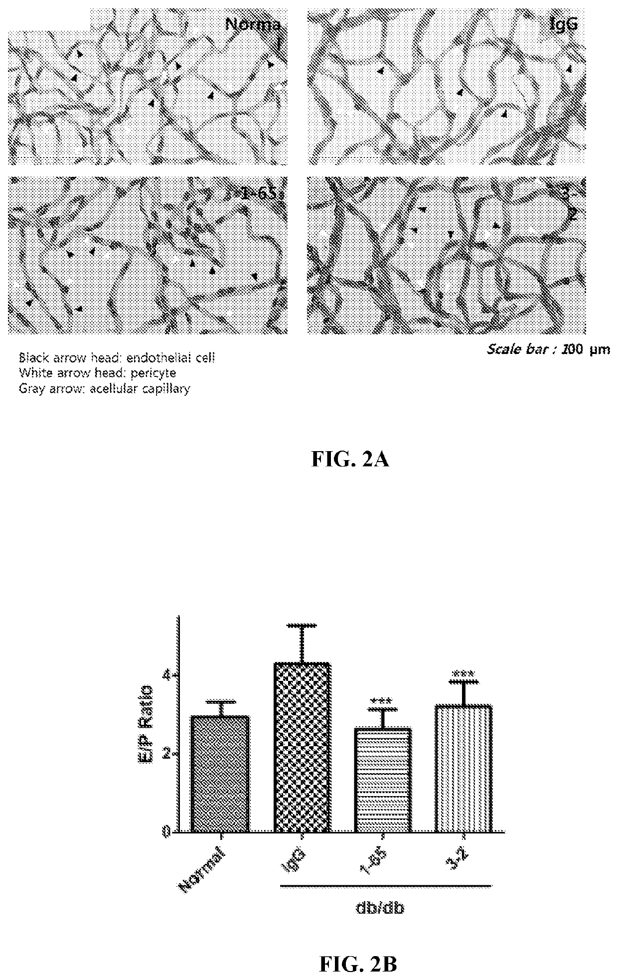
Evaluation of the Morphological Changes After Anti-FAM19A5 Antibody Treatment in a Mouse Model of Diabetic Retinopathy

[0352]To assess whether anti-FAM19A5 antibody treatment resulted in morphological changes of the retina, the type 2 diabetic db / db mice from Example 1 were sacrificed and both of the eyes were harvested from the animals.

[0353]Once harvested, one of the eyes from each of the animals were fixed in 10% neutral formalin for one day. Afterwards, the cornea, lens, and sclera were removed, and the remaining eye tissues were washed five time with distilled water for one hour while shaking. After the last wash, the distilled water was removed, and the eye tissues were incubated in 3% trypsin dissolved in 0.1 M Tris buffer for 2 hours at 37° C. Next, the retinal vessels were separated from the rest of the retinal tissue and attached to slides for staining with periodic acid-Schiff (PAS) to assess retinal microvessels for characteristic markers of early diabetic retinopathy (e....

example 3

Evaluation of the Effect of Anti-FAM19A5 Antibody on Choroidal Neovascularization in Age-Related Macular Degeneration Mouse Model

[0359]To assess the effect of anti-FAM19A5 antibody treatment on age-related macular degeneration, a mouse induced choroidal neovascularization (CNV) model was used. More specifically, the CNV model is an animal model of human wet age-related macular degeneration. Briefly, after stabilizing C57BL / 6 mice for one week, the pupils of the eyes of the animals were enlarged (with a topical treatment of 2.5% phenylephrine hydrochloride). Then, the animals were treated intraperitoneally with 2% Fluorescein (Sigma) to stain the blood vessels. Approximately 3-5 minutes after Fluorescein injection, the mice were anesthetized, and using a Phoenix green laser photocoagulator, wet AMD-like lesions were induced at four areas of the optic nerve with a laser (240 mW, 70 ms duration). FIG. 6. Afterwards, either human IgG control antibody or anti-FAM19A5 antibody were intrav...


PUM


Abstract

The present disclosure relates to the pharmaceutical use of antagonists (e.g., an antibody or antigen-binding portion thereof) that specifically bind to FAM19A5 to treat a retinopathy (e.g., diabetic retinopathy) and/or maculopathy (e.g., age-related macular degeneration) in a subject in need thereof.

Description

CROSS-REFERENCE TO RELATED APPLICATIONS[0001]This PCT application claims the priority benefit of U.S. Provisional Application No. 62 / 746,194, filed Oct. 16, 2018, which is incorporated herein by reference in its entirety.REFERENCE TO SEQUENCE LISTING SUBMITTED ELECTRONICALLY[0002]The content of the electronically submitted sequence listing in ASCII text file (Name: 3763.012PC01_SeqListing_ST25.txt; Size: 261,609 bytes; and Date of Creation: Oct. 13, 2019) filed with the application is incorporated herein by reference in its entirety.FIELD OF THE DISCLOSURE[0003]The present disclosure provides methods for the treatment of retinopathies (e.g., diabetic retinopathy) and / or macular degeneration (e.g., age-related macular degeneration) in a subject (e.g., a human) using antagonist against family with sequence similarity 19, member A5 (FAM19A5) (e.g., anti-FAM19A5 antibody) or a composition comprising such antagonists.BACKGROUND OF THE DISCLOSURE[0004]Retinopathies, such as diabetic retin...

Claims


Application Information

Patent Timeline
Patent Type & Authority Applications(United States)
IPC IPC(8): C07K16/24A61P27/02
CPCC07K16/24A61K2039/505A61P27/02C07K2317/76A61K9/0048C07K2317/33C07K2317/56C07K2317/92
Inventor KIM, BONGCHEOLKIM, DONG SIKKWON, SOON-GU
Owner NEURACLE SCI CO LTD
Why Eureka
Try Eureka