
Inhibition of retinal cell degeneration or neovascularization by cerium oxide nanoparticles

Inactive Publication Date: 2011-05-12
THE BOARD OF RGT UNIV OF OKLAHOMA +1
View PDF7 Cites 6 Cited by
  • Summary
  • Abstract
  • Description
  • Claims
  • Application Information

AI Technical Summary

Benefits of technology

[0026]The presently claimed and disclosed inventive concept(s) provides methods for reducing, reversing or inhibiting retinal cell degeneration, or neovascularization in tissues of a mammalian subject having a pathological condition involving neovascularization, by administration in vivo of nanoceria particles (cerium oxide nanoparticles) to the subject. The method of the presently claimed and disclosed inventive concept(s) is useful, for example, for reducing, treating, reversing or inhibiting degeneration of retinal cells such as rod and cone photoreceptor cells or neovascularization in ocu

Problems solved by technology

However, in response to an appropriate stimulus, the endothelial cells become activated and begin to proliferate and migrate into unvascularized tissue forming new blood vessels.
Such neovascularization in ocular tissues can induce corneal scarring, retinal detachment, fluid accumulation in the subretinal space, and macular edema, each of which can adversely affect vision and lead to blindness.
Excessive blood flow into neovascularized plaques can result in rupture and hemorrhage of the blood-filled plaques, releasing blood clots that can result in coronary thrombosis.
Such non-specific killing by chemotherapeutic agents results in side effects that are, at best, unpleasant, and can often result in unacceptable patient morbidity, or mortality.
In fact, the undesirable side effects associated with cancer therapies often limit the treatment a patient can receive.
For other pathological conditions associated with abnormal angiogenesis such as diabetic retinopathy, there are no effective treatments.
However, even if retinal transplantation is performed, the new retina would be subject to the same conditions that resulted in the original retinopathy.
Fluid can also leak into the center of the macula at any stage of diabetic retinopathy and cause macular edema and blurred vision.
Degeneration of the macula causes a decrease in acute vision and can lead to eventual loss of acute vision.
The wet form of macular degeneration is related to abnormal growth of blood vessels in the retina that can leak blood and can cause damage to photoreceptor cells.
These new blood vessels break and leak fluid, causing damage to the central retina.
Vision loss can result from the accumulation of deposits in the retina called druzen, and from the death of photoreceptor cells in the retina.
This process can lead to thinning and drying of the retina.
Age-related macular degeneration is a leading cause of presently incurable blindness, particularly in persons over 55 years of age.
Druzen may affect contrast sensitivity, and may reduce the eye's ability to see sufficiently to allow a person to read in dim light or to see sufficient detail to permit a person to drive an automobile safely at night.
Angiogenic blood vessels in the subretinal choroid can leak vision obscuring fluids, leading to blindness.
There are no cures for most forms of the other blinding diseases, the severity of which increases with age and dramatically decreases the quality of life for these patients.
About 2 million Americans over the age of 40 have significant vision loss due to AMD while an additional 8 million have a high risk of vision loss.
Further, because Native American Indians develop diabetes at a much higher rate than the general population, DR is becoming a progressively increasing problem in states such as Oklahoma which have large numbers of Native Americans.
The annual economic cost to the USA for adult vision loss is major at about $50 billion per year.
In the wet form, the presence of sub-macular neovascular vessels leads to retinal edema, ruptured blood vessels, the death of the cones in the macula and eventual blindness.
There are no treatments which have proven successful for dry AMD.
As indicated herein, there are many diseases which result in the programmed cell death of retinal photoreceptor cells thus leading to blindness.

Method used


Image

  • Inhibition of retinal cell degeneration or neovascularization by cerium oxide nanoparticles
  • Inhibition of retinal cell degeneration or neovascularization by cerium oxide nanoparticles
  • Inhibition of retinal cell degeneration or neovascularization by cerium oxide nanoparticles

Examples


example 1

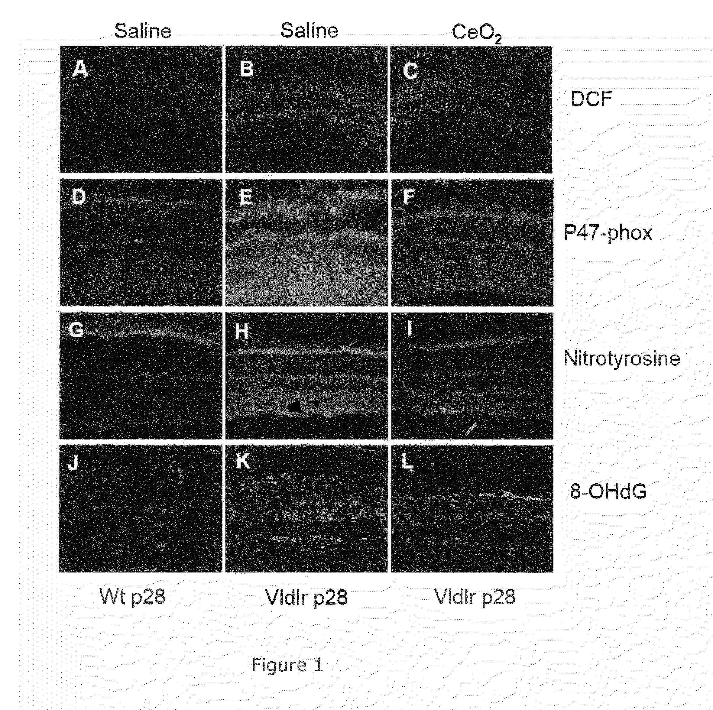
[0090]Effects on ROS and ROS-Induced Damage.

[0091]We first performed experiments to determine if there actually was an increase in ROS in the retina of the VLDLr KO mouse and if the nanoceria could reduce the ROS and the damage caused by them. In a cell, the three major sources of intracellular ROS are from 1) mitochondrial oxidative respiration, 2) NADPH-oxidase and 3) Nitrous oxide synthetase (NOS3).

[0092]The 2′,7′-dichloro-dihydro-fluorescein-diacetate (DCF) assay for detection of cellular oxidation by hydrogen peroxide, peroxynitrite and hydroxyl radicals was used with cryostat sections of eyes from normal and VLDLr− / −mice which had been injected intravitreally with either saline or nanoceria on post-natal day 7 (P7) and assayed on post-natal day 28 (P28). Fluorescence microscopy of cryostat sections from 28 day mice (FIG. 1A) detects some DCF signal in the wild type retina but very significant amounts in the VLDLr− / −retina (FIG. 1B). However, injection of the nanoceria on P7 gr...

example 2

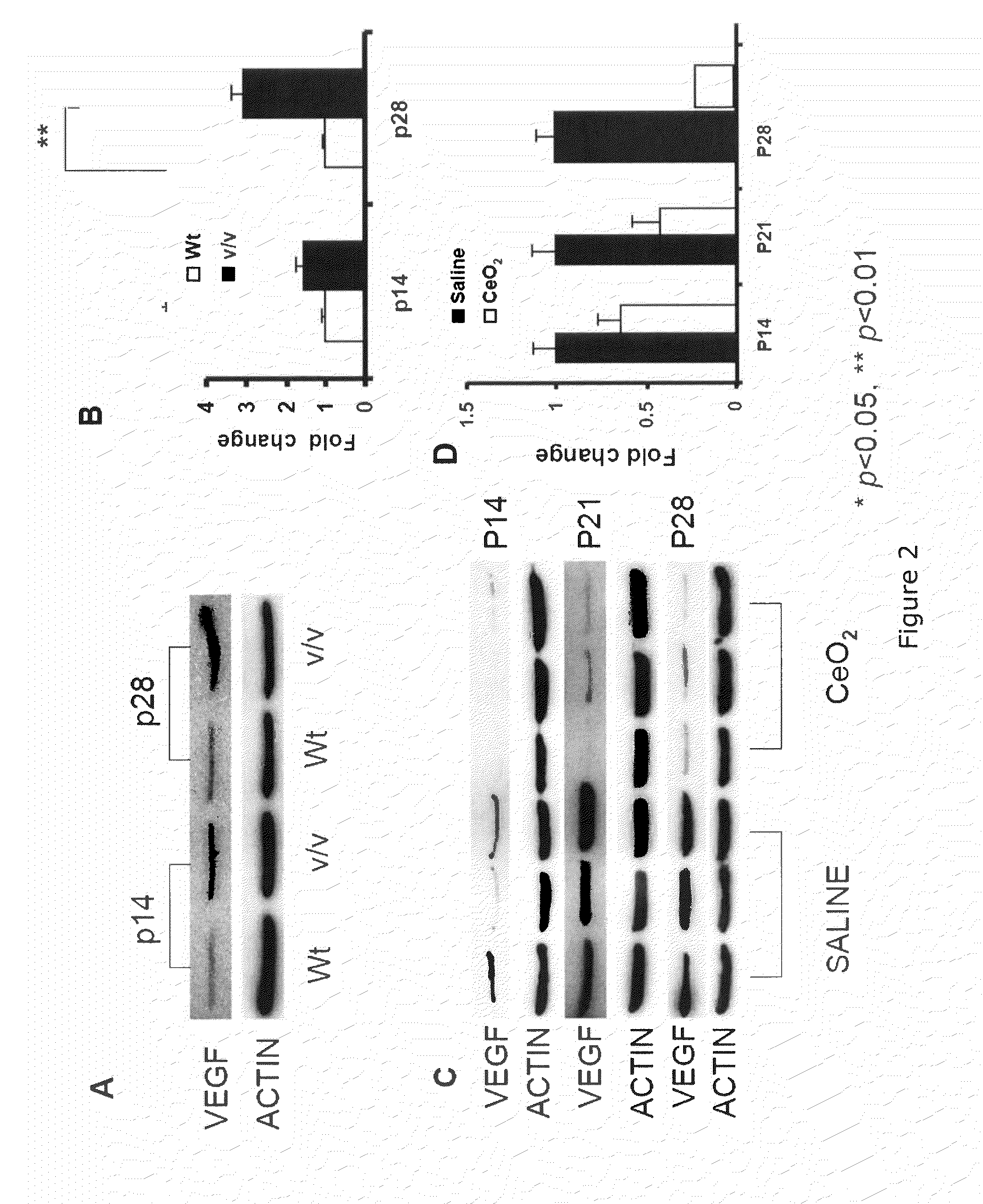
[0094]VEGF—Western Blot Data.

[0095]Mice were injected intravitreally with either 1 μl of PBS or 1 ul of PBS plus nanoceria at post-natal day 7P7 and the animals killed on P14, P21 or P28. Retinas were homogenized, subjected to SDS-PAGE, and blotted to nitrocellulose. The bands were detected with primary and secondary antibodies and visualized with an HRP-DAB assay (FIG. 2). Wild type (+ / +) retinas had barely detectable levels of VEGF with or without nanoceria. The VLDLr KO retinas had about an eightfold increase in VEGF compared to the + / + retinas. However, the nanoceria (“nanoparticle”) injected VLDLr KO mice had about 60% less VEGF at day 21. These data indicate that VEGF increased in the VLDLr KO retinas due to ROS and that it decreased because of the nanoceria mediated decrease in ROS.

[0096]The amount of VEGF, as determined by Western blots, is higher in the VLDLr− / −retina than in the wild type retina even at P14 and progressively increases (FIG. 2A). Densitometry (FIG. 2B) indi...

example 3

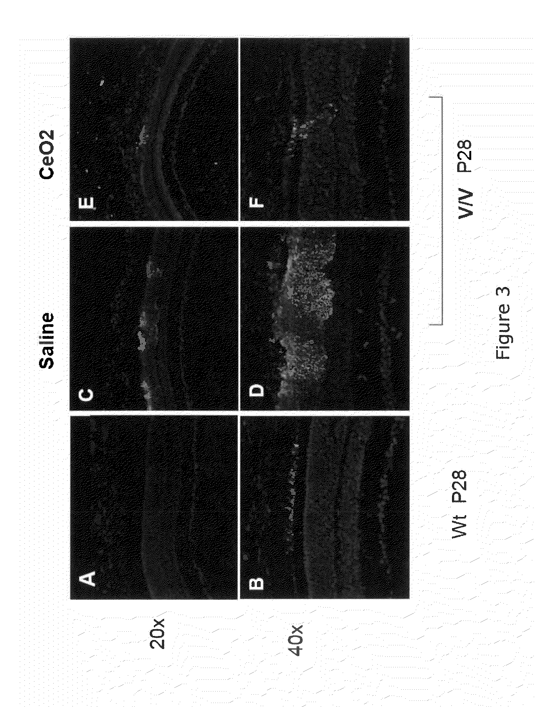
[0097]VEGF—Localization by Immunofluorescence.

[0098]To determine where VEGF was localized in the wild type and the VLDLr KO retinas and whether the nanoceria had any effect on the localization, Alexa green-conjugated secondary antibodies were used in combination with anti-VEGF primary immunoglobulins. The wild type mouse retinas (C57BL / 6J) (FIG. 3A,B) had very little VEGF and it was localized to the outer segments of the retina. However, the pattern of labeling with anti-VEGF in the VLDLr KO retina (FIG. 3C,D) was heavy but discontinuous; predominantly in the outer and inner segments of photoreceptors; and especially in their perinuclear regions in the ONL adjacent to vascular lesions. The intensity of labeling progressively diminished as the distance from the lesion increased. The age-matched VLDLr mice, which had received an intravitreal injection (1 ul of 1 mM) of nanoceria on P7, had fewer vascular lesions and exhibited greatly reduced staining surrounding the remaining lesions ...


PUM


Abstract

The presently claimed and disclosed inventive concept(s) provides methods for reducing, reversing or inhibiting retinal cell degeneration, or neovascularization in tissues of a mammalian subject having a pathological condition involving neovascularization, by administration in vivo of nanoceria particles (cerium oxide nanoparticles) to the subject. The method of the presently claimed and disclosed inventive concept(s) is useful, for example, for reducing, treating, reversing or inhibiting degeneration of retinal cells such as photoreceptor cells or neovascularization in ocular tissue such as the retina, macula or cornea; or other tissues such as, but not limited to, skin, synovial tissue, intestinal tissue, or bone. In addition, the method of the presently claimed and disclosed inventive concept(s) is useful for reducing or inhibiting neovascularization in a neoplasm (tumors), which can be benign or malignant and, where malignant, can be a metastatic neoplasm. As such, the presently claimed and disclosed inventive concept(s) is directed to using compositions containing nanoceria particles to reduce, treat, reverse or inhibit angiogenesis in a mammalian subject.

Description

CROSS-REFERENCE TO RELATED APPLICATIONS[0001]The present application is a continuation-in-part of U.S. Ser. No. 12 / 429,650, filed Apr. 24, 2009, which claims the benefit under 35 U.S.C. §119(e) of U.S. Provisional Application Ser. No. 61 / 125,602, filed Apr. 25, 2008.[0002]The present application also claims the benefit under 35 U.S.C. §119(e) of U.S. Provisional Application Ser. No. 61 / 174,678, filed May 1, 2009.[0003]The present application is also a continuation-in-part of U.S. Ser. No. 11 / 142,665, filed Apr. 27, 2006, which claims the benefit under 35 U.S.C. §119(e) of U.S. Provisional Application Ser. No. 60 / 716,630, filed Sep. 13, 2005, and U.S. Provisional Application Ser. No. 60 / 676,043, filed Apr. 29, 2005.[0004]The entireties of each of the applications listed herein are hereby expressly incorporated herein by reference.STATEMENT REGARDING FEDERALLY SPONSORED RESEARCH OR DEVELOPMENT[0005]Not applicable.BACKGROUND[0006]Angiogenesis (also referred to herein as neovascularizat...

Claims


Application Information

Patent Timeline
IPC IPC(8): A61F2/14A61K33/24A61P27/02A61P27/06A61P29/00A61P19/02A61P1/00A61P17/00A61K9/14B82Y5/00
CPCA61K33/00A61P1/00A61P17/00A61P19/02A61P27/02A61P27/06A61P29/00
Inventor MCGINNIS, JAMES F.ZHOU, XIAOHONGWONG, LILY L.SEAL, SUDIPTA
Owner THE BOARD OF RGT UNIV OF OKLAHOMA
Learn More