

Anti-pd-l1 antibody and use thereof
a technology of anti-pdl1 and antibody, which is applied in the field of anti-pdl1 antibody, can solve the problems of increasing treatment costs, exhaustion or tolerance to heterogeneous antigens, and inconvenience for patients
- Summary
- Abstract
- Description
- Claims
- Application Information
AI Technical Summary
Benefits of technology
Problems solved by technology
Method used
Image

Examples
example 1
of Anti-PD-L1 Antibody by Use of Phage Antibody Library
[0300]Human PD-L1-Fc protein (Origincell Therapeutics Co., Ltd.) as antigen was used to sort the phage natural human antibody library (Origincell Therapeutics Co., Ltd.). The ELISA tube was coated with a CBS buffer containing 20 μg / ml (the first and the second rounds) or 10 μg / ml (the third and the fourth rounds) of PD-L1 protein at 4° C. overnight. The tube was then washed with a PBS buffer. 10% skimmed milk powder was added to block the ELISA tube, and then 1 ml of blocked phage was added and incubated at room temperature (20±5° C.) for 1 hour. After washing thoroughly with PBST, 800 μl of a Gly-HCl buffer solution at pH 2.2 was added for elution, and then 400 μl of a Tris-HCl buffer solution at pH 8.0 was added immediately for neutralization. Then, the mixture was added to 20 ml of E. Coli SS320 in the logarithmic growth phase with an OD value of about 0.8, mixed well and stood at 37° C. for 1 hour. 500 μl of microbial soluti...
example 2
n and Purification of Anti-PD-L1 Fully Human Intact Antibody
[0303]A primer was designed for PCR amplification of the VH of the phage antibody 1B10, and the PCR product was cloned by recombination into the pCMV-IgG1NDL vector which was double digested with AgeI and SalI. A design primer was designed for PCR amplification of the VL of the phage antibody 1B10, and the PCR product was cloned by recombination into a pCMV-λ, vector which was double digested with AgeI and BsiWI. After sequencing exactly, the heavy chain and the light chain expression vectors were co-transfected into 293F cells for transient expression, and purified by ProteinA column to obtain an intact IgG1,λ antibody of the phage antibody 1B10. This anti-PD-L1 fully human antibody was named YN-002.
[0304]The sequencing results show that the nucleotide sequence of the VH encoding the antibody YN-002 is shown in SEQ ID NO: 1, and the amino acid sequence of VH of the antibody YN-002 is shown in SEQ ID NO: 2. The nucleotide s...
example 3
Version of Anti-Pd-L1 Fully Human Antibody YN-002
[0305]By comparing the antibody YN-002 heavy chain immunoglobulin sequence with the known human germline immunoglobulin heavy chain sequence, it was confirmed that the antibody YN-002 heavy chain used the VH segment from human germline IGHV1-69*09, the D segment from human germline IGHD5-18*01, and JH segment from human germline IGHJ4*02.
[0306]By comparing the light chain immunoglobulin sequence of the antibody YN-002 with the known human germline immunoglobulin light chain sequence, it was confirmed that the light chain of the antibody YN-002 used the VL segment from human germline IGLV2-14*01, and JL segment from human germline IGLJ2*01.
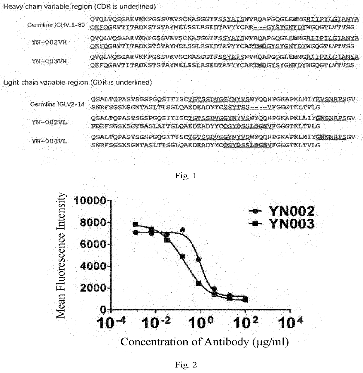
[0307]The sequence of the CDR region of the antibody YN-002 was analyzed by the Kabat system (see FIG. 1).
[0308]The sequencing results show that the amino acid sequences of LCDR1-3 of the antibody YN-002 are shown in SEQ ID NO: 54, SEQ ID NO: 60 and SEQ ID NO: 63, respectively; and the amino acid seq...
PUM
| Property | Measurement | Unit |
|---|---|---|
| Angle | aaaaa | aaaaa |
| Angle | aaaaa | aaaaa |
| Angle | aaaaa | aaaaa |
Abstract
Description
Claims
Application Information