Medical lead conductor fracture visualization method and apparatus

a technology of lead conductor and fracture visualization, which is applied in the field of medical lead conductor fracture visualization method and apparatus, can solve the problems of increasing the number of separate polarity and insulated coiled wire conductors, increasing the number of separate polarity and insulating coiled wire conductors that are difficult to accommodate, and conductor breaking

Inactive Publication Date: 2001-09-25
MEDTRONIC INC
View PDF20 Cites 160 Cited by
  • Summary
  • Abstract
  • Description
  • Claims
  • Application Information

AI Technical Summary

Benefits of technology

and the advantages and features thereof have been presented here simply to point out some of the ways that the invention overcomes difficulties presented in the prior art and to distinguish the invention from the prior art and is not intended to operate in any ma

Problems solved by technology

The increased number of separate polarity and insulated coiled wire conductors is difficult to accommodate in the conventional coaxial coiled wire conductor winding arrangement having a desired, small, lead body outer diameter.
The transvenous pathway can include a number of twists and turns, and the lead body can be forced against bony structures of the body that apply stress to It, causing the lead body to be crushed and/or causing the lead conductor to break.
Over the years of implantation, the lead conductors and insulation are subjected to such cumulative mechanical stresses that can result in degradation of the insulation or fractures of the lead conductors with untoward effects on device performance and patient well being.
However, despite these improvements, not all causes of lead fracture can be overcome by lead body design, and lead conductor fractures still occur from time to time.
Lead conductivity may decre

Method used


Image

  • Medical lead conductor fracture visualization method and apparatus
  • Medical lead conductor fracture visualization method and apparatus
  • Medical lead conductor fracture visualization method and apparatus

Examples


Embodiment Construction

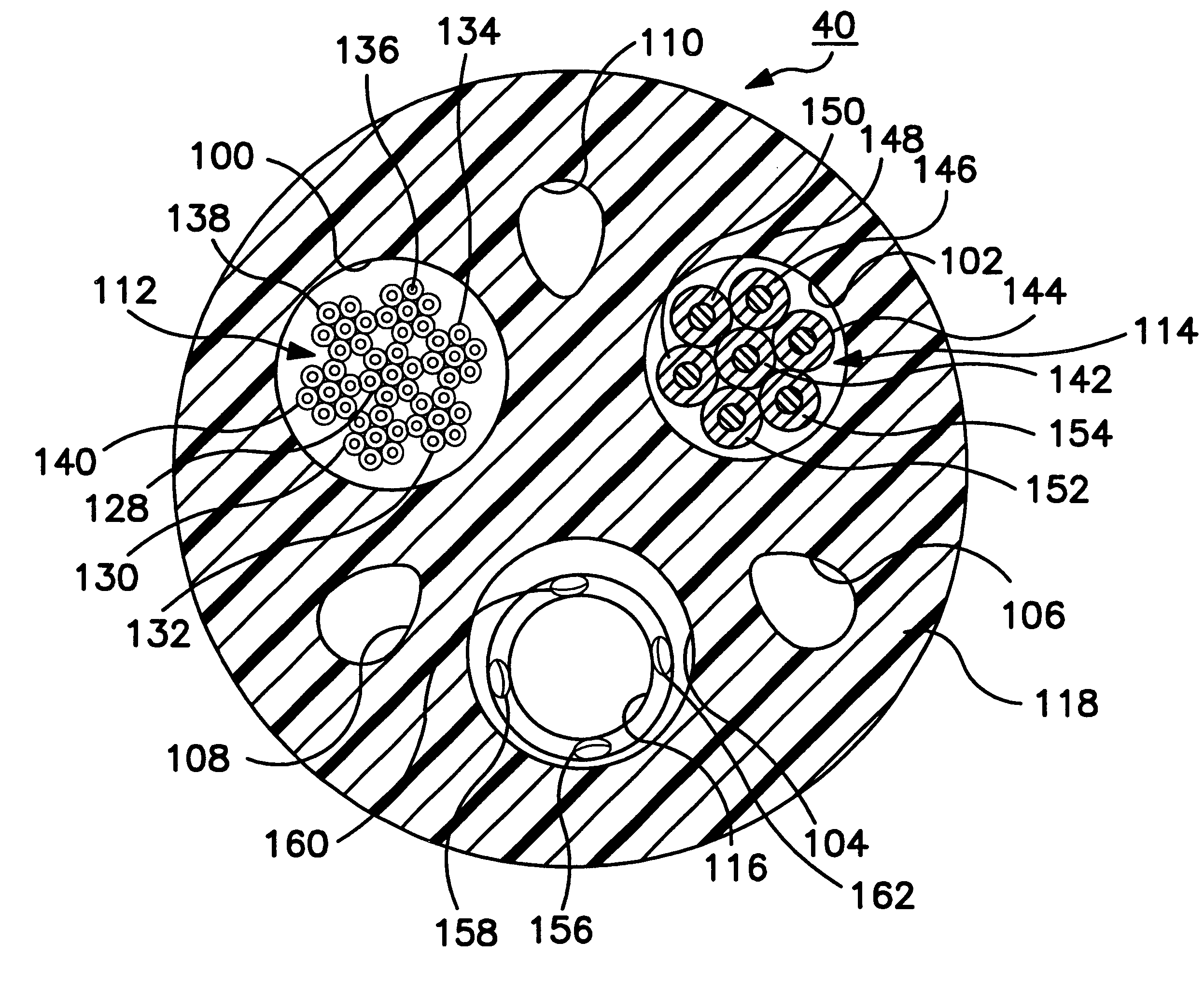
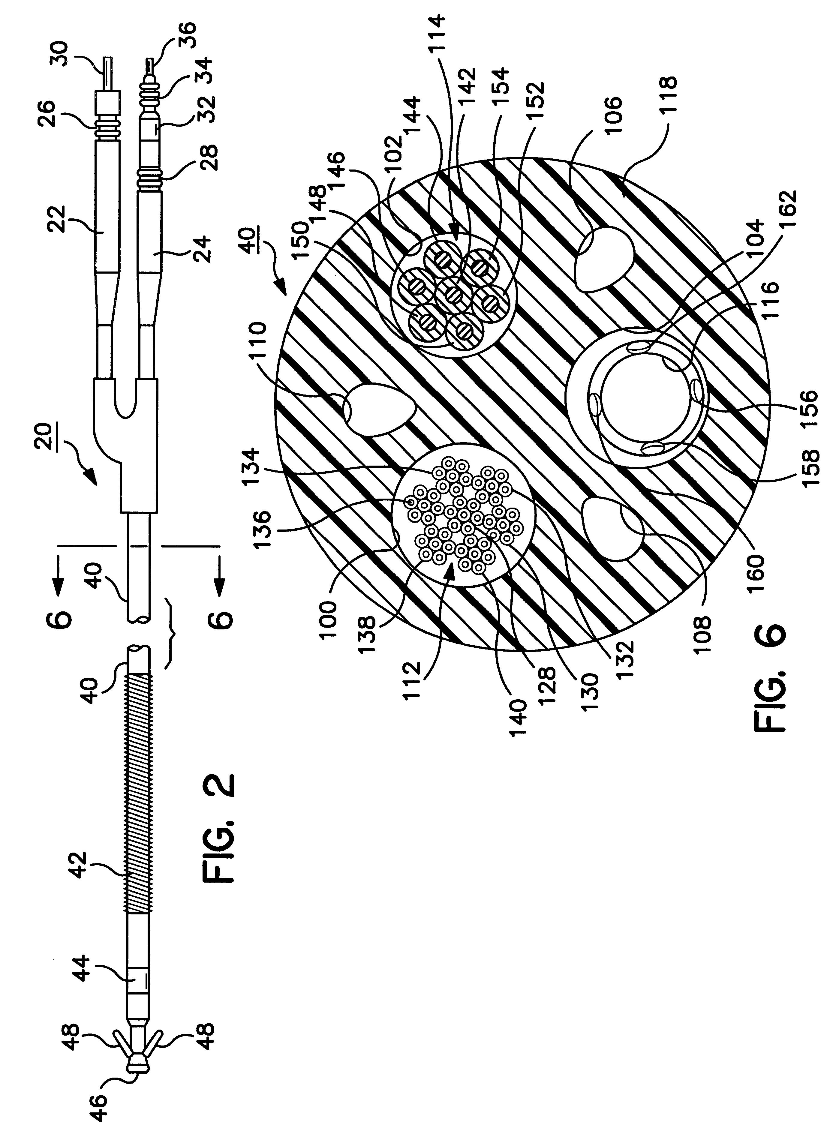
The present invention finds particular utility in the fabrication and implantation of cardiac leads, e.g., atrial and / or ventricular pacing leads and / or cardioversion / defibrillation leads having elongated lead bodies and lead conductors that are subject to fracture. Preferred embodiments of such lead conductor fabrications and implantations of such endocardial cardiac leads that are implanted transvenously will be described in detail. But, it is to be understood that the present invention is not limited to the same. The present invention can be implemented in the fabrication and use of other epicardial cardiac leads that are implanted subcutaneously and in electrical leads intended to be disposed within the patient's body, including nerve, brain, organ, and muscle stimulation leads.

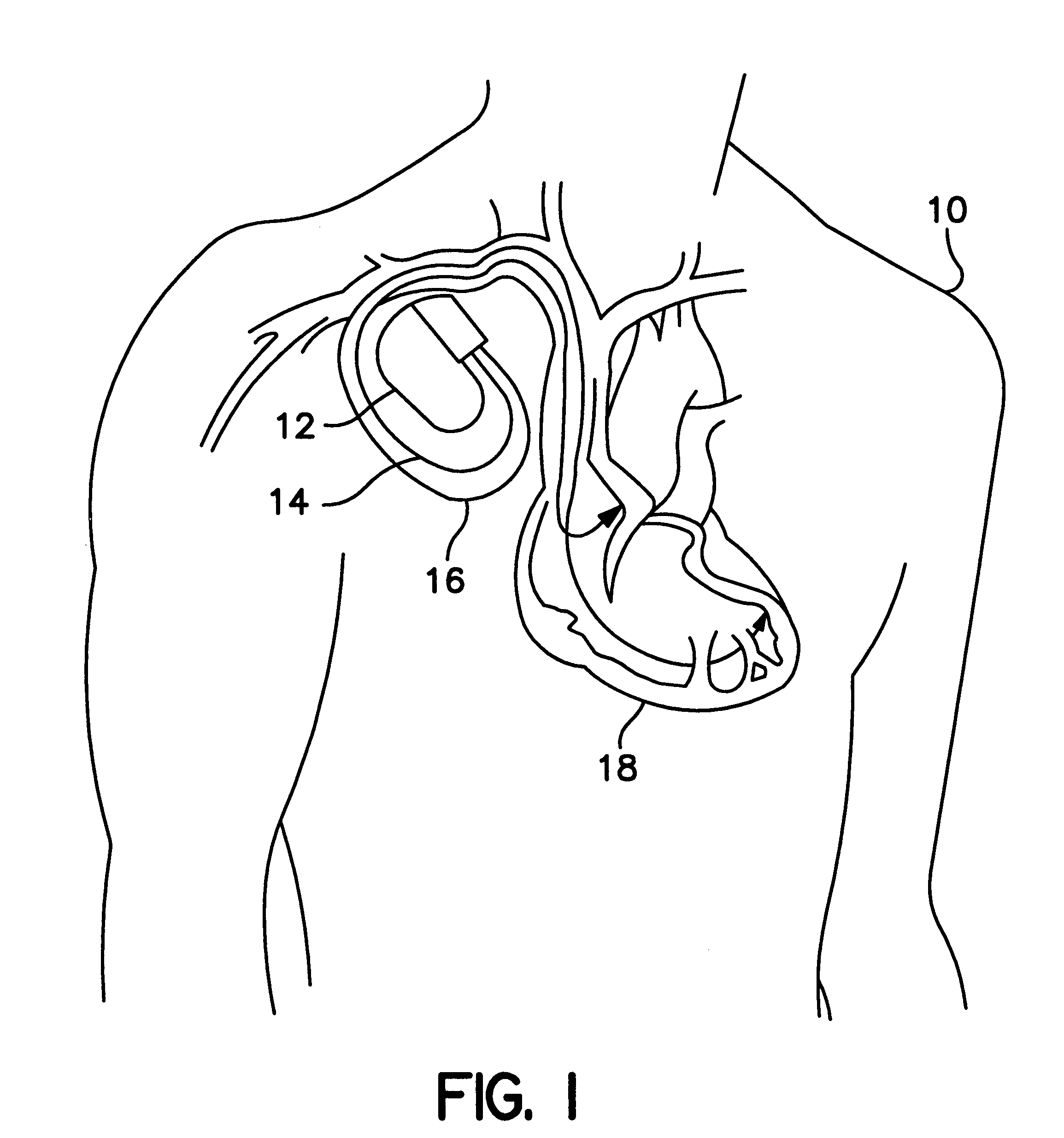
FIG. 1 depicts a typical arrangement of a pacing or implantable cardioverter / defibrillator (ICD) system implanted in a patient 10, the system comprising a subcutaneously disposed implantable pulse generat...


PUM


Abstract

Methods for sensing or electrical stimulation of body organs or tissues are disclosed wherein a lead conductor wire or filament of a stranded lead conductor generates a radioactive emission when it is fractured sufficiently or is completely broken. The conductor wire or filament is formed of an inner core and an outer sheath surrounding the inner core, wherein the inner core is irradiated or is formed of a radioactive isotope in an alloy that provides an enhanced radiopaque aura when the sheath is fractured and the inner core is exposed. When the conductor wire or filament is intact, the radioactive inner core is fully encased within the outer sheath, and the outer sheath blocks or reduces radioactive emission along its length to a constant, relatively low level. In use, the emission is detected externally to the body, and the detection signifies that a fracture or break has occurred. Such leads preferably comprise cardiac leads for delivering electrical stimulation to the heart, e.g., pacing pulses and cardioversion/defibrillation shocks, and/or sensing the cardiac electrogram, having multiple lead conductors encased in a lead body subject to fracture under stress. The lead conductors can comprise mono-filar or multi-filar, parallel wound, coiled wires that arranged in a co-axial manner or in a side-by-side arrangement within the lead body. Or the straight or coiled lead conductors can be formed of a strand comprising a plurality of outer filaments wound helically about a central core filament or of a cable comprising a plurality of such peripheral strands wound helically about a central core strand. At least the outer filaments of a stranded conductor and peripheral strands of a conductor cable are formed with the radioactive core.

Description

The present invention relates to medical electrical leads for sensing or electrical stimulation of body organs or tissues and their method of fabrication and use, such leads having multiple electrical conductors encased in a lead body subject to fracture under stress, and particularly to implantable cardiac leads for delivering electrical stimulation to the heart, e.g., pacing pulses and cardioversion / defibrillation shocks, and / or sensing the cardiac electrogram or other physiologic data.Implantable medical electrical stimulation and / or sensing leads are well known in the fields of cardiac stimulation and monitoring, including cardiac pacing and cardioversion / defibrillation, and in other fields of electrical stimulation or monitoring of electrical signals or other physiologic parameters of the body. A pacemaker or cardioverter / defibrillator implantable pulse generator (IPG) or a cardiac monitor is typically coupled to the heart through one or more of such endocardial leads. The prox...

Claims


Application Information

Patent Timeline
IPC IPC(8): A61N1/05
CPCA61N1/056
Inventor SCHAENZER, DAVID G.
Owner MEDTRONIC INC
Why Eureka
Try Eureka