Method of isolating a peptide which immunologically mimics microbial carbohydrates including group B streptococcal carbohydrates and the use thereof in a vaccine

Inactive Publication Date: 2002-09-03
MONTANA STATE UNIVERSITY
View PDF5 Cites 5 Cited by
  • Summary
  • Abstract
  • Description
  • Claims
  • Application Information

AI Technical Summary

Benefits of technology

(2) contacting a phage-display library, having phage, with one or more of said protective antibodies identified in step

Problems solved by technology

However, the data set forth below contradicts thi

Method used


Image

  • Method of isolating a peptide which immunologically mimics microbial carbohydrates including group B streptococcal carbohydrates and the use thereof in a vaccine
  • Method of isolating a peptide which immunologically mimics microbial carbohydrates including group B streptococcal carbohydrates and the use thereof in a vaccine
  • Method of isolating a peptide which immunologically mimics microbial carbohydrates including group B streptococcal carbohydrates and the use thereof in a vaccine

Examples


example 1

Selection of Phage

A phage-display library expressing a random nine AA sequence was selected for binding to one of two different anti-GBS monoclonal antibodies: S9, a protective monoclonal antibody that binds to the type III capsular polysaccharide, and S7, specific for the group B carbohydrate. (Pincus, S. H. et al., J. Immunol. 140:2779). The latter was used primarily as a specificity control. Within each selection, two separate desorption protocols were used to identify two populations of phage: 0.1 M glycine pH 2.2 or 0.5M NH.sub.4 OH pH 11. Phage with binding specificity for the selecting antibody were identified by immunoblots of plaques. Forty clones were selected, ten from each elution condition and selecting antibody, and amplified to a titer of 10.sup.13 -10.sup.14 pfu / ml.

The DNA encoding the displayed peptide from the two different pools of S9-selected phage was sequenced. Within each pool, the sequence of each clone was identical, but two very different sequences were see...

example 2

Specificity of Antibody Binding to Displayed Peptides

To show the specificity of phage binding, ELISA plates were coated with antibodies. The immobilized antibodies were incubated with representative phage clones from each selection (10.sup.10 pfu per well) and binding measured. The results are shown in Table II, below. The parental phage (M13KBst) bound to no antibody. Phage selected with antibody S7 or S9 bound only to the selecting antibody. Phage that were first absorbed on antibody S10, prior to the selection on S7 or S9, bound to all IgM antibodies, indicating that within the library there is a population of phage that bind to all IgMs.

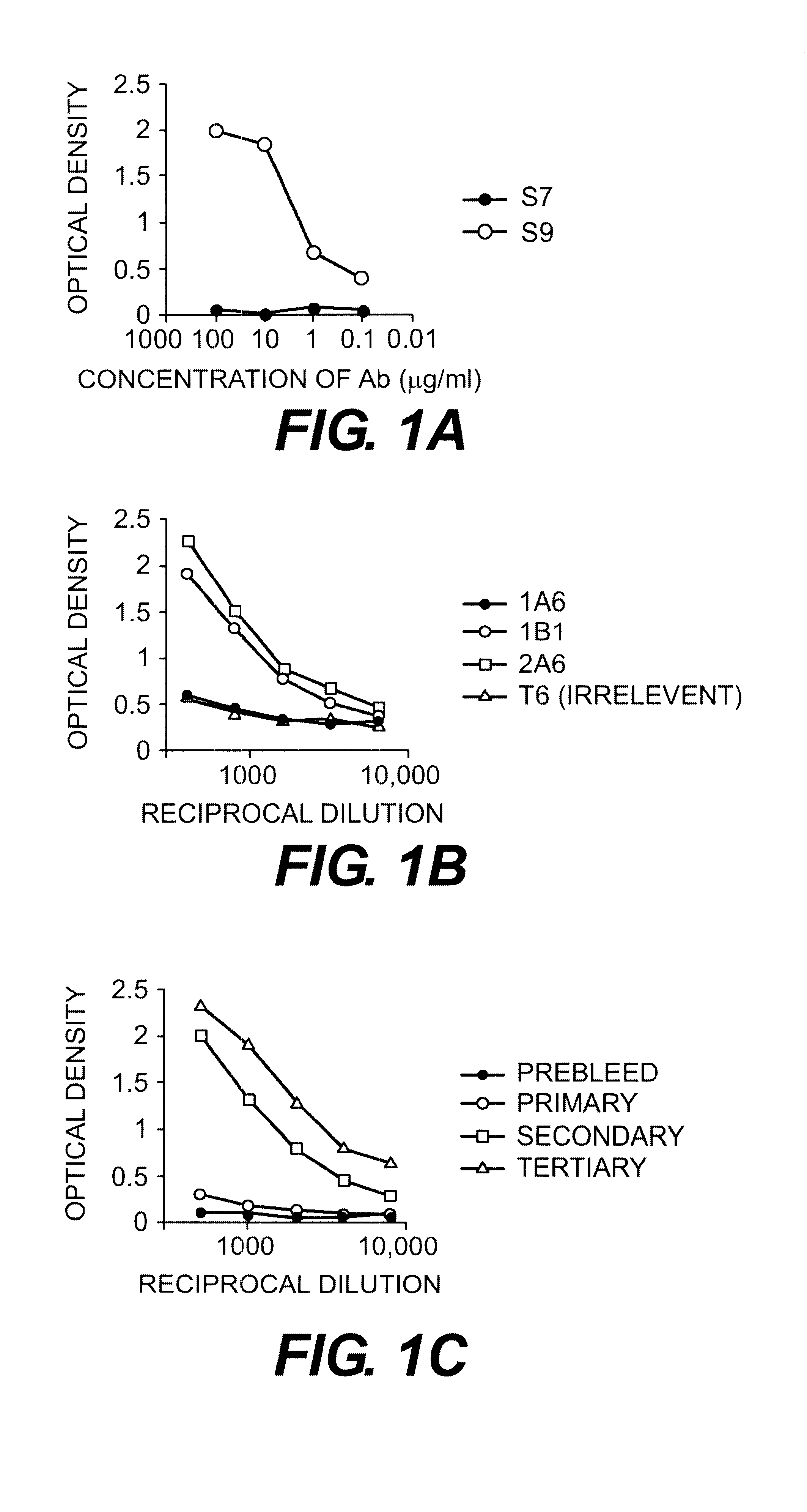
Binding of antibodies to the synthetic peptide FDTGAFDPDWPAC (SEQ ID NO:2) was also demonstrated by ELISA. FIG. 1 shows the binding of the antibodies to peptide. Panel A shows binding of the monoclonal antibodies S7 and S9. Binding to the peptide was seen only with antibody S9. Panel B shows that two of three other IgG anti-GBS type III monoclona...

example 3

Phage and Peptides Block the Binding of Antibody to GBS

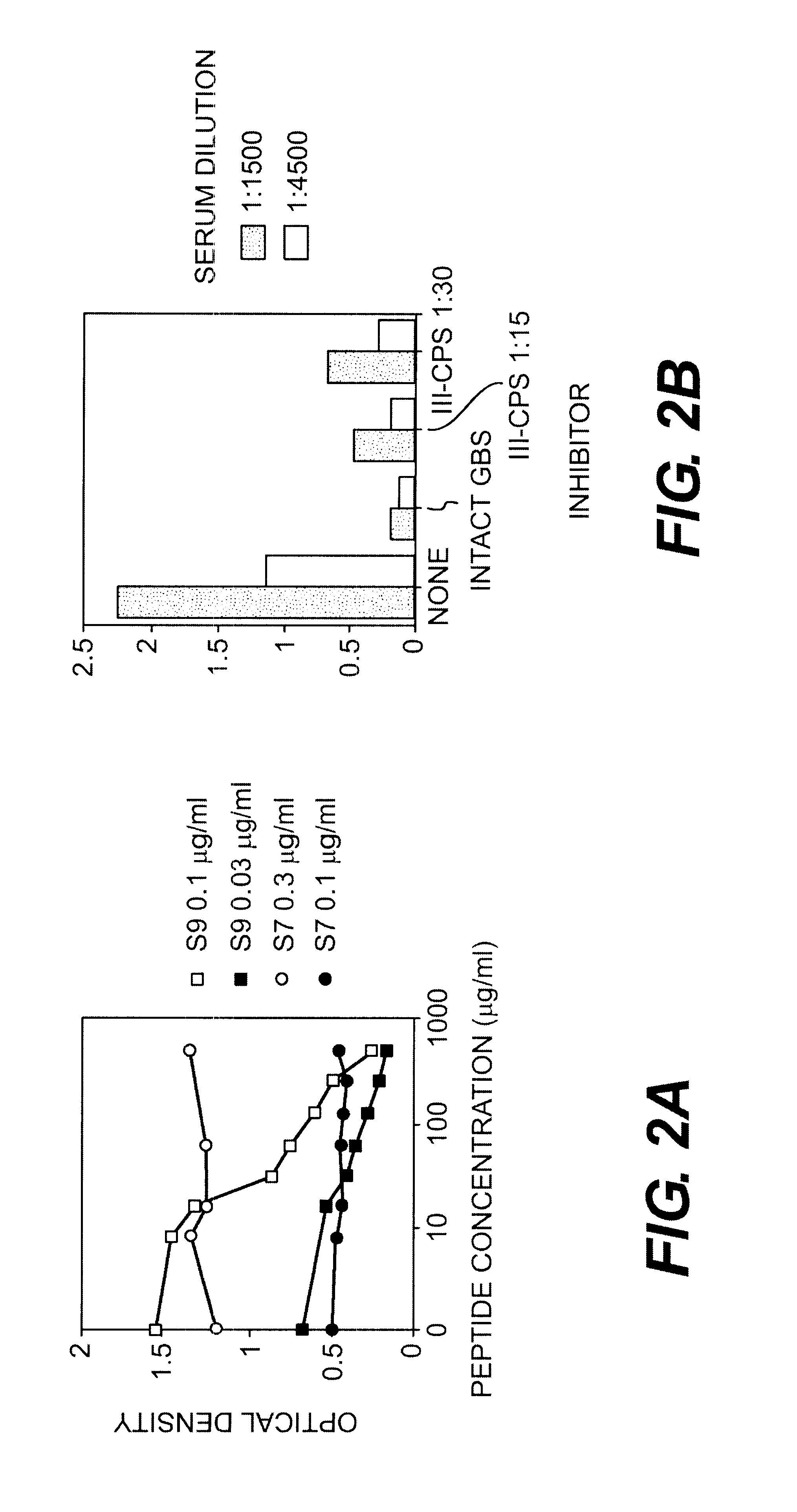
The demonstration of specific recognition of the peptide sequence by the selecting antibody is a good indication that the phage bind to the variable regions. However to demonstrate that the displayed sequence actually resembles the carbohydrate epitope of GBS, blocking of antibody binding to GBS antigens must be shown. To perform these experiments, ELISA was used where antibody and inhibitor (phage or peptide) were premixed, incubated for one hour, and then plated onto the microtiter plates with GBS. Inhibition of the antibody's binding to GBS indicated that the phage or peptide was successfully competing with the GBS for antibody binding.

In Table III, below, intact phage were used to inhibit the binding of antibodies S7 and S9 to GBS. The concentrations of S7 used were slightly higher than those of S9 because there are fewer antigenic determinants recognized by S7 on the surface of GBS. (Pincus, S. H. et al., J. Immunol. 140:27...


PUM

PropertyMeasurementUnit
Timeaaaaaaaaaa
Timeaaaaaaaaaa
Angleaaaaaaaaaa

Abstract

This invention relates to new vaccines against microorganisms based on antigenically mimetic peptides. The invention also relates to methods of discovering such mimetic peptides by first screening peptide-display phage libraries with antibodies against the microbial carbohydrates(s) of interest to locate antigenically mimetic peptides. Vaccines against Group B Streptococcus, or Streptococcus Agalactiae, are preferably produced using this method.

Description

II. FIELD OF THE INVENTIONThis invention relates to new vaccines against microorganisms based on antigenically mimetic peptides. The invention also relates to methods of discovering such mimetic peptides by screening peptide-display phage libraries with antibodies against the microbial carbohydrates(s) of interest to locate antigenically mimetic peptides. Vaccines against Group B Streptococcus, or Streptococcus Agalactiae, can be produced using this method. Vaccines against other microbial pathogens may also be produced using this method.III. BACKGROUND OF THE INVENTIONVaccines protect against disease by harnessing the body's innate ability to protect itself against foreign invading agents. During vaccination, the patient is injected with antigens, or DNA encoding antigens, which generate protective antibodies but which typically cannot cause severe disease themselves. For example, vaccination for bacterial diseases such as typhoid fever consists of injecting a patient with the bact...

Claims


Application Information

Patent Timeline
IPC IPC(8): C07K7/00C07K1/04C07K7/06C07K1/00C07K7/08C07K16/12C12N15/10A61K39/00C40B40/02
CPCC07K1/047C07K7/06C07K7/08C07K16/1275C12N15/1037C40B40/02A61K39/00
Inventor PINCUS, SETH H.
Owner MONTANA STATE UNIVERSITY
Why Eureka
Try Eureka