
Ultrasound system for disease detection and patient treatment

a technology of ultrasound and patient treatment, applied in the field of cancer detection and treatment, can solve the problems of bleeding complications, not being able to test prostate cancer in absolute, and neither of these physicians is necessarily right or wrong

Inactive Publication Date: 2003-07-08
RGT UNIV OF CALIFORNIA
View PDF4 Cites 46 Cited by
  • Summary
  • Abstract
  • Description
  • Claims
  • Application Information

AI Technical Summary

Benefits of technology

In the case of diagnostics, the present invention uses ultrasound to extract a marker, such as a cancer tumor marker, into the coupling medium and provides advantages over existing methods: i) it is non-invasive with tumors in accessible locations, such the prostate gland, colon, or cervix; ii) it measures a marker associated with interstitial fluid associated with the tumor, e.g., PSA concentration in the interstitial fluid of the prostate, as opposed to systemic levels, which provides in increased diagnostic sensitivity; and iii) it is convenient to use.
In the case of treatment, the present invention uses ultrasound to deliver one or more drugs from the coupling medium directly to the site of the disease and provides advantages over existing methods: i) it is non-invasive with disease sites in accessible locations, as above; ii) it delivers drugs locally to the site, thus limiting side effects; and iii) it is painless.

Problems solved by technology

Although a highly experienced physician can make a relatively good diagnosis using digital rectal examinations ("DRE"), it is a "subjective" technique.
The problem with using DREs to make decisions about what to do with particular patients is that two different experienced physicians may think that they feel quite different things when they carry out a DRE on the same patient.
Neither of these physicians is necessarily right or wrong in what they think.
Although PSA tests have revolutionized prostate cancer diagnosis, it is not an absolute test of prostate cancer.
This procedure is painful, uncomfortable, and may lead to bleeding complications.
Hormone therapy is limited by significant side effects.
All forms of surgery for removal of the prostate are associated with complications.
These include lack of bladder control (urinary incontinence), urethra stricture (difficulty in urination), impotence, and the normal risks associated with anesthesia and major surgical procedures.
However, even the best surgeons have patients with unexpected complications.
Any form of prostatectomy is a major operation and has risks attached.
The known side effects of cryotherapy can include impotence (in about 80% of patients), scarring of the urethra and urinary dysfunction (which are relatively unusual), and irritation of the bladder, the urethra, the rectal wall, and the genitalia.
Thus, existing methods of diagnostics and treatment for prostate cancer suffer from several limitations.
Specifically, the existing methods of diagnostics are invasive and inconvenient.
In addition, this method does not acquire a sample directly from the prostate.
The existing methods of treatment also suffer from several limitations.
Specifically, surgery and hormone therapy result in severe side effects.
In addition, chemotherapy is limited by severe side effects of cytotoxic agents.
Occasionally, however, late stage aggressive tumors stop producing PSA.
PSMA is a membrane antigen that is easily detected on the cell surface but is harder to detect in plasma.
However, chemotherapy has not been at the forefront of prostate cancer treatment since many (indeed most) of these cytotoxic pharmaceuticals also have potentially severe side effects.
Hence, if given systemically, they are likely to cause undesirable side effects.

Method used


Image

  • Ultrasound system for disease detection and patient treatment
  • Ultrasound system for disease detection and patient treatment
  • Ultrasound system for disease detection and patient treatment

Examples


Embodiment Construction

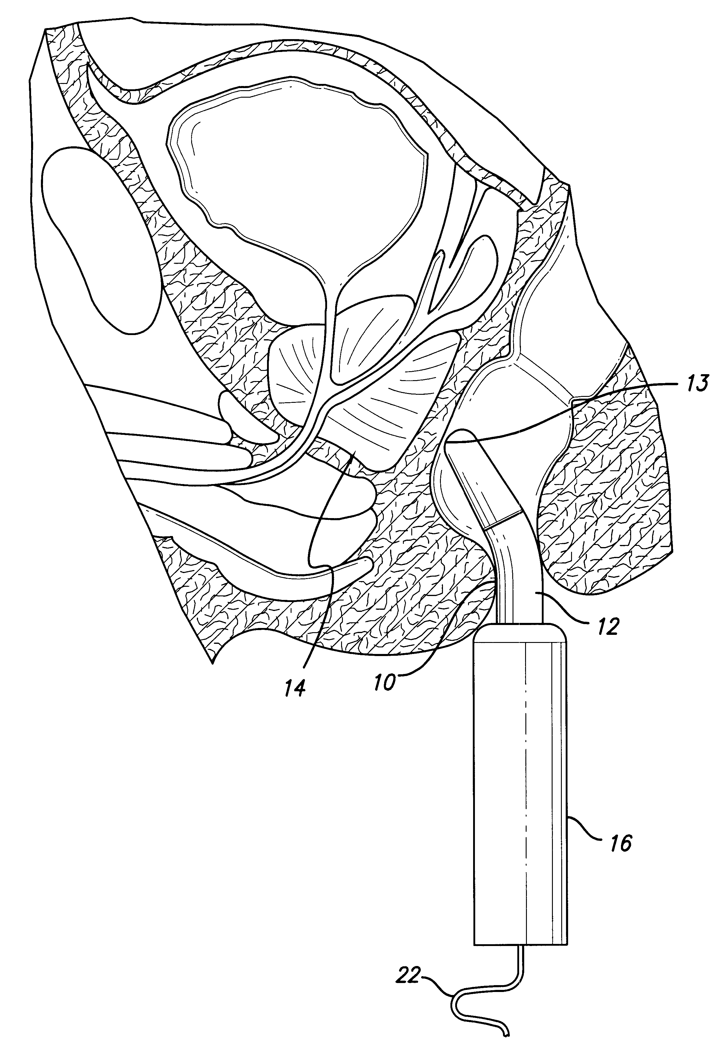
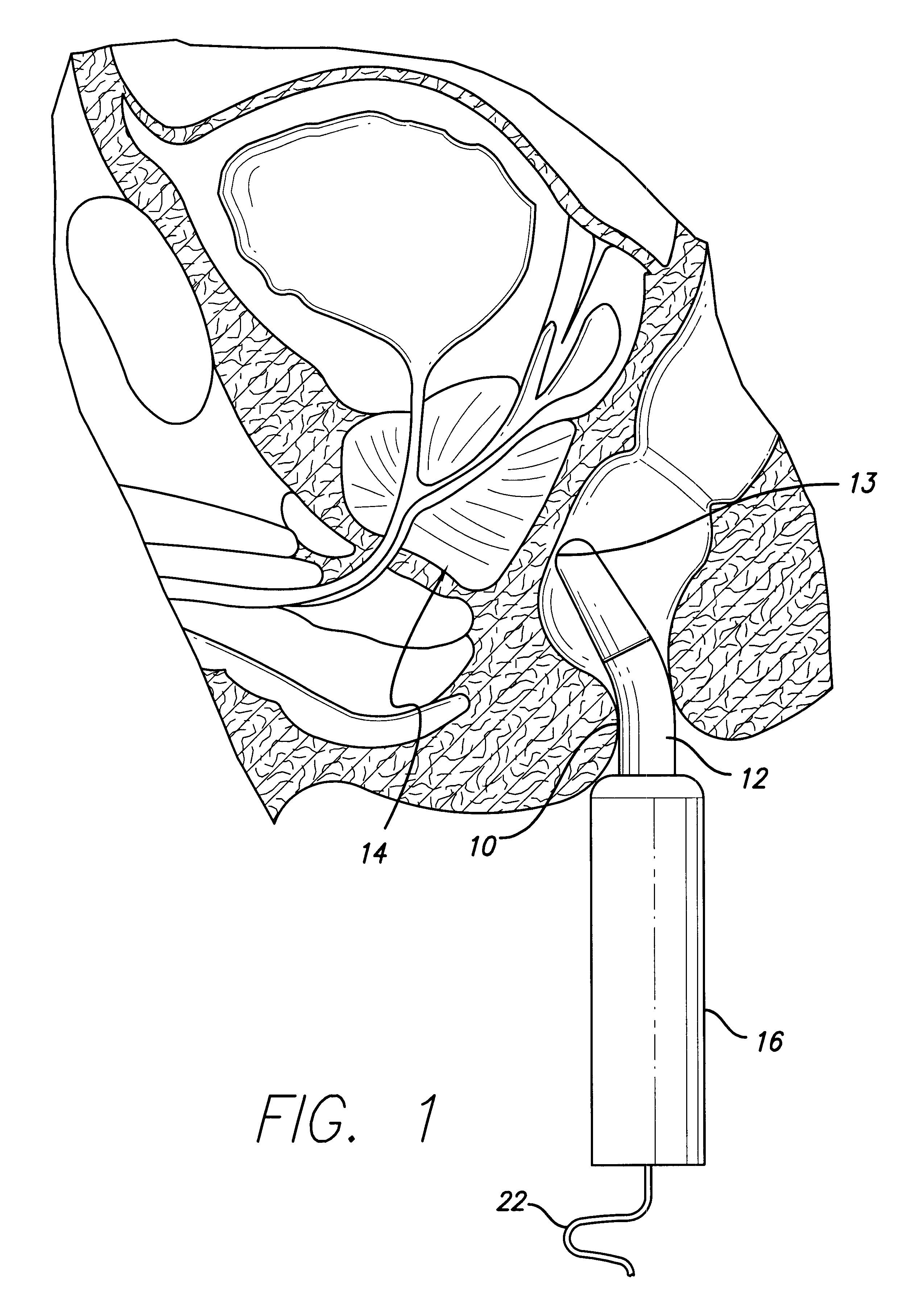
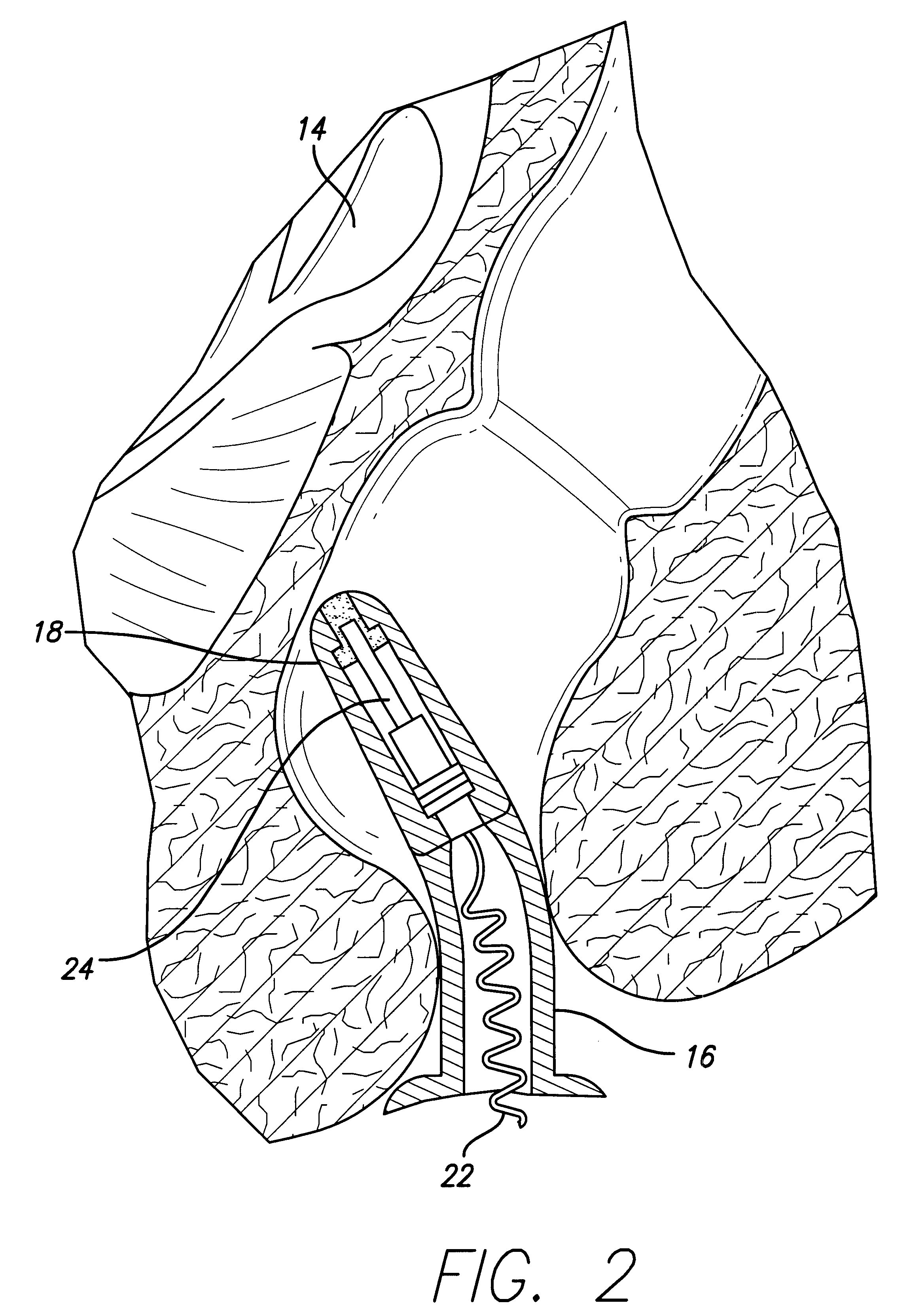
In one configuration, the physician loads a cartridge 10 into the handle 16 of the ultrasound probe and the probe is then inserted through the rectum such that the cartridge 10 is placed against the prostate, as shown in FIGS. 1 and 2. Ultrasound is activated for a predetermined time and the probe is then removed. The cartridge 10 is then removed from the probe and is placed into a device where the concentration of the aforementioned cancer markers in the cartridge is measured. A diagnosis of cancer is then made based on the test results. The same device may also be used for drug delivery. In that case, the physician will load a different cartridge 10 that contains one or more of the above drugs. The probe is placed against the prostate and is activated for a predetermined time. The device may be used by the physician only once (during the routine check-up) or multiple times (to deliver drugs or follow-up the treatment). Thus, this method will provide a non-invasive device for diagn...


PUM


Abstract

A device and method of detection and treatment of a solid cancer tumor, for example prostate cancer. In a diagnostic method, ultrasound is used to extract a cancer tumor marker from interstitial fluid into a coupling medium. In a method for treating a solid tumor, ultrasound is used to deliver one or more drugs from the coupling medium directly to the site of the tumor.

Description

The field of the invention is a system for detection and treatment of cancer.BACKGROUND AND STATE OF THE ARTCurrently, existing methods of diagnosing prostate cancer involve a series of steps and procedures; see Petrylak, infra; Potter,et al., infra; Naitoh, et al., infra; Hegarty et al., infra; Selley, et al., infra. The initial detection of signs that the patient may have prostate cancer is most commonly the result of some regular form of check-up carried out by a primary care physician, which includes a digital rectal examination. In this examination the physician inserts his finger into the rectum in order to be able to feel the size, shape, and texture of the prostate and other nearby organs. Although a highly experienced physician can make a relatively good diagnosis using digital rectal examinations ("DRE"), it is a "subjective" technique. The problem with using DREs to make decisions about what to do with particular patients is that two different experienced physicians may t...

Claims


Application Information

Patent Timeline
IPC IPC(8): A61N7/00A61B17/00A61M37/00
CPCA61B8/00A61N7/00A61B2017/00274A61B2018/00547A61M37/0092
Inventor MITRAGOTRI, SAMIR
Owner RGT UNIV OF CALIFORNIA
Learn More