Method for obtaining a picture of the internal structure of an object using x-ray radiation and device for the implementation thereof

a technology of internal structure and x-ray radiation, applied in the field of intravision means, can solve the problems of poor signal to noise ratio, inability to quantitatively compare individual local fragments by density, and difficulty in obtaining a picture of the internal structure of an obj

Inactive Publication Date: 2004-06-22
KUMAKHOV MURADIN ABUBEKIROVICH
View PDF19 Cites 145 Cited by
  • Summary
  • Abstract
  • Description
  • Claims
  • Application Information

AI Technical Summary

Problems solved by technology

In the above mentioned known methods and devices, the image of a real three-dimensional structure is acquired as the said two-dimensional shadow projection, which interpretation requires from the specialist who carries out object analysis, in particular, technical or medical diagnostics, respective experience and qualification and, in a number of cases, is problematic.
The reasons for this are low contrast, poor signal to noise ratio, inevitable overlapping of the images of structural elements, impossibility of quantitative comparison between individual local fragments by density.
In this case, the dose of radiation absorbed by the substance is high

Method used


Image

  • Method for obtaining a picture of the internal structure of an object using x-ray radiation and device for the implementation thereof
  • Method for obtaining a picture of the internal structure of an object using x-ray radiation and device for the implementation thereof
  • Method for obtaining a picture of the internal structure of an object using x-ray radiation and device for the implementation thereof

Examples


Embodiment Construction

The suggested method is embodied with the help of the suggested device as following.

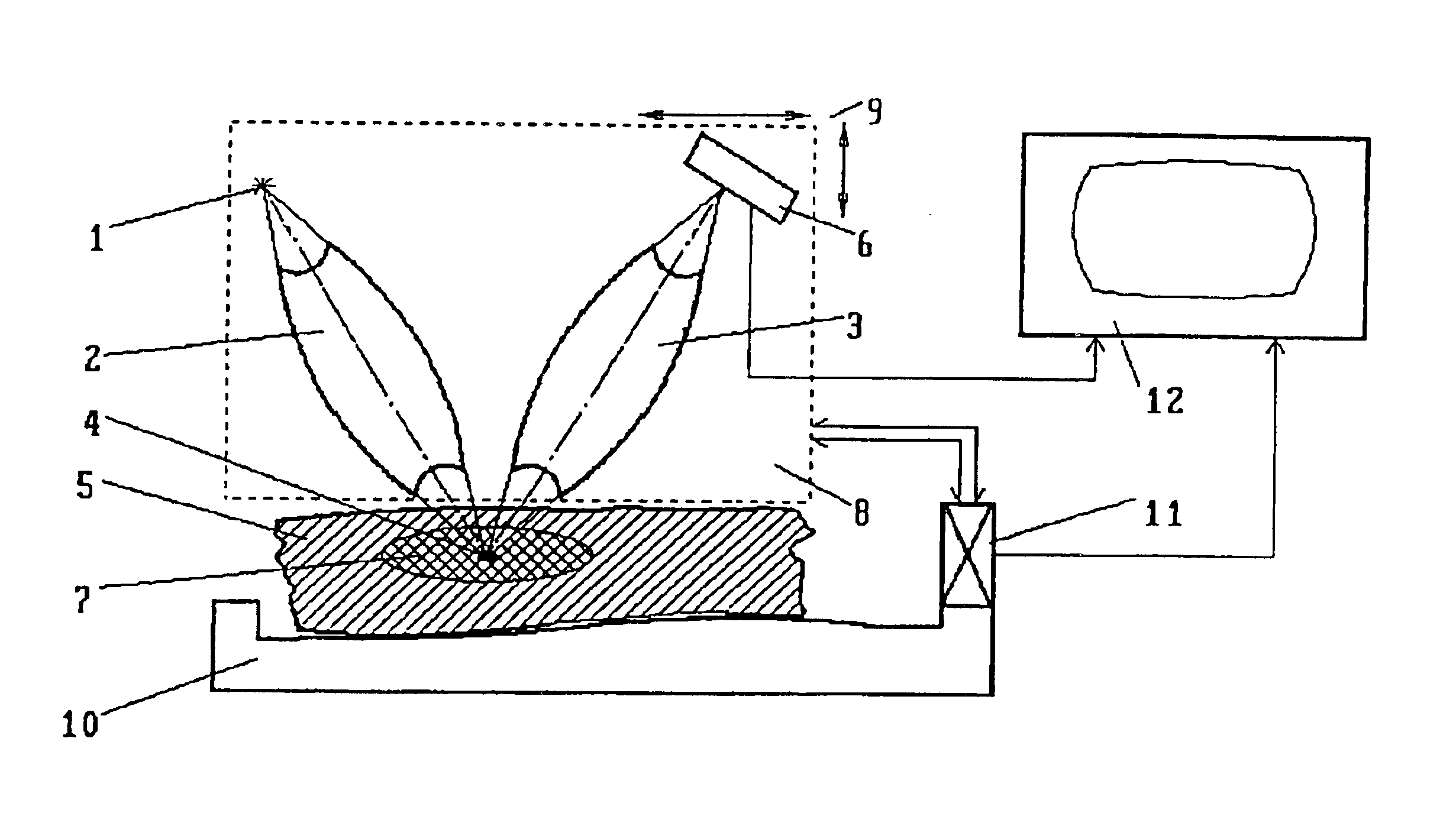
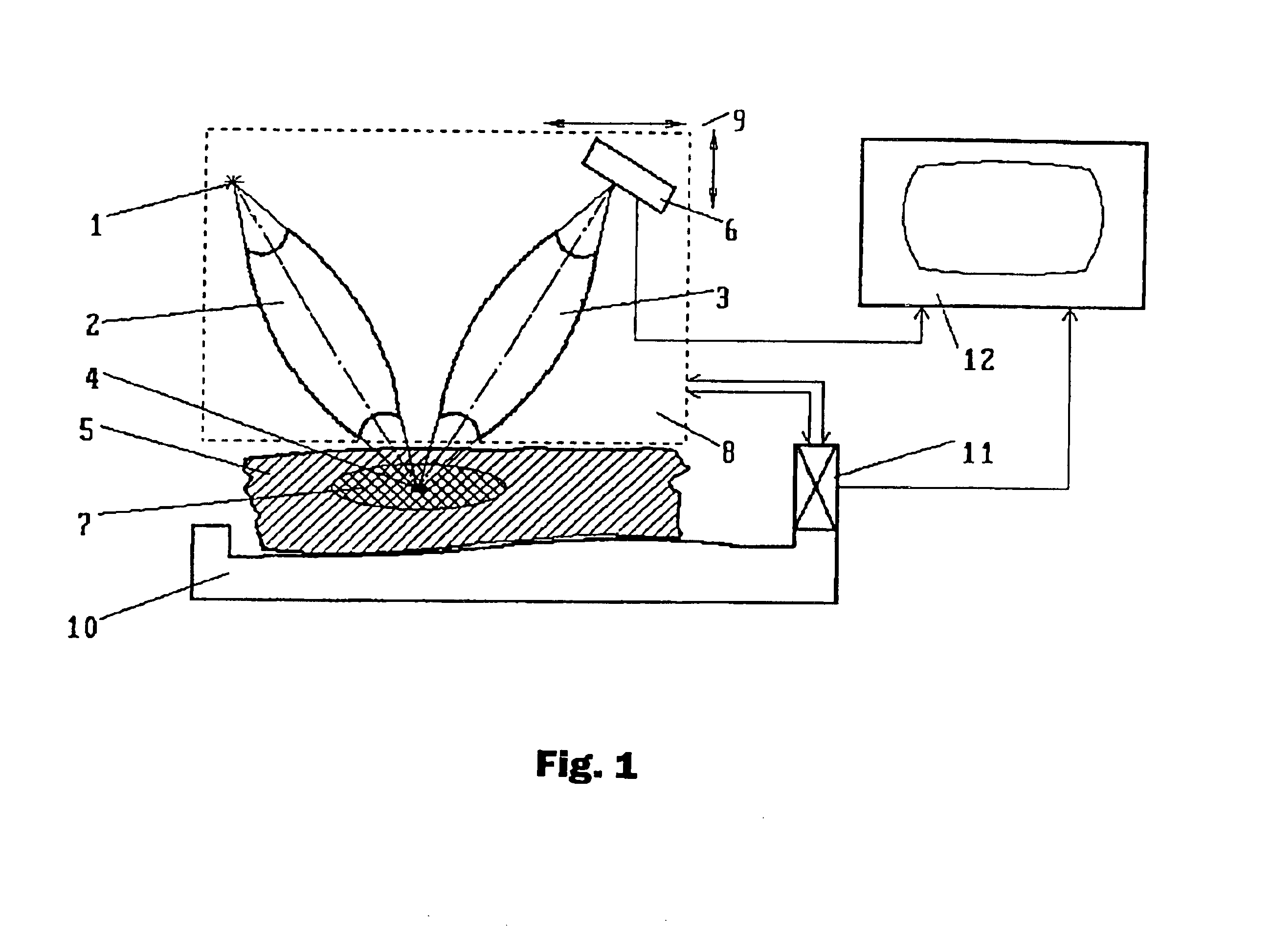
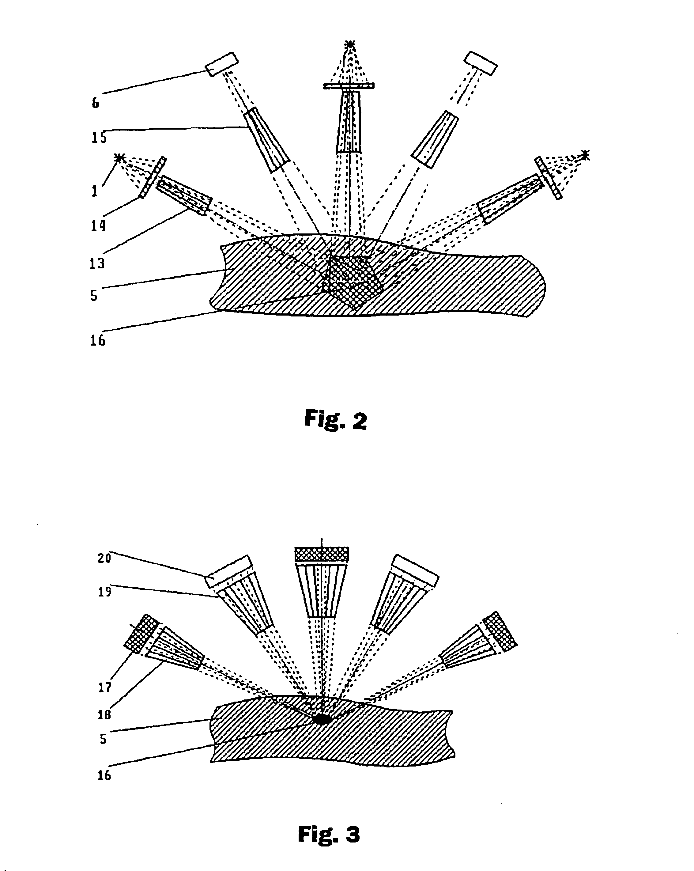
Divergent X-rays from a quasi-point source (FIG. 1) is focused by the X-ray lens 2 in the specified current point 4 within the target area 7 of an object 5 (for example, a biological object). The latter is positioned as necessary with the help of the means 10 for positioning. Focused in current point 4, radiation excites secondary scattered radiation in the substance of object 5 (coherent and non-coherent Compton radiation, fluorescent radiation). The intensity of secondary radiation is proportional, with the accuracy of the fluctuations due to the stochastic nature of the process of secondary radiation excitation, to the density of the substance where it is excited. The focus of the second X-ray lens 3 is located in the same current point 4. This second lens focuses the scattered radiation that it has captured onto detector 6, which converts it into an electric signal that is input to the means 12 f...


PUM


Abstract

A method and device for producing visually sensed images of the internal structure of, particularly, a biological object. When used for diagnostic purposes in medicine the x-ray dosage of tissues surrounding the spot that is being examined is decreased.

Description

BACKGROUND OF THE INVENTIONS1. Field of the InventionsThe suggested inventions relate to the intra-vision means and are designed for producing visually sensed images of the internal structure of an object, in particular, of a biological object, with X-rays. The preferential applications include defectoscopy and medical diagnostics.2. Description of the Prior ArtVarious methods and devices of the said intended use are known, where traditional principles of projection roentgenoscopy are embodied. In such methods and devices, the visible image of the object's internal structure, for example, tissues of a biological object, is obtained as a shadow projection. Density of the acquired image in each of its points is determined by the total attenuation of X-rays that passed through the object on their way from the source to the detection means. The latter is either a fluorescent screen or an X-ray film, which should be chemically treated to get the image visualized (see Polytechnical Dictio...

Claims


Application Information

Patent Timeline
IPC IPC(8): G01N23/22G01N23/223A61B6/00A61B6/03G01N23/18G01T1/00G21K1/02G21K1/06G21K5/02
CPCG01N23/223G21K2201/06G01N2223/076G01N2223/601G01N2223/316G01N2223/50
Inventor KUMAKHOV, MURADIN ABUBEKIROVICH
Owner KUMAKHOV MURADIN ABUBEKIROVICH
Why Eureka
Try Eureka