
Functionalized TGF-beta fusion proteins

a fusion protein, functional technology, applied in the direction of peptides, peptide/protein ingredients, peptide sources, etc., can solve the problems of limited clinical use value, 1 antibody available, and high cost of radioactive iodination process

Inactive Publication Date: 2004-06-29
GOVERNMENT OF THE UNITED STATE OF AMERICA AS REPRESENTED BY THE SEC OF THE DEPT OF HEALTH & HUMAN SERVICES THE
View PDF17 Cites 35 Cited by
  • Summary
  • Abstract
  • Description
  • Claims
  • Application Information

AI Technical Summary

Benefits of technology

FIG. 7C illustrates the quantitative results of a sandwich ELISA to detect tagged TGF-.beta.1 ligands in conditioned media. Solid bars, FLAG-tagged TGF-.beta.1; stippled bars, HA-tagged TGF-.beta.1; gray bars, untagged (WT) TGF-.beta.1 in a FLAG-TGF-.beta.1 SELISA; open bars, untagged (WT) TGF-.beta.1 in a HA-TGF-.beta.1 SELISA. "No 1 st," negative control where primary antibody (anti-FLAG or anti-HA) was omitted. Dilutions refer to dilutions of conditioned media.

Problems solved by technology

There are some TGF-.beta.1 antibodies available, but their usefulness in a clinical setting is limited at least in part because they often display some degree of cross-reactivity to other TGF-.beta. family proteins (see, e.g., U.S. Pat. No. 5,571,714).
Radioactive iodination is an expensive and hazardous process, and it usually would be inappropriate to use .sup.125 I labeled proteins for in vivo experimentation, for instance in clinical trials.
The ability to track the distribution of any exogenously administered, recombinant forms of TGF-.beta. family proteins has been restricted by the inability to distinguish between the endogenous forms of the protein produced in treated cells or tissues.
There have been a few reports of TGF-.beta. fusions in the literature, but the described molecules have been essentially biologically non-functional.
Serious difficulties were encountered in refolding the denatured fusion protein, and full biological activity was not retained using this system.
It is believed that all prior efforts to fuse a TGF-.beta. family protein to a peptide or protein have resulted in biologically non-functional molecules.

Method used


Image

  • Functionalized TGF-beta fusion proteins
  • Functionalized TGF-beta fusion proteins
  • Functionalized TGF-beta fusion proteins

Examples


example 1

Construction and Expression of FLAG-TGF-.beta.1 and 11 / 12FLAG-TGF-.beta.1

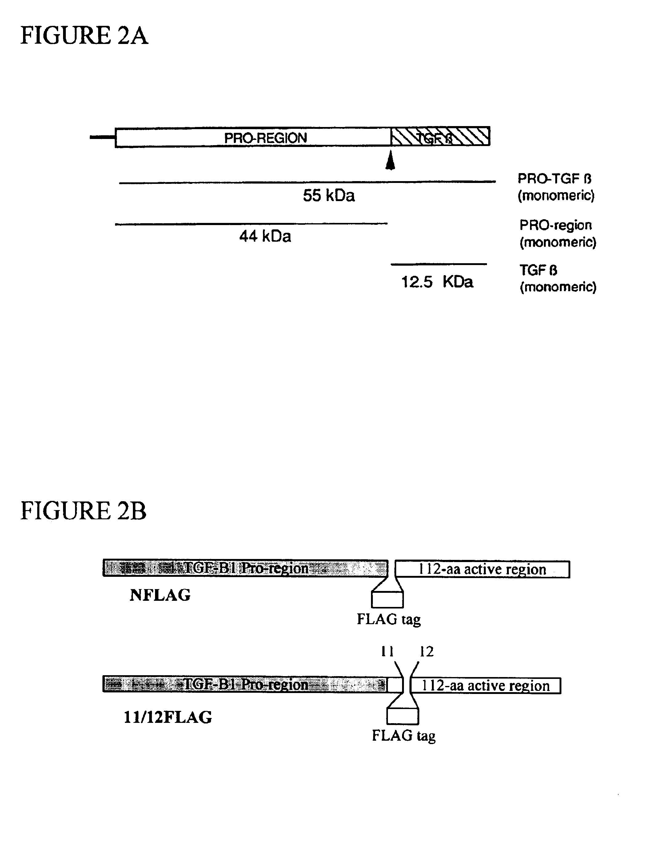
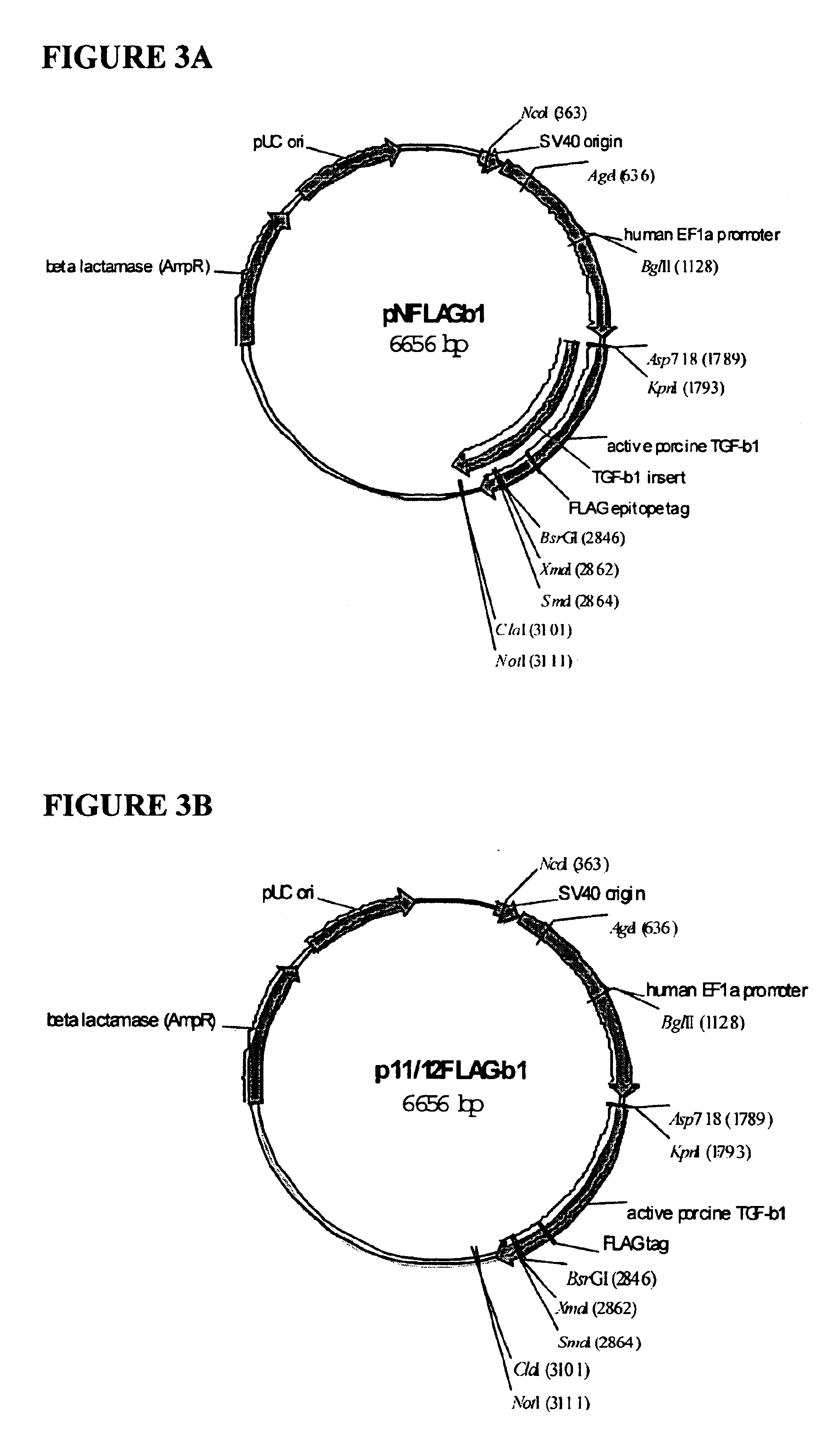
This example describes the construction and expression of two biologically functional TGF-.beta. fusion proteins, each comprising the FLAG epitope tag near the N-terminus of the mature TGF-.beta. protein. The disclosed epitope-tagged TGF-.beta. differs fundamentally from earlier attempts to tag TGF-.beta. in that the inventors have succeeded in expressing epitope-tagged biologically active TGF-.beta. in a mammalian host.

Expression of the FLAG-TGF-.beta. fusions described in this example is driven by the human elongation factor 1-alpha (EF1-.alpha.) gene promoter, a strong promoter capable of driving expression in virtually any type of cell. Two FLAG-tagged TGF-.beta.1 constructs, differing only in the location of insertion of the FLAG tag, are described. In one, the FLAG tag is inserted immediately following the cleavage site (N-terminal in the mature, processed TGF-.beta.1 molecule). In the second construct, F...

example 2

Biological Activity of FLAG-TGF-.beta.1 and 11 / 12FLAG-TGF-.beta.1

This example provides assays that can be used to test the biological activity of TGF-.beta. fusion molecules. In particular, this example demonstrates that representative functionalized TGF-.beta. fusion proteins have TGF-.beta. biological activity at least as great as native TGF-.beta., in spite of the addition of the FLAG epitope tag. Biological activity is demonstrated by two independent methods: (1) growth inhibition of CCL64 cells; and (2) phosphorylation of smad 2 in NMuMG cells.

Smad2 Phosphorylation Studies.

NmuMG cells were plated in 60-cm dishes, incubated in medium containing 0.5% FBS for three hours. COS cell supernatants from cells transfected with various TGF-.beta.1 plasmids were added to these cells (after first washing extensively with PBS). The cells were then incubated with the supernatants for 30 minutes. Cells were washed with PBS and scraped off the dishes. Cell pellets were dissolved in NP-40 lysis...

example 3

Use of FLAG-TGF-.beta.1 and 11 / 12FLAG-TGF-.beta.1 in Experimental Systems.

This example demonstrates that the functionalizing peptide portion of the subject fusion proteins is functional in biological and test systems. FLAG-TGF-.beta.1 has been successfully used in a number of different assays. FLAG-TGF-.beta.1 was used to (1) detect expression of tagged ligand in transfected cells; (2) detect cell surface expression of TGF-.beta. receptor complexes by flow cytometry; and (3) measure cell surface levels of receptor complexes in a non-radioactive cross-linking assay. The tagged ligand is a safer, faster and cheaper alternative to the use of [.sup.125 I] radiolabeled TGF-.beta.1 ligand and expands the repertoire of techniques that can be used to look at TGF-.beta. receptor expression levels.

A. Flow Cytometry: Detection of receptor Molecule T.beta.RI Using FLAG-TGF-.beta.1

Cells were incubated with FLAG-TGF-.beta.1 in FACS buffer. Next, cells were incubated with a FITC-conjugated goat an...


PUM

PropertyMeasurementUnit
Tmaaaaaaaaaa
total volumeaaaaaaaaaa
concentrationaaaaaaaaaa

Abstract

This disclosure relates to TGF-beta family protein fusions that display substantial native TGF-beta family protein function while also having an additional functionality conveyed by the addition of a functionalizing peptide domain. Such functionalizing peptide domain can be a tag peptide (e.g., an epitope tag, a purification tag, a molecular size differentiation tag, etc.) or a passenger or targeting protein. Also provided are methods of making these fusions, as well as methods of using them for diagnosis and diagnosis of various conditions, in measuring and monitoring levels of the fusion molecule in experimental systems and subjects, and in measuring and detecting receptor proteins.

Description

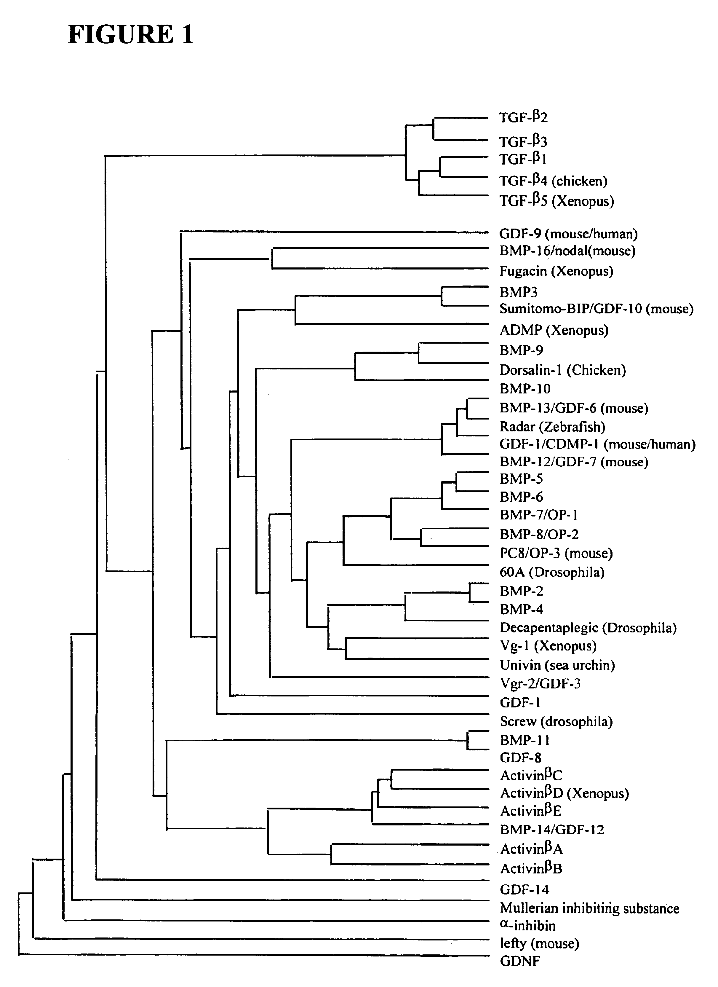
FIELDThe disclosure is in the field of protein labeling and detection. More specifically, the disclosure relates to the use of molecular tags to identify and track recombinant molecules, such as small proteins, in a subject.TGF-.beta. Protein FamilyTransforming growth factor-.beta.1 (TGF-.beta.1) is a growth factor and immunomodulatory cytokine that is secreted from cells and acts through specific binding interactions with a collection of different cell-surface localized receptors. TGF-.beta.1 is the prototype for a large family of secreted polypeptides that includes the three mammalian TGF-.beta. isoforms (TGF-.beta.1, TGF-.beta.2, and TGF-.beta.3), bone morphogenesis proteins (BMPs), activins, and Mullerian inhibitory substance (MIS). More distantly related members of this protein family include murine nodal gene products, Drosophila decapentaplegic complex gene products, and Vg1 from Xenopus.In general, TGF-.beta. family proteins are homodimers, wherein each functional protein co...

Claims


Application Information

Patent Timeline
Patent Type & Authority Patents(United States)
IPC IPC(8): C07K14/435C07K14/495
CPCC07K14/495C07K2319/20
Inventor WOLFRAIM, LAWRENCE A.LETTERIO, JOHN J.
Owner GOVERNMENT OF THE UNITED STATE OF AMERICA AS REPRESENTED BY THE SEC OF THE DEPT OF HEALTH & HUMAN SERVICES THE
Learn More