System and method for tissue biopsy using ultrasonic imaging

a tissue biopsy and ultrasonic imaging technology, applied in the field of diagnostic imaging, can solve problems such as unsatisfactory side effects

Inactive Publication Date: 2005-03-01
ADVANCED IMAGING TECH
View PDF25 Cites 46 Cited by
  • Summary
  • Abstract
  • Description
  • Claims
  • Application Information

AI Technical Summary

Problems solved by technology

This technique may provide sufficient guidance for the biopsy procedure, but has the undesirable side affect of exposing the patient and the physician to undesirable radiation.

Method used


Image

  • System and method for tissue biopsy using ultrasonic imaging
  • System and method for tissue biopsy using ultrasonic imaging
  • System and method for tissue biopsy using ultrasonic imaging

Examples


Embodiment Construction

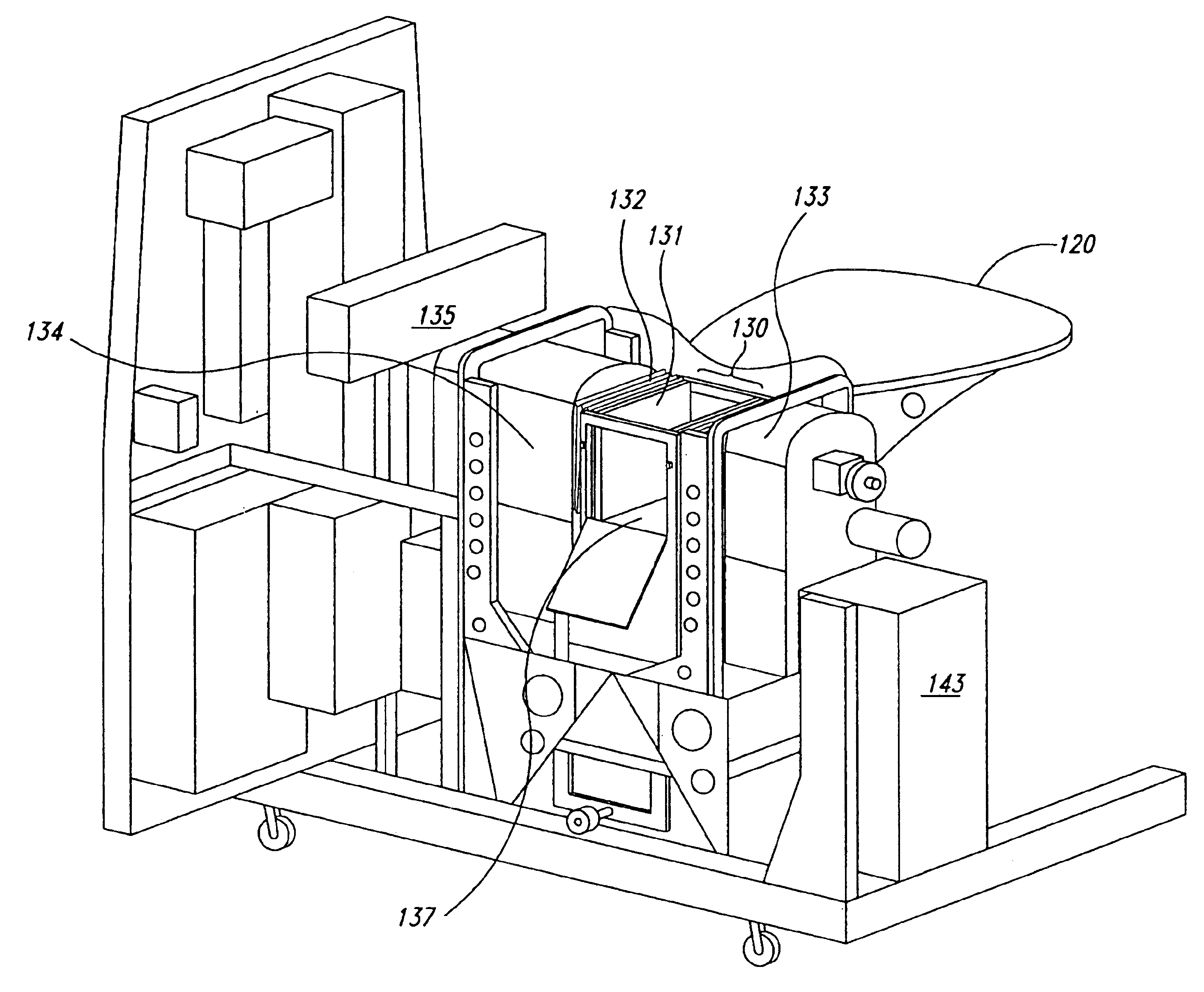
The present invention is directed to an apparatus and method to perform a needle biopsy and localization interface between commercially available biopsy probes and an imaging system. In one embodiment, the intended biopsy probes are handheld devices that are guided manually by the operator or may be mechanically coupled to the imaging system. Alternatively, the system may be automated such that three-dimensional location information is provided to a robotic arm or similar device which automatically guides the biopsy probe to the intended location.

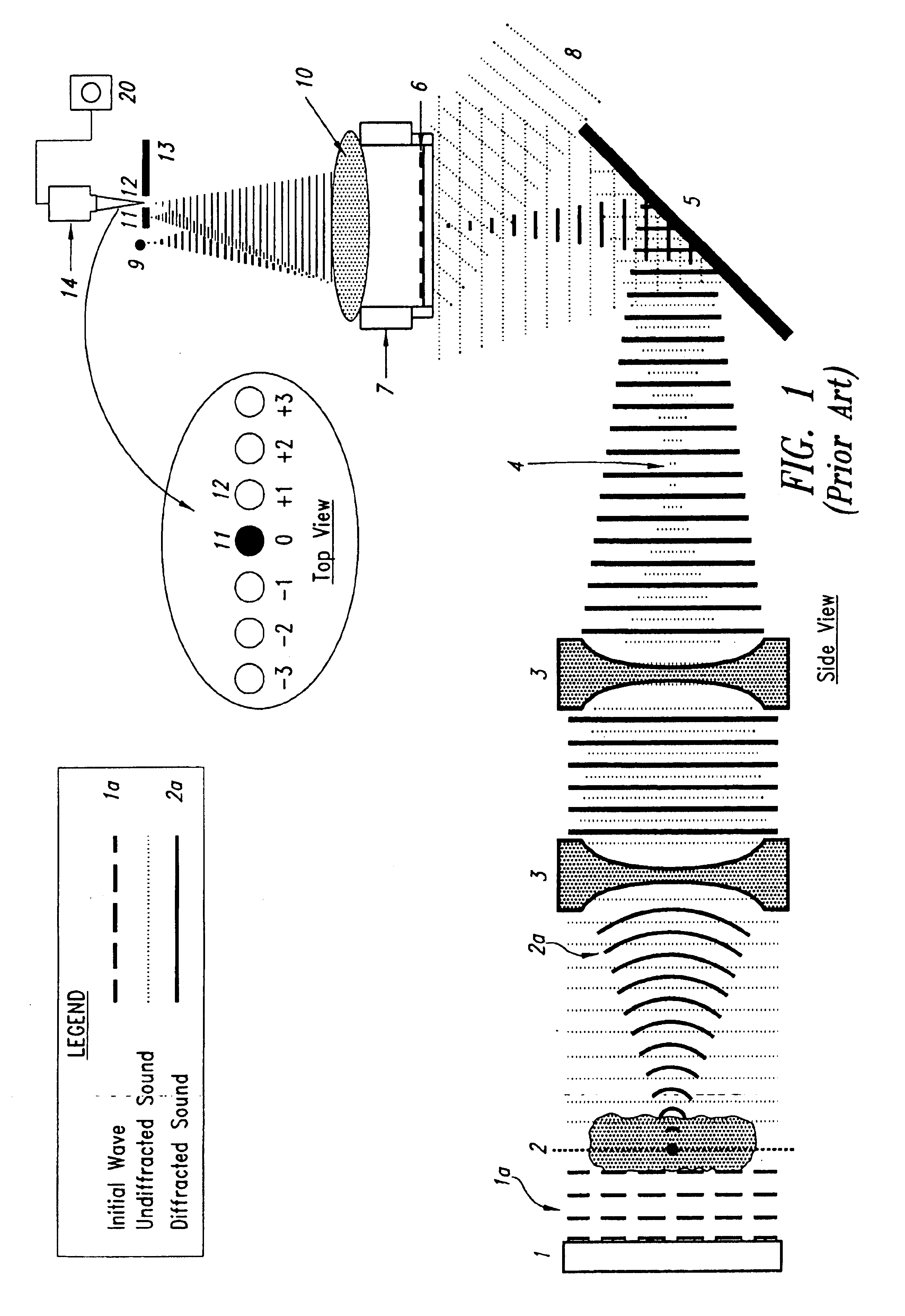
One embodiment of the imaging process can be that of through transmission acoustical holography. One such imaging system is described in U.S. Pat. No. 6,432,053 entitled “Process for Non-Invasively Determining the Dimensions of a Lesion,” which is assigned to the assignee of the present invention and which is incorporated herein by reference in its entirety. Ultrasonic holography as typically practiced is illustrated in FIG. 1. A plane wave...


PUM


Abstract

A tissue biopsy device uses ultrasonic imaging to guide the biopsy needle. An ultrasonic imaging device comprises three acoustically coupled chambers with an ultrasound transducer in a first chamber, at least a portion of an ultrasound detector in the second chamber and the portion of patient anatomy to be imaged placed in the third chamber, which is intermediate the first and second chambers. The three chambers are filled with an acoustically transmissive liquid. One or more of the end walls dividing the first and third chambers and second and third chambers may be movable to form compression plates that are used to retain the patient anatomy in a fixed position during the imaging and biopsy process. When a structure, such as a lesion, has been located, the imaging may be used to determine the precise location of the lesion in three dimensions. The ultrasonically transmissive fluid is drained from the central third chamber with ultrasonic coupling occurring through the ultrasonically transmissive compression plates and the imaged patient anatomy. This permits real-time imaging of the patient anatomy during the biopsy process. The three-dimensional coordinates are used to provide a manual guide for insertion of the biopsy needle. Light bars may be projected onto the external anatomy of the patient to indicate the desired point of entry of the biopsy needle. The physician may use the real-time imaging to view both the lesion and the biopsy needle. In an alternative embodiment, a biopsy needle may be automatically positioned at the location of the lesion by a three-dimensional positioning system.

Description

BACKGROUND OF THE INVENTION1. Field of the InventionThe present invention is directed generally to diagnostic imaging and more particularly, to a system and method for diagnostic imaging as a tool for performing biopsy.2. Description of the Related ArtIn biopsy or localization procedures it is necessary to ensure accurate placement and deployment of a biopsy needle, localization wire, or clip at the site of a lesion. Knowledge of lesion location prior to needle insertion and real-time image information to ensure probe trajectory along a direct line approach through the tissue to the targeted lesion is important. Conventional techniques, such as x-ray imaging, use radiation imaging techniques to guide the biopsy device to the targeted lesion. This technique may provide sufficient guidance for the biopsy procedure, but has the undesirable side affect of exposing the patient and the physician to undesirable radiation. Therefore, it can be appreciated that there is a significant need fo...

Claims


Application Information

Patent Timeline
Patent Type & Authority Patents(United States)
IPC IPC(8): A61B19/00A61B8/08G01S15/89G01S15/00A61B10/00A61B10/02
CPCA61B8/0833G01S15/897A61B19/20A61B10/0233A61B2019/5276G01N2291/02881A61B90/10A61B2090/378
Inventor SHELBY, JEROD O.FECHT, BARBARA A.O'CONNOR, DENIS A.
Owner ADVANCED IMAGING TECH
Why Eureka
Try Eureka