

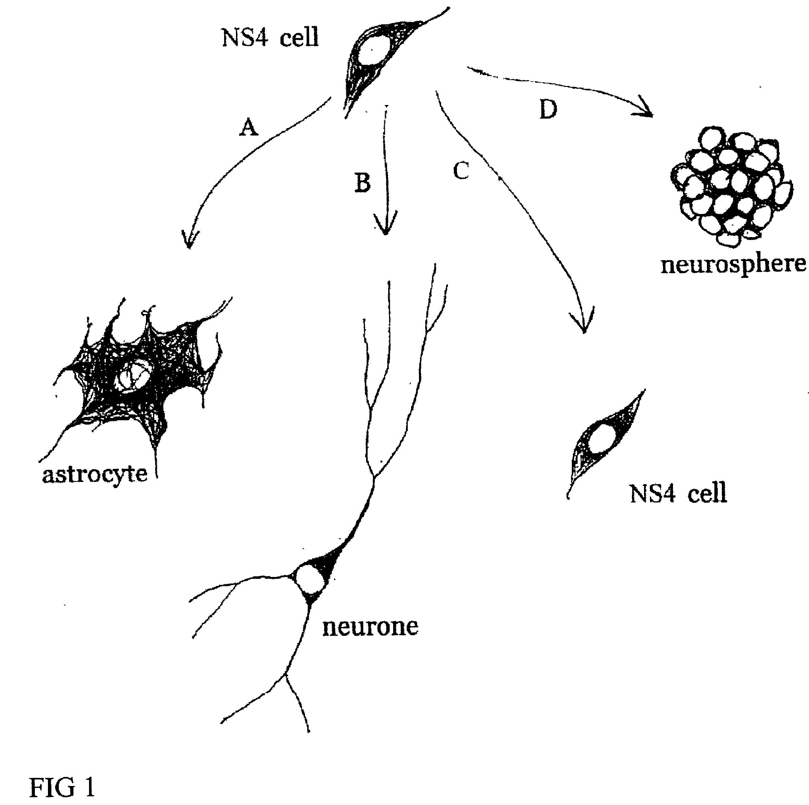
Cultures of GFAP+ nestin+ cells that differentiate to neurons
a technology of gfap+ nestin+ cells and culture, applied in the field of glial cell cultures, can solve the problem that cells can make a significant number of astrocytes
- Summary
- Abstract
- Description
- Claims
- Application Information
AI Technical Summary
Benefits of technology
Problems solved by technology
Method used
Image

Examples
example 1
DISSOCIATION OF MURINE EMBRYONIC NEURAL TISSUE AND PROLIFERATION OF MURINE NS4 CELLS
Separate embryonic (E12-15) primary neural cultures were established by mechanical or enzymatic dissociation from the striatal anlage (lateral ganglionic eminence and medial ganglionic eminence) and grown in DMEM, 10% FCS, N2 supplement and EGF (20 ng / ml) for 4-20 passages. The cells grew adherently and phenotypes were analyzed using morphology and immunocytochemistry. For immunocytochemistry analysis, cells were fixed in 4% 10 paraformaldehyde for 10 min. and exposed to primary and secondary antibodies according to well-established protocols. For neuronal differentiation, cells were switched to serum-free medium without EGF. After 1-7 days, cells were fixed and evaluated by morphology and immunocytochemistry. To evaluate the paternal origin of the differentiated progeny, cultures were established from embryonic transgenic mouse lateral ganglionic eminence and MGE expressing the receptor of the avian...
example 2
DISSOCIATION OF HUMAN EMBRYONIC NEURAL TISSUE AND PROLIFERATION OF HUMAN NS4 CELLS
Human first trimester CNS tissue was collected and the LGE and MGE were dissected out and mechanically dissociated and cultured in DMEM, N2 supplement, 10% FCS (“NS4 Complete Medium”), and EGF (20 ng / ml) or EGE and bFGF (20 ng / ml each). The tissues were incubated in 0.1% trypsin and 0.05% DNase in DMEM for 15-20 min at 37° C. Tissue was mechanically dissociated with a fire polished Pasteur pipette. Dissociated cells were plated at high density in tissue culture treated flasks without additional coating in “NS4 Complete Medium” and either EGF (20 ng / ml) or EGF and bFGF (20 ng / ml each). Cell cultures were housed in an incubator at 37° C., 100% humidity, 95% air / 5% CO2. When the cultures were confluent, they were passaged 1:3.
After several passages (>4), parental cells were investigated for their morphology and expression of GFAP, nestin, and, beta-tubulin III. Similar to the mouse cultures (EXAMPLE 1), h...
example 3
GLIAL METHODS
Lateral ganglionic eminence and medial ganglionic eminence sections were dissected from E13.5 or E15.5 embryos. The tissue pieces were incubated in 0.1% trypsin and 0.05% DNase in DMEM for 15-20 min at 37° C. before mechanical dissociation and plating, at high density in tissue culture treated flasks without additional coating. Cells were expanded in DMEM F12 with N2 supplement (Gibco), glutamine (2 mM), antibiotics, 10% fetal calf serum (FCS), and EGF (20 ng / ml). When the cultures were confluent, they were passaged 1:3. Both neurons and glia were present in the initial cultures. However, by the 4th passage (P4), or after freezing and thawing, the cultures were devoid of cells possessing neuronal morphologies or expressing neuronal markers (i.e., beta-tubulin III). These cultures were highly enriched in cells expressing nestin as well as glial phenotypes (i.e. GFAP and RC2). In the case of LGE glial cultures, the cells were expanded extensively (passaged >25 times). The...
PUM
| Property | Measurement | Unit |
|---|---|---|
| temperatures | aaaaa | aaaaa |
| temperatures | aaaaa | aaaaa |
| concentrations | aaaaa | aaaaa |
Abstract
Description
Claims
Application Information