Detection of methylated CpG rich sequences diagnostic for malignant cells

a technology of methylated cpg and rich sequences, applied in the direction of instruments, biochemistry apparatus and processes, material analysis, etc., can solve the problem of still uncertain methylation status of multiple cpg islands

Inactive Publication Date: 2005-05-17
THE OHIO STATE UNIV RES FOUND
View PDF2 Cites 77 Cited by
  • Summary
  • Abstract
  • Description
  • Claims
  • Application Information

AI Technical Summary

Problems solved by technology

However, at present it is still uncertain whether the methylation status of multiple CpG islands in the genomic DNA of subjects suspected of having cancer can be used as a diagnostic tool for determining whether or not tissue obtained from such subjects contain malignant cells.

Method used


Image

  • Detection of methylated CpG rich sequences diagnostic for malignant cells
  • Detection of methylated CpG rich sequences diagnostic for malignant cells
  • Detection of methylated CpG rich sequences diagnostic for malignant cells

Examples


example 1

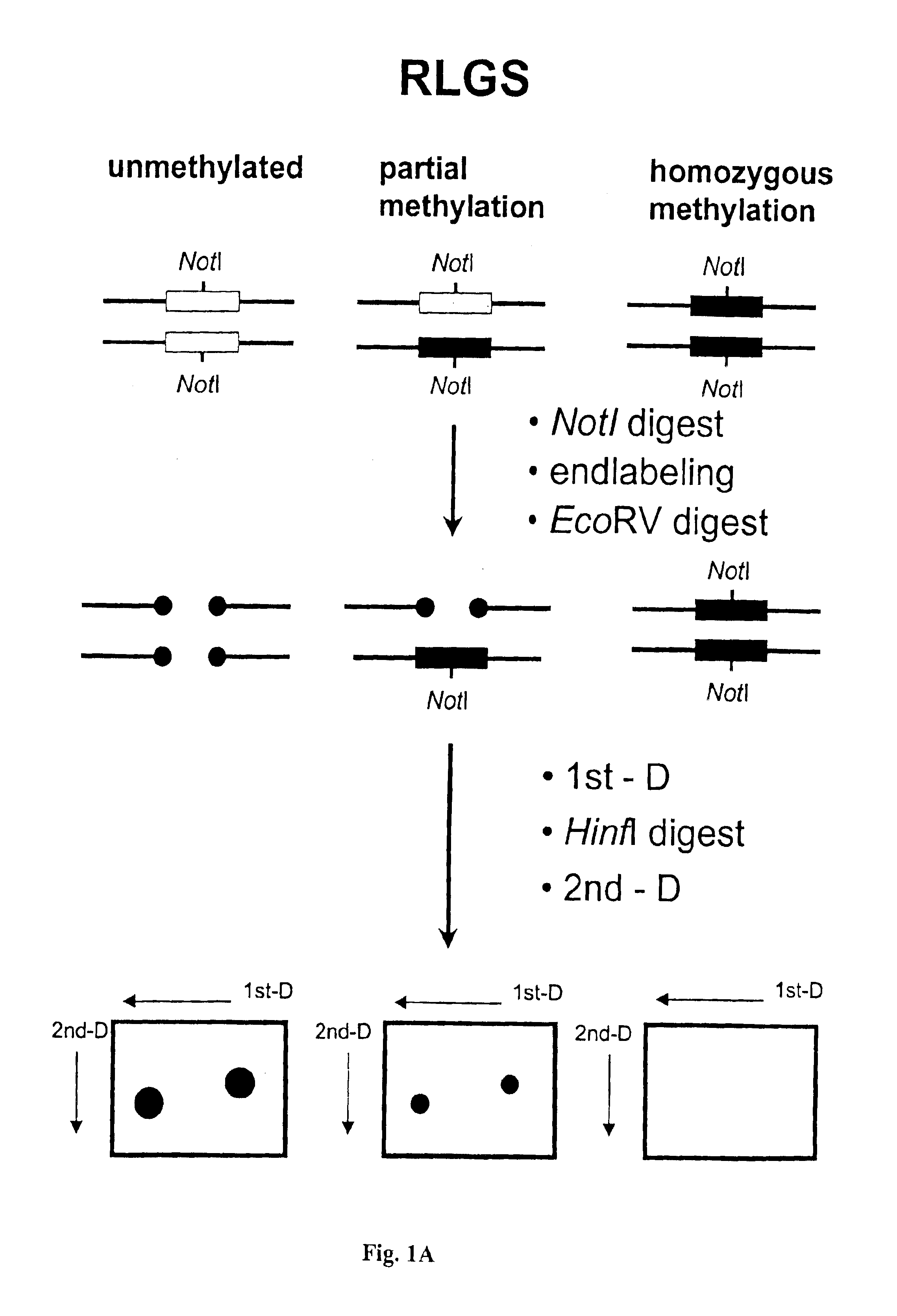
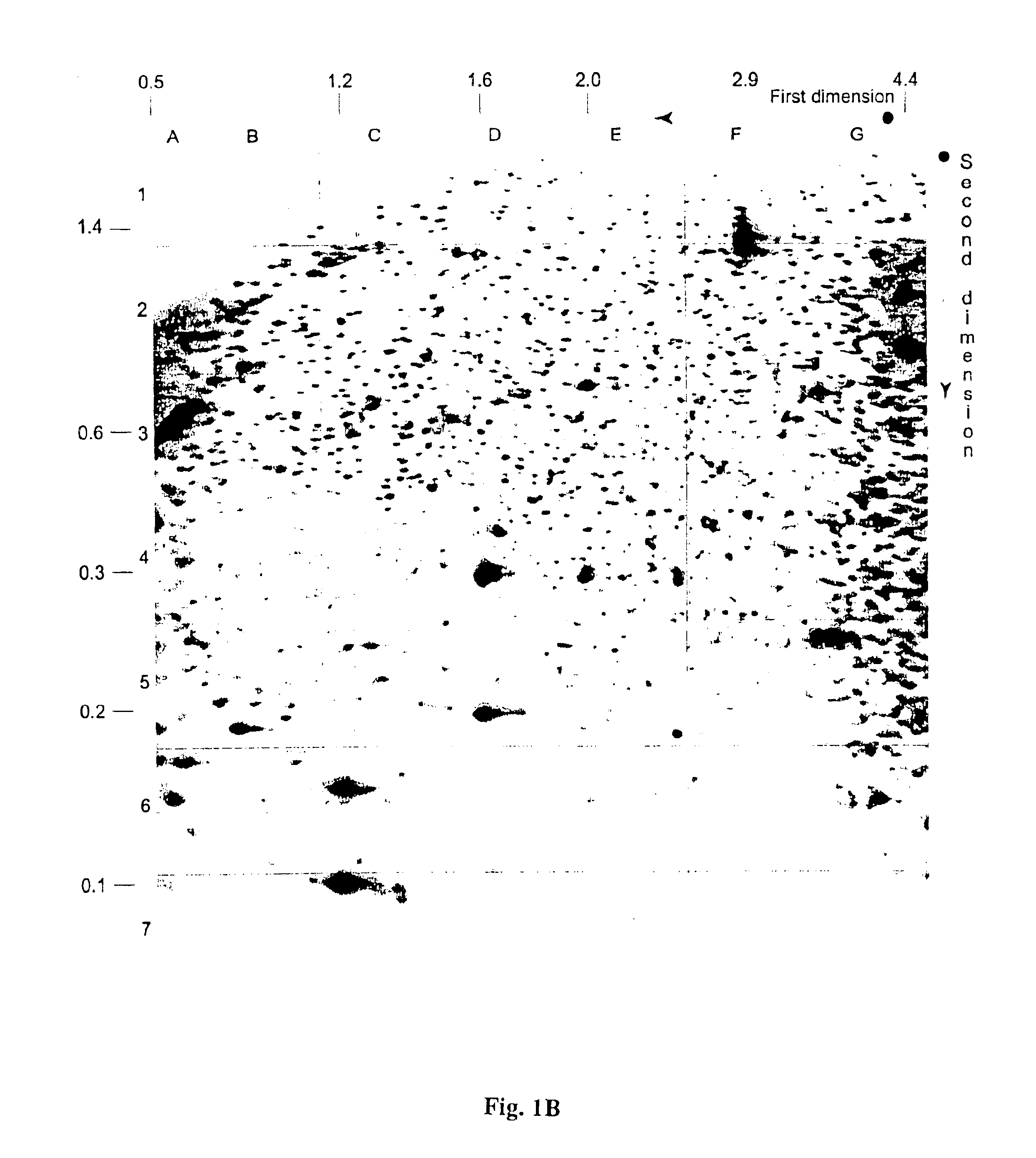
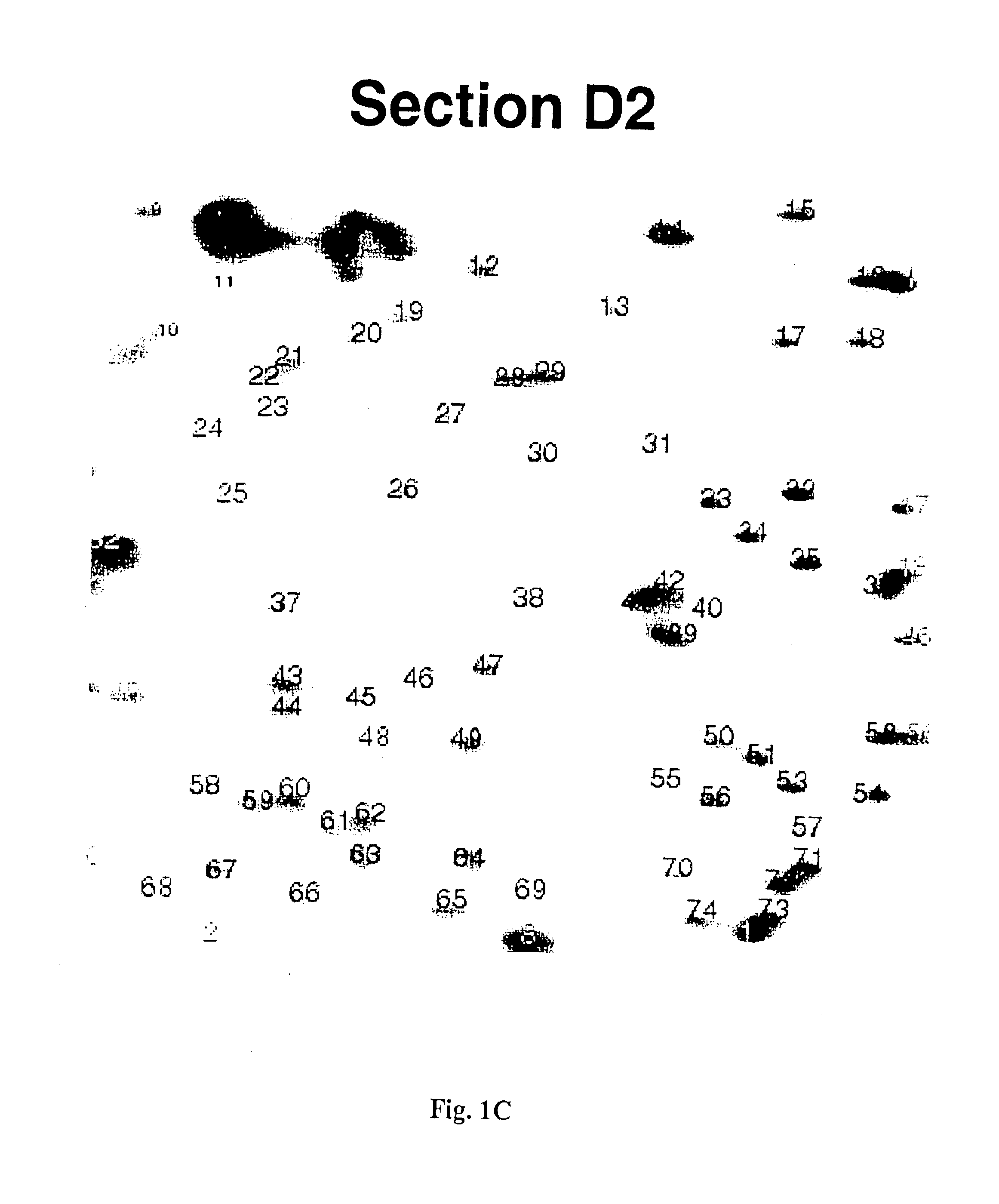
Identification of Diagnostic Markers Using NotI and RLGS

A. Isolation and Enzymatic Processing of Genomic DNA

[0053]Tissue from solid tumors was obtained as surgical tissue samples. Where possible, surrounding non-tumor tissue was taken and used as a control. Where it was not possible to obtain patient-matched normal tissue, normal tissue from multiple patients was used. Tissue samples from patients with acute myelogenous leukemis (AML) consisted of either bone marrow aspirates or peripheral blood. Normal samples were obtained from the same patients who were in remission after chemotherapy.

[0054]The surgically removed tissues were quickly frozen in liquid nitrogen and stored a −80° C. prior to isolation of DNA. When DNA was ready to be isolated, 2 ml of lysis buffer (10 mM Tris, pH 8.0; 150 mM EDTA, 1% sarkosyl) was added to 100-300 mg of tissue in a 50 ml Falcon tube and frozen in liquid nitrogen. The frozen mixture was then removed from the tube, wrapped in aluminum foil, and quickl...

example 2

Identification of Diagnostic Markers for Lung Cancer Using AscI and RLGS

[0091]Tissue from lung tumors was obtained as surgical tissue samples. Where possible, surrounding non-tumor tissue from the same patient was obtained and used as a control. DNA was isolated from the tissue as described in Example 1. In preparation for RLGS analysis, the ends of the DNA were blocked as described in Example 1. The DNA was then digested with AscI followed by digestion with EcoRV. The AscI restriction enzyme recognizes the sequence 5′GGCGCGCC3′ and does not cleave said sequence if cytosines within the sequence are methylated. First dimension gel electrophoresis, in-gel digestion with HinfI, second dimension gel electrophoresis and autoradiography were performed as described in Example 1.

[0092]RLGS profiles from lung tumor DNA were compared with RLGS profiles obtained from healthy, non-tumor tissue DNA. Spots which were lost or present at reduced intensity in tumor tissue RLGS profiles as compared t...

example 3

Design of Primers for Cancer Diagnosis

[0095]Primers are designed for diagnosis of cancer using methylation-specific PCR (MSR). The primers are designed to amplify regions of the human genome whose sequences are contained within the library clones disclosed in this application. Two sets of primers are needed for each library clone whose DNA sequence is to be used for diagnosis of cancer. Each primer set is designed to amplify the same region of the genome, said region beginning at the end of a library clone containing the methylation-sensitive restriction enzyme recognition site (i.e., the NotI site for the library described in Example 1; the AscI site for the library described in Example 2) and ending at a region contained within the clone up to 200 nucleotides from the methylation-sensitive restriction enzyme recognition site.

[0096]The first set of primers is designed to amplify template genome DNA whose cytosine residues are not methylated and, after bisulfite treatment, the cytos...


PUM

PropertyMeasurementUnit
lengthaaaaaaaaaa
pHaaaaaaaaaa
concentrationaaaaaaaaaa

Abstract

The present invention provides methods for determining the methylation status of CpG-containing dinucleotides on a genome-wide scale using infrequent cleaving, methylation sensitive restriction endonucleases and two-dimensional gel electrophoretic display of the resulting DNA fragments. Such methods can be used to diagnose cancer, classify tumors and provide prognoses for cancer patients. The present invention also provides isolated polynucleotides and oligonucleotides comprising CpG dinucleotides that are differentially methylated in malignant cells as compared to normal, non-malignant cells. Such polynucleotides and oligonucleotides are useful for diagnosis of cancer. The present invention also provides methods for identifying new DNA clones within a library that contain specific CpG dinucleotides that are differentially methylated in cancer cells as compared to normal cells.

Description

[0001]This invention was conducted, at least in part, with government support under National Institutes of Health Grants No: P30 CA16058 and CA80912 awarded by the National Cancer Institute. The U.S. government has certain rights in the inventionBACKGROUND OF THE INVENTION[0002]Diagnosis of cancer, classification of tumors, and cancer-patient prognosis all depend on detection of properties inherent to cancer, or malignant cells, that are absent in normal, nonmalignant cells. Since cancer is largely a genetic disease, resulting from and associated with changes in the DNA of cells, one important method of diagnosis is through detection of related changes within the DNA of cancer cells. Such changes can be of two types. The first type of change is a genetic change that occurs when the sequence of nucleotide bases within the DNA is changed. Base changes, deletions and insertions in the DNA are examples of such genetic changes. The second type of change in the DNA is an epigenetic change...

Claims


Application Information

Patent Timeline
Patent Type & Authority Patents(United States)
IPC IPC(8): C12Q1/68G01N33/53C12N15/09G01N27/447G01N33/574
CPCC12Q1/6827C12Q1/6886C12Q2565/125C12Q2521/331C12Q2521/301C12Q2600/154C12Q2600/158
Inventor PLASS, CHRISTOPH
Owner THE OHIO STATE UNIV RES FOUND
Why Eureka
Try Eureka