Method for stitching partial radiation images to reconstruct a full image

a partial radiation and image technology, applied in the field of digital radiography, can solve the problems of limited size of digital detectors, affecting the quality of digital images, and imposing shadows on cassettes, and achieve the effect of improving image quality and high degree of geometric accuracy

Inactive Publication Date: 2005-05-17
CARESTREAM HEALTH INC
View PDF24 Cites 97 Cited by
  • Summary
  • Abstract
  • Description
  • Claims
  • Application Information

AI Technical Summary

Benefits of technology

[0015]The invention has the following advantages.
[0016]1. Enables the generation of a composite full image from a plurality of sub-images without requiring the use of external reference markers to perform geometric distortion corrections.
[0017]2. Preserves a high degree of geometric accuracy in the stitched image.
[0018]3. Results in improved image quality in a reconstructed (composite) image by minimizing the presence of shadows of non-anatomic structures superimposed on the anatomic regions of interest.

Problems solved by technology

As medical institutions are migrating from analog screen-film systems to digital modalities, such as computed radiography (CR), these types of exams impose a significant challenge.
This is because the size of digital detector is limited.
The drawback of this approach is that the metallic frames of the front (closer to the x-ray source) cassettes impose shadows in the image recorded in the back cassettes.
The shadows are not removable and therefore may hinder diagnostic interpretation of the acquired images.
However, in practice, this method requires that the storage phosphor screens be removed from the cassettes before imaging, and to be placed back into the cassettes in a darkroom after the x-ray exposure, which is cumbersome in the clinical environment.
The drawback of this method is that a precisely fabricated pattern of reference markers must be imaged simultaneously with the patient in order to achieve precise geometric registration of the sub-images.

Method used


Image

  • Method for stitching partial radiation images to reconstruct a full image
  • Method for stitching partial radiation images to reconstruct a full image
  • Method for stitching partial radiation images to reconstruct a full image

Examples


Embodiment Construction

[0030]In general, the present invention relates to the radiographic imaging of an elongated object such as the full spine, e.g., for diagnosing scoliosis, or leg of a human subject.

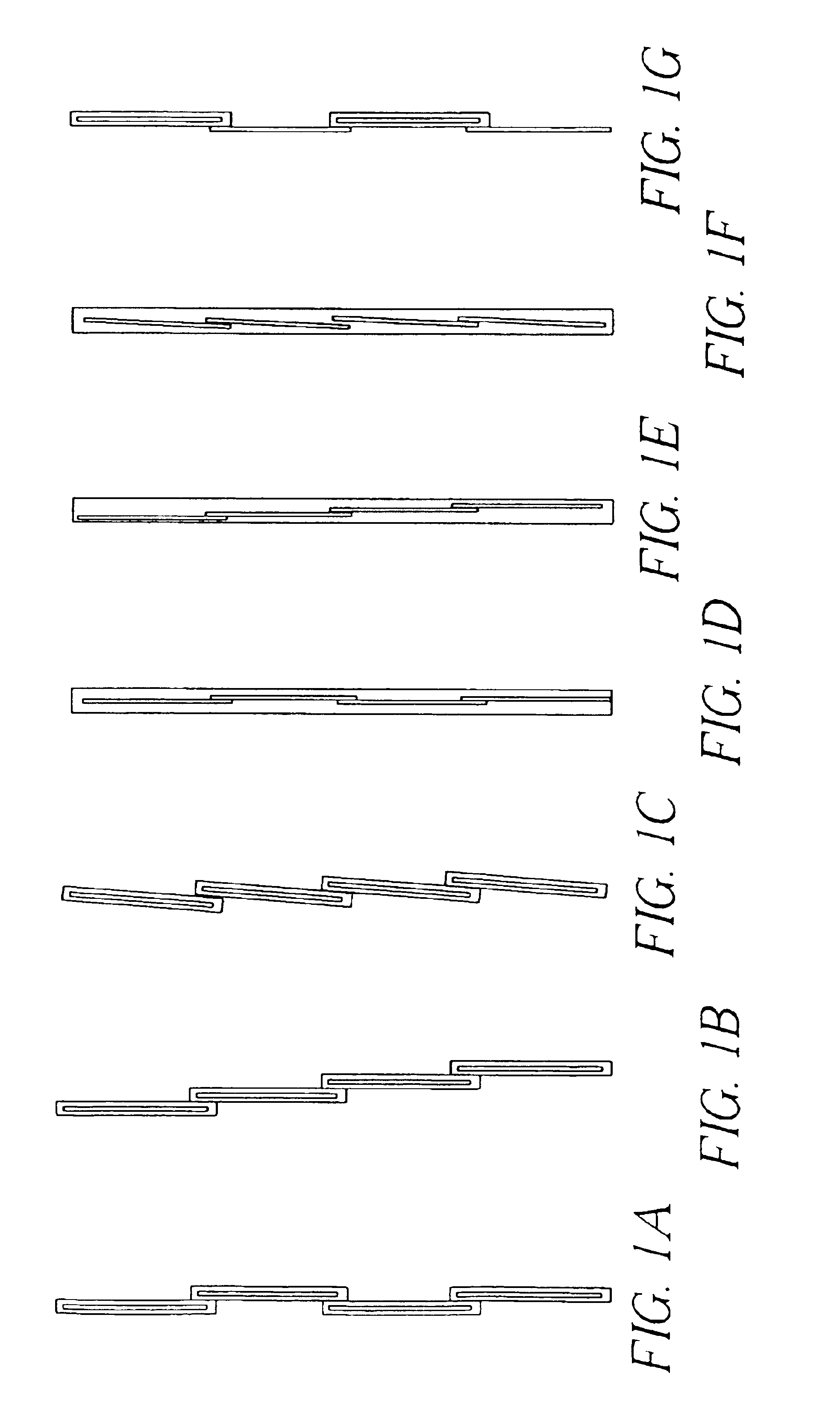
[0031]US Patent Application by Brahm, Odea, Rogers, Wang proposes a method that is a hybrid between the cassette stacking and storage phosphor screen stacking methods. As shown in FIG. 1G, cassettes and storage phosphor screens are placed in a partially overlapping and alternating arrangement with the screens always positioned in front of the cassettes. This method eliminates the cassette metallic frame shadow from the acquired images, and reduces the number of storage phosphor screens that need to be removed out of and to be replaced back into cassettes.

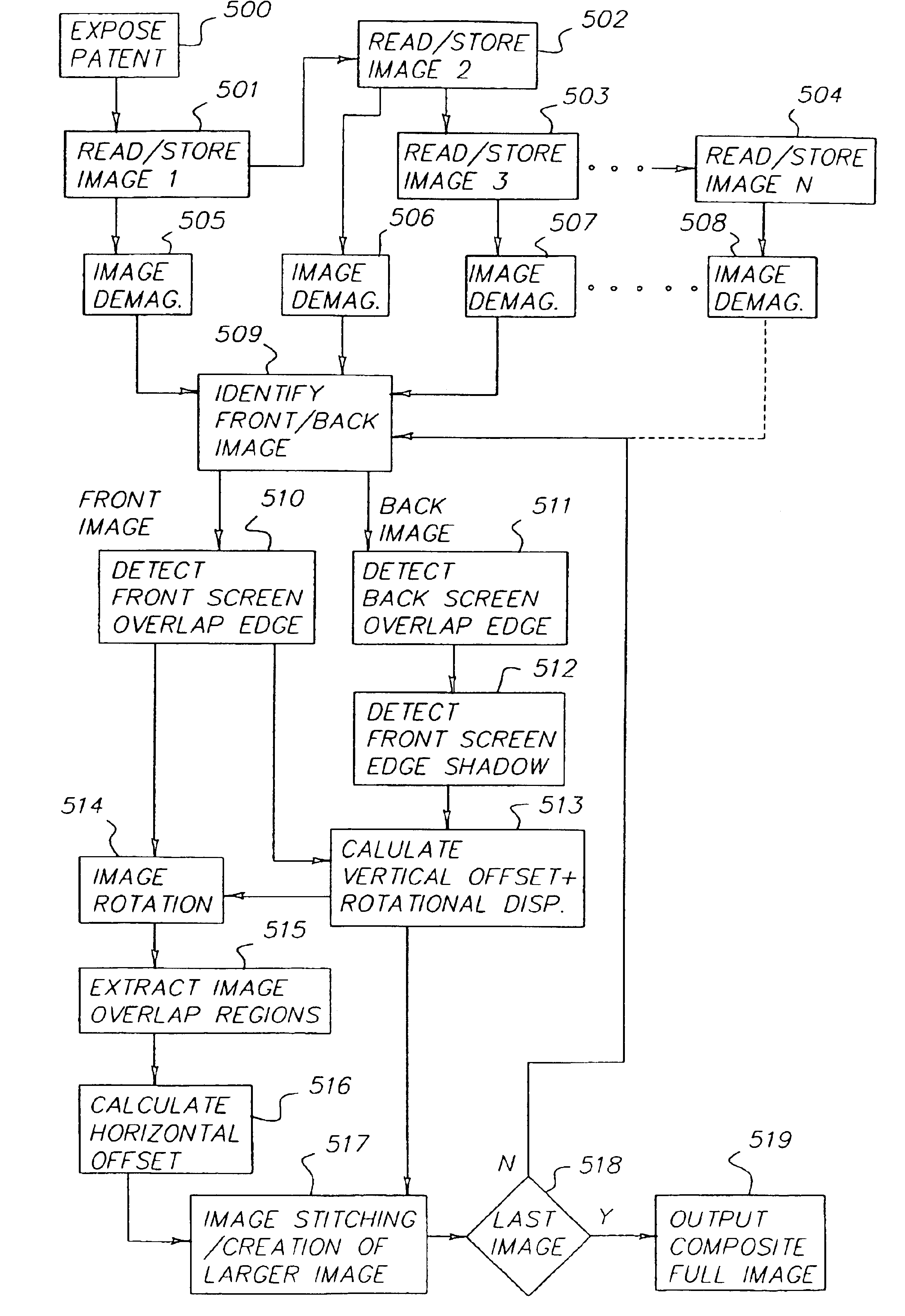
[0032]When an x-ray exposure is taken with any of the cassette / phosphor screen setups shown in FIGS. 1A-1G, a plurality of sub-images is obtained, each of which bears a partial image of the elongated object. Because the phosphor screens are the fundamenta...


PUM


Abstract

A method of forming a composite digital image comprising: providing 1-N digital images formed from 1-N contiguous segments of a larger radiographic image recorded in 1-N overlapping storage phosphor screens wherein N is equal to or greater than 2 and wherein the image content in the overlapped region of contiguous images is the same, and the end edge of a screen nearest an x-ray source is present in both contiguous images; selecting a pair of contiguous digital images, wherein one image is closer to said x-ray source than said other image; correcting for any geometric distortion if applicable in said pair of digital images based on the distance between x-ray source and said storage phosphor screen; determining any rotational displacement and any vertical displacement between said pair of digital images by matching said end edge of said one image present in said pair of images correcting for image orientation if applicable based on any said rotational displacement; determining any horizontal displacement between said pair of digital images by correlating the image content in said overlapping regions of said pair of digital images; stitching said pair of digital images together to form a larger composite digital image along said one image edge based on any said horizontal and vertical displacement; and repeating said correcting for any geometric distortion to said stitching with the larger composite image and the next consecutive overlapping digital image until all N digital images are stitched together to form a full composite image.

Description

FIELD OF THE INVENTION[0001]This invention relates in general to digital radiography, and in particular to the imaging of a long human body part, such as the spine or legs, using a storage phosphor-based computed radiography system.BACKGROUND OF THE INVENTION[0002]When a long segment of the human body is imaged using the conventional screen-film technique, special cassettes and films of extended length are used, such as 30×90 cm and 35×105 cm. As medical institutions are migrating from analog screen-film systems to digital modalities, such as computed radiography (CR), these types of exams impose a significant challenge. This is because the size of digital detector is limited. For example, the largest CR storage phosphor cassette from several major CR vendors is limited to 35×43 cm, which can only image a portion of the long body part at a time. To address this problem, several methods have been proposed. European Patent EP0919856A1 discloses a way of staggering several storage phos...

Claims


Application Information

Patent Timeline
Patent Type & Authority Patents(United States)
IPC IPC(8): G01T1/24G01T1/29G01T1/00A61B6/00G06T1/00H04N1/04
CPCG01T1/2014A61B6/5241
Inventor WANG, XIAOHUIFOOS, DAVID H.DORAN, JAMESROGERS, MICHAEL K.
Owner CARESTREAM HEALTH INC
Why Eureka
Try Eureka