Tissue analysis and kits therefor

a tissue analysis and tissue technology, applied in the field of tissue analysis, can solve the problems of dilution of malignant cells, less accurate methods that require tissue disaggregation, and inability to correlate malignant cells, and the loss of tissue architecture precludes the ability to correlate malignant cells, and the use of serial sections in this type of analysis

Inactive Publication Date: 2005-06-14
GENENTECH INC +1
View PDF15 Cites 77 Cited by
  • Summary
  • Abstract
  • Description
  • Claims
  • Application Information

AI Technical Summary

Problems solved by technology

However, most of the methodologies available for evaluation of tissue for the presence of genes associated with or predisposing an individual to a malignancy have well-known drawbacks.
For example, methods that require disaggregation of the tissue, such as Southern, Northern, or Western blot analysis, are rendered less accurate by dilution of the malignant cells by the normal or otherwise non-malignant cells that are present in the same tissue.
Furthermore, the resulting loss of tissue architecture precludes the ability to correlate malignant cells with the presence of genetic abnormalities in a context that allows morphological specificity.
This issue is particularly problematic in tissue types known to be heterogeneous, such as in human breast carcinoma, where a significant percentage of the cells present in any area may be non-malignant.
Use of serial sections in this type of analysis increases the probability of error, especially in heterogeneous tissue such as breast tissue.

Method used


Image

  • Tissue analysis and kits therefor
  • Tissue analysis and kits therefor
  • Tissue analysis and kits therefor

Examples


example 1

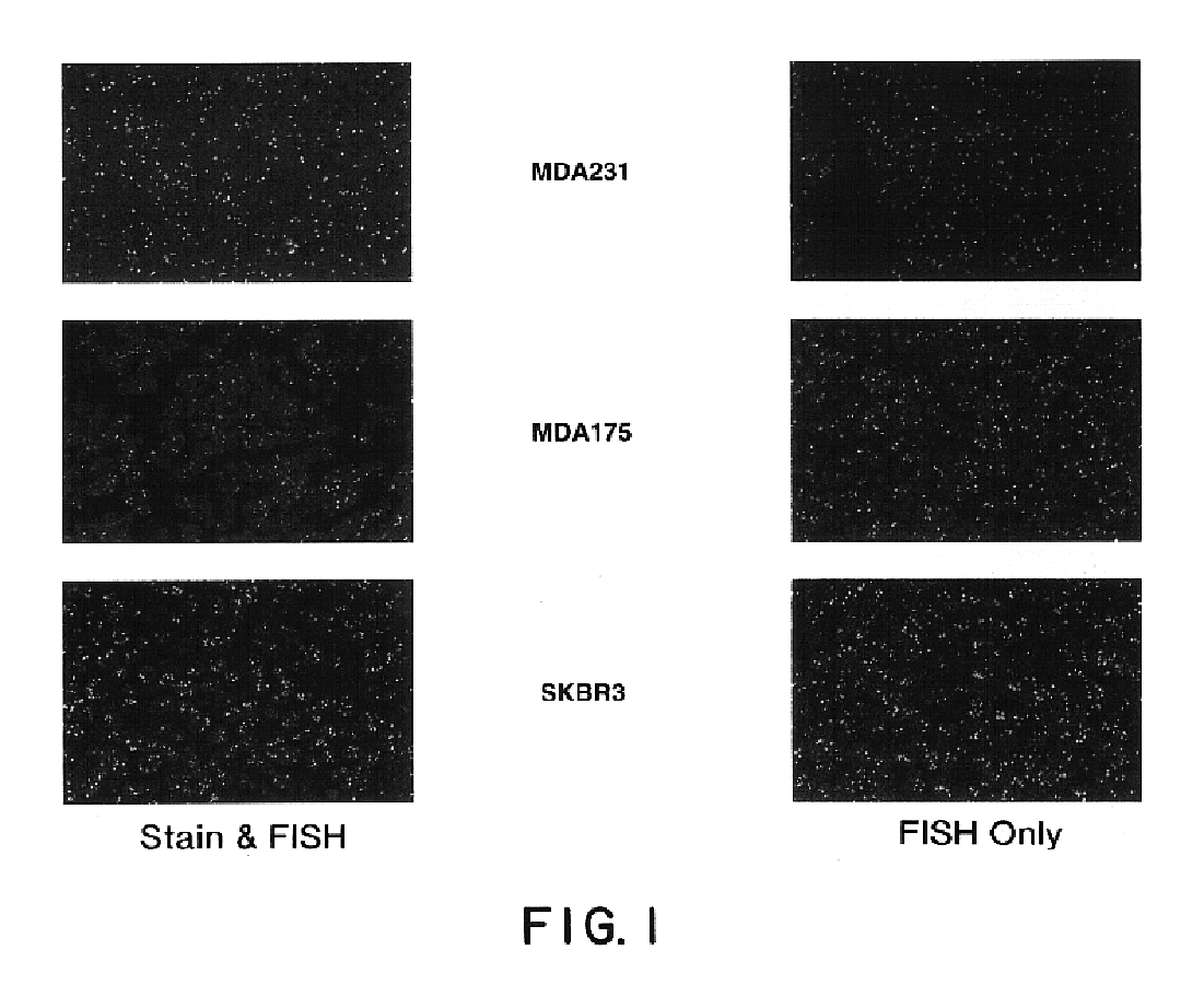
Evaluation of Morphological Stains on FISH Analysis

[0098]A sensitive and specific evaluation of breast tumors for the amplification of HER2 / neu by FISH requires definitive identification and scoring of invasive ductal carcinoma cells as distinct from other stromal and inflammatory elements found in the biopsy. Thus, it was necessary to identify a stain which would allow for complete morphological evaluation of the tissue, and which also allowed for easy quantification of nuclear signals upon subsequent hybridization with fluorescently labeled nucleic acid probes. Many morphological stains fluoresce when illuminated by light of the same wavelength required to visualize the probe fluorophores of interest. When this autofluorescence is of a color similar to that of the probe fluorophore, signal quantification is made difficult.

[0099]Results from the evaluation of six commonly used morphological stains may be found in Table 2.

[0100]

TABLE 2Evaluation of Morphological StainsAutofl.Autofl....

example 2

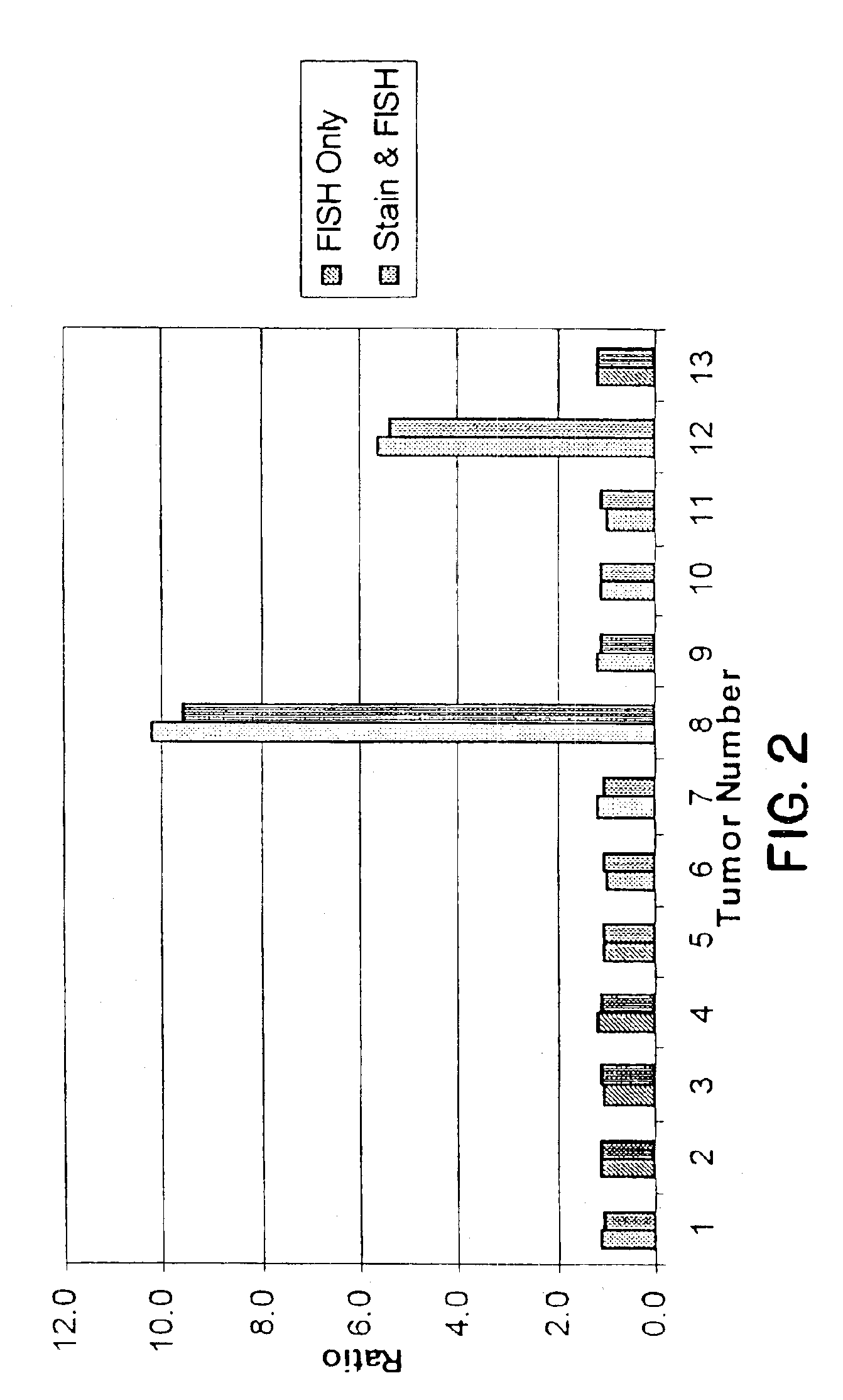
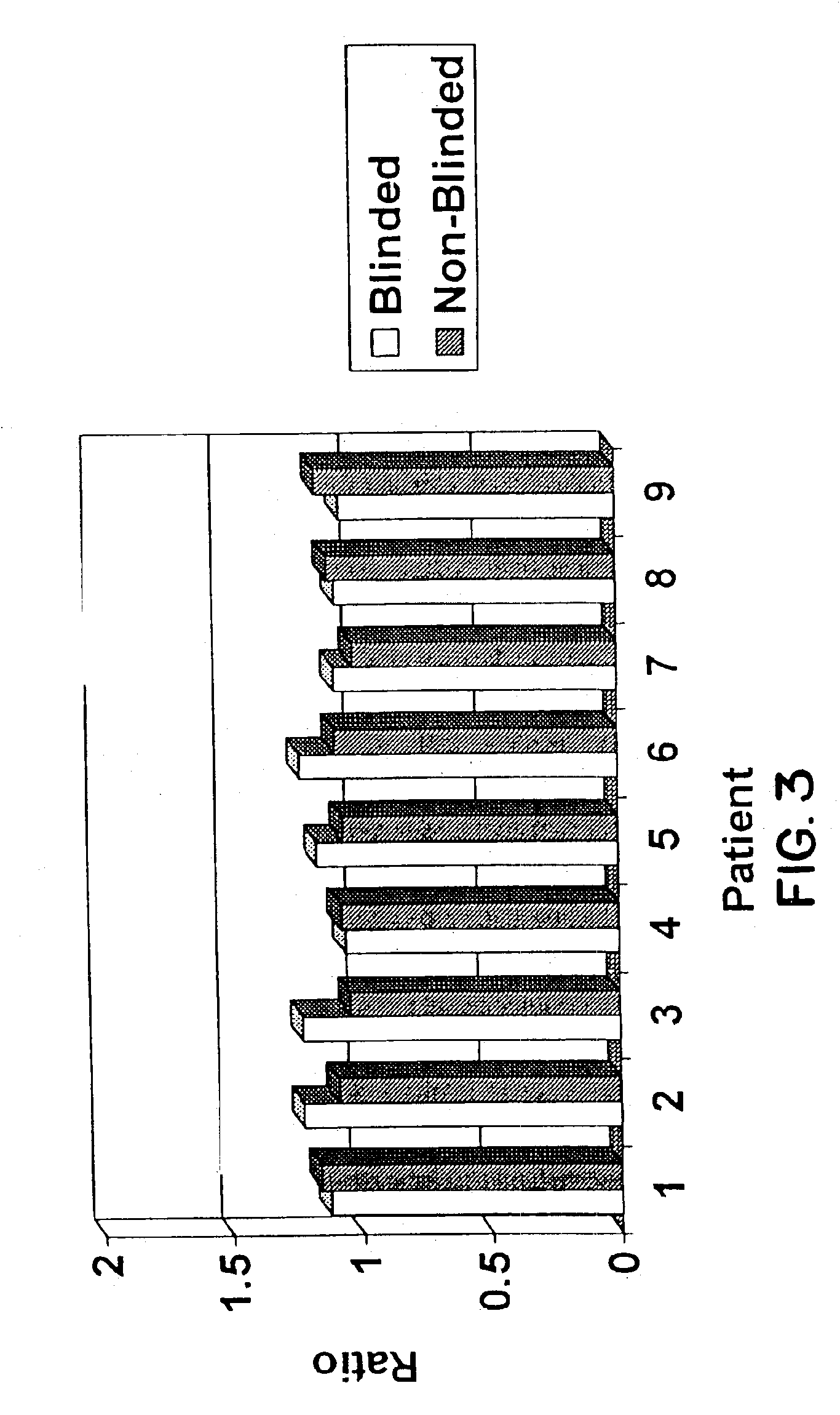
Detection and Quantification of HER2 / neu Amplification in Breast Tissue

Materials and Methods

Tissue Specimens and Preparation for FISH

[0102]Five micron sections were cut from breast tissue which had been fixed in buffered formalin and embedded in paraffin. Sections were placed on positively charged Superfrost Plus slides (CMS, Houston, Tex.) which had also been treated with poly-L-lysine or were mounted on positively charged slides which had no additional treatment prior to the mounting of the slides. Some material had been archived for up to fourteen years. Slide mounted tissue sections were heated on a 65° C. slide warmer for approximately 3 seconds, placed on the bench top for 2 seconds, and deparaffinized in Hemo-De (CMS, Houston, Tex.) for 10 minutes, ×3, followed by immersion in 100% ethanol (EtOH) for 5 minutes, ×2. Slides were air dried in a vertical position. Those slides to be stained for morphologic evaluation were dipped in HEMA 3® Solution I (CMS, Houston, Tex.) for one ...


PUM

PropertyMeasurementUnit
thicknessaaaaaaaaaa
thicknessaaaaaaaaaa
pHaaaaaaaaaa

Abstract

This invention relates to methods of analyzing a tissue sample from a subject. In particular, the invention combines morphological staining and / or immunohistochemistry (IHC) with fluorescence in situ hybridization (FISH) within the same section of a tissue sample. The analysis can be automated or manual. The invention also relates to kits for use in the above methods.

Description

CROSS-REFERENCE TO RELATED APPLICATIONS[0001]This application is a continuation of U.S. application Ser. No. 09 / 167,691, filed on Oct. 7, 1998, U.S. Pat. No. 6,573,043, which application is incorporated herein by reference and to which application priority is claimed under 35 U.S.C. 120.FIELD OF THE INVENTION[0002]The present invention relates generally to the field of tissue analysis. Specifically, the invention combines morphological staining and / or immunohistochemistry (IHC) with fluorescence in situ hybridization (FISH) within the same section of a tissue sample thereby allowing for accurate and simplified prognostic, diagnostic, or research applications on a subject's tissue sample. In addition, the invention provides kits for analysis of a tissue sample utilizing the present methods.BACKGROUND OF THE INVENTION[0003]Advancements in the understanding of genetics and developments in technology and epidemiology have allowed for the correlation of genetic abnormalities with certain...

Claims


Application Information

Patent Timeline
Patent Type & Authority Patents(United States)
IPC IPC(8): C12Q1/68G01N33/574G01N33/58C12Q1/6841C12Q1/6886
CPCC12Q1/6841C12Q1/6886G01N33/57415G01N33/57492G01N33/582G01N33/6872C12Q1/6804G01N2333/71C12Q2563/131C12Q2600/156
Inventor COHEN, ROBERT L.GARDINER, MARY BETHSLIWKOWSKI, MARK X.STELZER, GREGORY T.
Owner GENENTECH INC
Why Eureka
Try Eureka