Method and apparatus to automatically determine tissue cancellation parameters in X-ray dual energy imaging

Inactive Publication Date: 2005-07-12
GE MEDICAL SYST GLOBAL TECH CO LLC
View PDF10 Cites 16 Cited by
  • Summary
  • Abstract
  • Description
  • Claims
  • Application Information

AI Technical Summary

Benefits of technology

[0008]In accordance with at least one preferred embodiment, a method is provided to automatically determine a structure cancelled image in a dual energy decomposition system. A high energy and a low energy level image of internal anatomy formed of at least first and second types of structure are acquired. A gradient mask is computed using an edge cancellation parameter and a gradient threshold. A localization mask is computed from a localization image based on either the low or the high energy image and compared with an intensity threshold automatically selected from a set based upon a selected diagnostic application. A characteristic mask is computed using the gradient mask and localization mask. A first cancellation parameter is evaluated against the char

Problems solved by technology

There is an inherent difficulty associated with producing a clear resultant image, however.
The effect of anatomic noise on the resultant image is to produce clutter, shadowing, and other obscuring effects that render the resultant image much less intelligible than the ideal clear resultant image.
However, in the past, users of the previously mentioned decompo

Method used


Image

  • Method and apparatus to automatically determine tissue cancellation parameters in X-ray dual energy imaging
  • Method and apparatus to automatically determine tissue cancellation parameters in X-ray dual energy imaging
  • Method and apparatus to automatically determine tissue cancellation parameters in X-ray dual energy imaging

Examples


Embodiment Construction

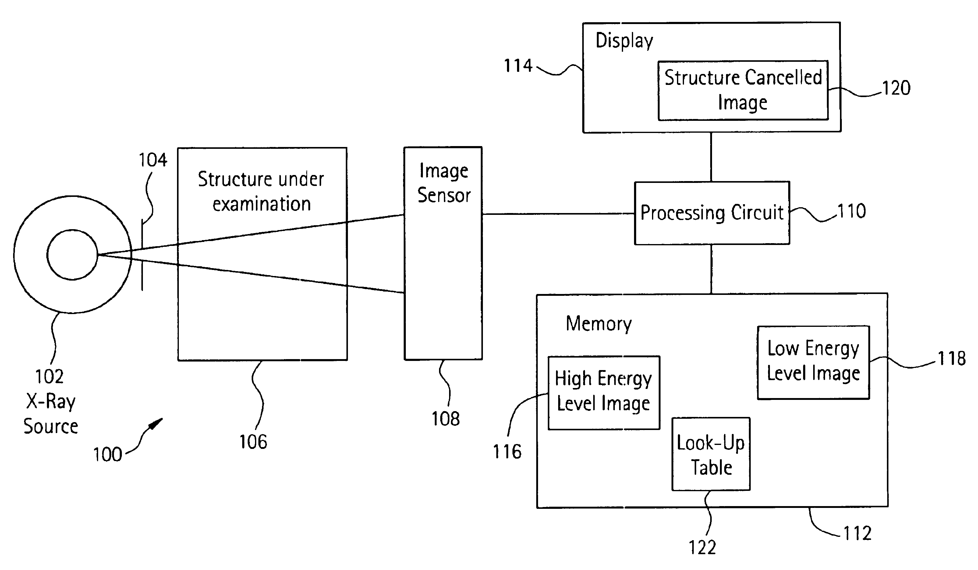
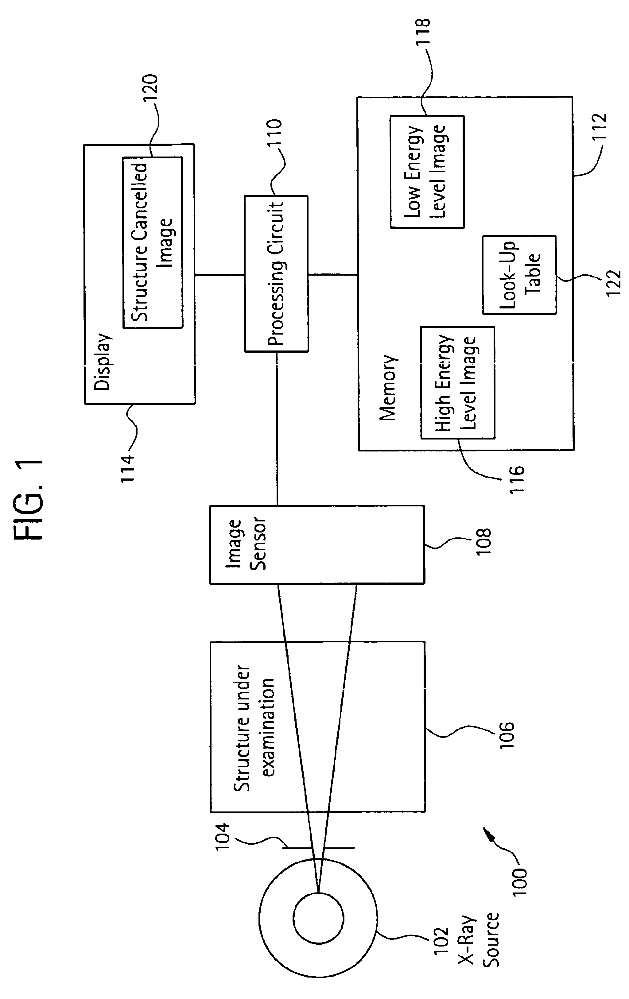
[0022]With initial reference to FIG. 1, the figure illustrates an X-ray imaging system 100. The imaging system 100 includes an X-ray source 102 and a collimator 104 which subject structure under examination 106 to X-ray photons. By way of example, the X-ray source 102 may be an X-ray tube, and the structure under examination 106 may be a human patient, test phantom or other inanimate object under test.

[0023]The X-ray imaging system 100 also includes an image sensor 108, such as a flat panel solid state detector, coupled to a processing circuit 110. The processing circuit 110 (e.g., a microcontroller, microprocessor, custom ASIC, or the like) couples to a memory 112 and a display 114. The memory 112 (e.g., including one or more of a hard disk, floppy disk, CDROM, EPROM, and the like) stores a high energy level image 116 (e.g., an image read out from the image sensor 108 after 110-140 kVp exposure) and a low energy level image 118 (e.g., an image read out after 60-90 kVp exposure). Th...


PUM


Abstract

A method is provided to automatically determine a structure cancelled image in a dual energy decomposition system. A high energy and a low energy level image of internal anatomy formed of at least first and second types of structure are acquired. A characteristic mask is computed using a gradient mask and a localization mask. A first cancellation parameter is evaluated against the characteristic mask, and a second cancellation parameter is computed based upon the first cancellation parameter. The first or second cancellation parameter is selected from a range in a look-up table determined by an effective kVp used to acquire the high level image and an effective kVp used to acquire the low level image. An image of soft structure and an image of hard structure are obtained from the first and second energy level images according to a cancellation equation using the first and second cancellation parameters.

Description

BACKGROUND OF INVENTION[0001]The preferred embodiments of the present invention relate to medical diagnostic X-ray imaging. In particular, the preferred embodiments of the present invention relate to dual energy decomposition for tissue specific imaging using a look-up table to obtain a range of cancellation parameters.[0002]Today, doctors and technicians commonly have access to very sophisticated medical diagnostic X-ray imaging devices. Typically during the operation of an X-ray imaging device, an X-ray source emits X-ray photons under very controlled circumstances. The X-ray photons travel through a region of interest (ROI) of a patient under examination and impinge upon a detector. In the past, X-ray imaging devices employed rudimentary film based detectors. However, recent developments have led to solid state detectors comprised of a grid of discrete detector elements that individually respond to exposure by X-ray photons.[0003]Regardless of the detector used, however, the goal...

Claims


Application Information

Patent Timeline
IPC IPC(8): A61B6/00G06T5/00G06T5/50G06T1/00
CPCA61B6/00A61B6/405A61B6/482A61B6/505G06T5/50G06T2207/10116G06T2207/30008G06T2207/30061
Inventor AVINASH, GOPAL B.SABOL, JOHN MICHAELNICOLAS, FRANCOIS SERGE
Owner GE MEDICAL SYST GLOBAL TECH CO LLC
Why Eureka
Try Eureka