

Reagents and methods for automated hybridization
a technology of automated hybridization and reagents, applied in the fields of genetics, molecular biology, medicine, etc., can solve the problems of insufficient development of situ hybridization applications for use with existing automated instruments, large loss of potential information, and laborious and time-consuming manual manipulation of microarrays
- Summary
- Abstract
- Description
- Claims
- Application Information
AI Technical Summary
Benefits of technology
Problems solved by technology
Method used
Image

Examples
example 1
Automated In situ Hybridization with a Riboprobe
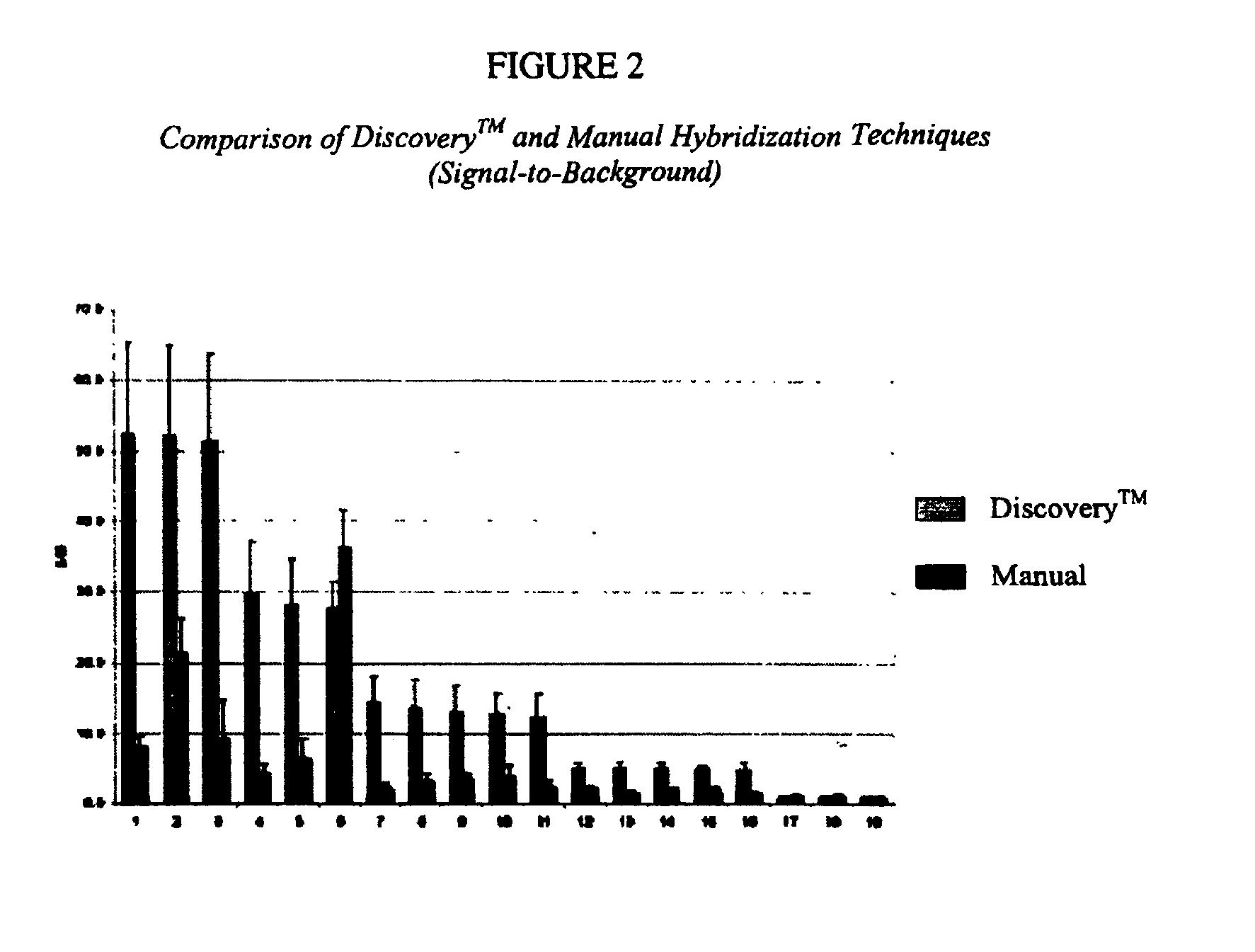
[0098]A tissue sample is fixed, either in neutral-buffered formalin (NBF) or paraformaldehyde (PFA), embedded in paraffin and cut into 5 mm sections. A paraffin section is then placed onto a microscope slide and stained for one or more targets using ISH techniques. Traditional ISH protocols are very time consuming and technically very involved. However, once the slide is prepared, the remaining ISH protocol is automated on the DISCOVERY™ system.
[0099]DISCOVERY™ ISH protocols include deparaffinization, various pretreatment steps, hybridization, post-hybridization stringency washes, and chromogenic signal detection. The RIBOMAP™ kit supplies the reagents for optimal pretreatment steps. Detection is performed using biotin-labeled antibody recognition of DIG molecules, followed by streptavidin-alkaline phosphatase (SA-AP) binding to the antibody. Colorimetric detection using the V-BLUE™ substrate (Ventana Product No. 760-062) is catalyzed ...
example 2
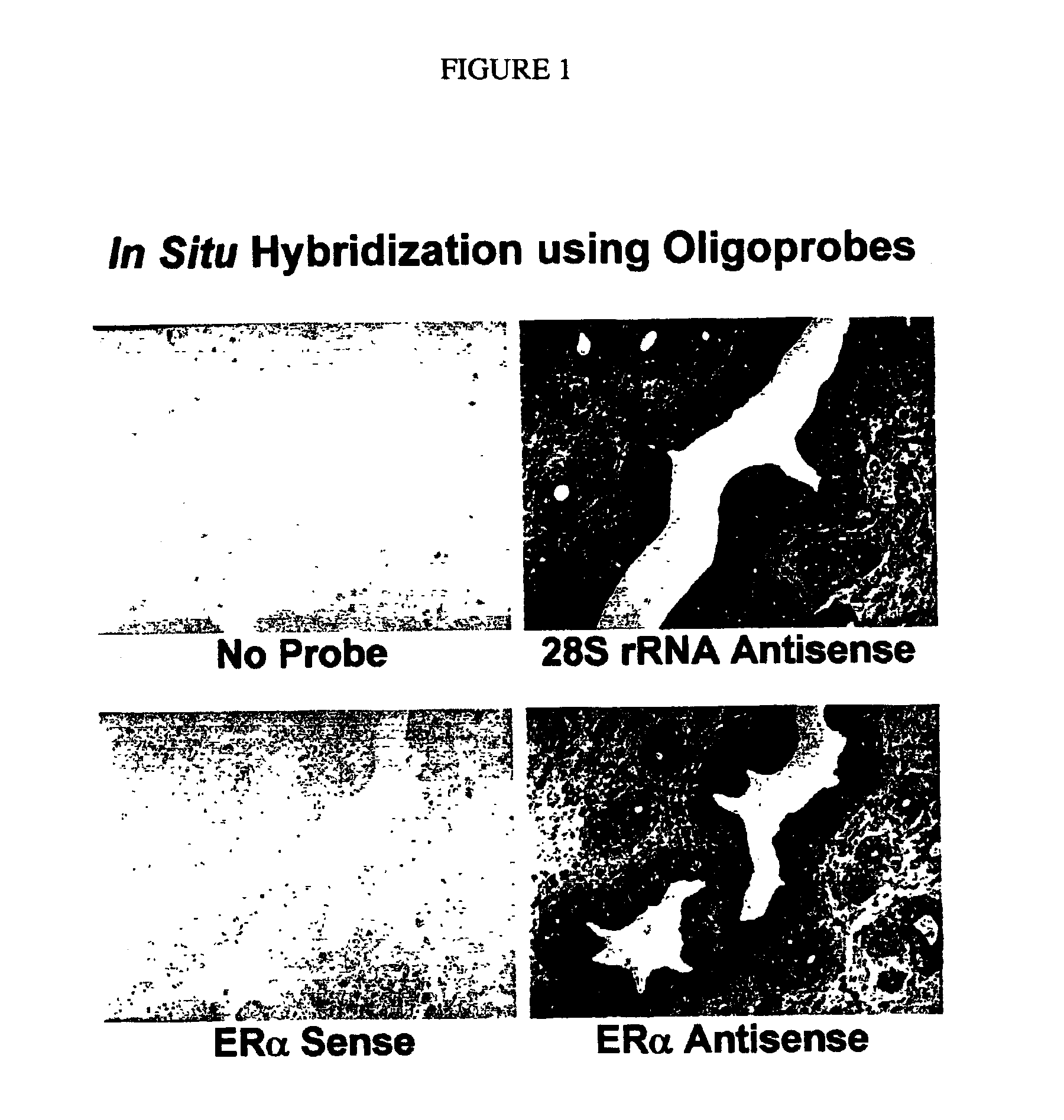
Automated In situ Hybridization with an Oligoprobe
[0125]Formalin-fixed, paraffin-embedded sections of the mouse oviduct are processed in pretreatment steps with the following RIBOMAP™ reagents: RIBOPREP™, RIBOCLEAR™, and RIBOCC™. In addition, Protease, I, II, or III (Ventana Medical Systems, Inc.; Catalog Nos. 760-2018, 760-2019, and 760-2020, respectively) may be used in the protocol.
[0126]Next, the tissue sections are hybridized with DIG-labeled estrogen receptor a (ERα) sense or antisense oligoprobe using the hybridization buffer of the CHIPMAP™ system, CHIPHYBE™ (Ventana Medical Systems). As a positive control for RNA preservation in the tissue sections, parallel tissue sections are hybridized with DIG-labeled 28S rRNA antisense oligoprobe (Yoshii et al. (1995) J. Histochem. Cytochem. 43: 321). Oligoprobes are synthesized by conventional methods, and labeled with a tailing kit made by Roche Diagnostics (Indianapolis, Ind.).
[0127]After hybridization for five hours, the slides are...
example 3
Protocol for In situ Hybridization Using the DISCOVERY™
[0130]DISCOVERY™ ISH protocols may be created on DISCOVERY™ ISH software on the computer unit of the DISCOVERY™ System, as follows:[0131]1. Open NEXES® software.[0132]2. To create a protocol, click on the “Protocols” button on the main screen. A window appears on the screen with “Create / Edit Protocol” and “Delete Protocol”. The user clicks on “Create / Edit Protocol” to open the “NEXES® Protocol Editor-DISCOVERY™ Staining Module” window.[0133]3. Select the “Research ISH Blue Plus” procedure under the “Procedure” filed.
[0134]To set the pretreatment steps:[0135]1. Click on the check box next to “Deparaffinization”.[0136]2. Click on the check box next to “Fixative”. New fields appear on the screen. In the “Low Temperature” field, select “37 Deg C.”. In the “Fixative” field, select “RIBOPREP™”. Under the “Plus Incubation Time” select “20 minutes”.[0137]3. Click on the check box next to “Pretreatment #1”. Two new check boxes appear on ...
PUM
| Property | Measurement | Unit |
|---|---|---|
| Fraction | aaaaa | aaaaa |
| Fraction | aaaaa | aaaaa |
| Fraction | aaaaa | aaaaa |
Abstract
Description
Claims
Application Information