
Method and apparatus for enhanced fat saturation during MRI

a magnetic resonance imaging and enhanced fat technology, applied in the field of enhanced fat saturation during mri, can solve the problems of reducing the sensitivity of mri for certain disease processes, affecting and affecting the sensitivity of mri for certain diseases, so as to improve the homogeneity of magnetic field

Inactive Publication Date: 2005-08-23
GE MEDICAL SYST GLOBAL TECH CO LLC
View PDF4 Cites 3 Cited by
  • Summary
  • Abstract
  • Description
  • Claims
  • Application Information

AI Technical Summary

Benefits of technology

[0015]The present invention provides for a method for using a Silicon Dioxide (SiO2) material that is equally effective in improving magnetic field homogeneity during MR imaging. The present invention further provides for using silicon dioxide in its granular state. The granular silicon dioxide can then be poured inside a flexible container and the flexible container can be formed to fit any contour of the body.
[0016]The silicon dioxide employed in the present invention is well-suited for use in MR imaging because it does not generate an MR signal and has diamagnetic properties similar to water. Silicon dioxide also has a dielectric constant that minimizes interaction with the RF field. Lastly, silicon dioxide is not electrically conductive, thus preventing any possible eddy currents.
[0017]The present invention also provides for use of other granular materials having properties similar to silicon dioxide. For example, Al2O3 and MgO also have similar diamagnetic properties and can be used in powder form. Powder form is ideal so that the powder conforms to body parts. Powder form is also ideal in that it is easier to clean up after a spill.

Problems solved by technology

One disadvantage to the abundance of hydrogen in the human body, however, is that signal from tissues of little or no interest may obscure signal from more diagnostically relevant adjacent tissues.
This decreases the sensitivity of MRI for certain disease processes and creates a problem for the diagnostician.
These pretreated hydrogen atoms have, in a sense, been briefly deactivated and are not able to emit a useful signal when the actual imaging portion of the pulse sequence commences.
This fat saturation process is unreliable if the inhomogeneity obscures the precessional difference between fat and water.
This problem is further compounded by the fact that the local magnetic environment of tissues tends to change based upon their position relative to the coil; position in the magnetic bore; and position with respect to organs or tissues with different magnetic susceptibilities (e.g. tissue next to bone or tissue next to air).
Further, interpretive problems can arise in several ways.
First, if the fat is not saturated effectively, then pathology can be obscured.
Second, if the fat is saturated in only portions of the body part being imaged, then the areas not saturated may be misinterpreted as pathologic tissue.
Third, drastic alteration in geometry and magnetic susceptibility which naturally occur in the neck, shoulders and ankle, for example, can lead to inappropriate saturation of non-fatty tissues which are the subject of the examination.
The bright background is a serious disadvantage for this procedure because it is distracting and counteracts the improved visualization produced by using water-filled bags with fat saturation sequences.
It also causes a pseudo increase in the intensity dynamic range of the display device, leading to poor contrast in the pathologically relevant part of the image.
Unfortunately, fluorocarbons that are acceptable for use in the MRI imaging field are generally very expensive.
Some forms of fluorocarbons have very low viscosity and will leak out of any enclosure rapidly and cause a slippery mess.
Slippery fluid on the exterior of the container also prevents adhesion, thus making the enclosures difficult to seal initially and difficult to repair in the event of a puncture.
Also, material options for containing fluorocarbons are limited because, in addition to proton imaging, many high field scanners also do multinuclear spectroscopy.
Therefore, the presence of perflourocarbon would be very disruptive to Fluorine spectroscopy.

Method used


Image

  • Method and apparatus for enhanced fat saturation during MRI
  • Method and apparatus for enhanced fat saturation during MRI
  • Method and apparatus for enhanced fat saturation during MRI

Examples


Embodiment Construction

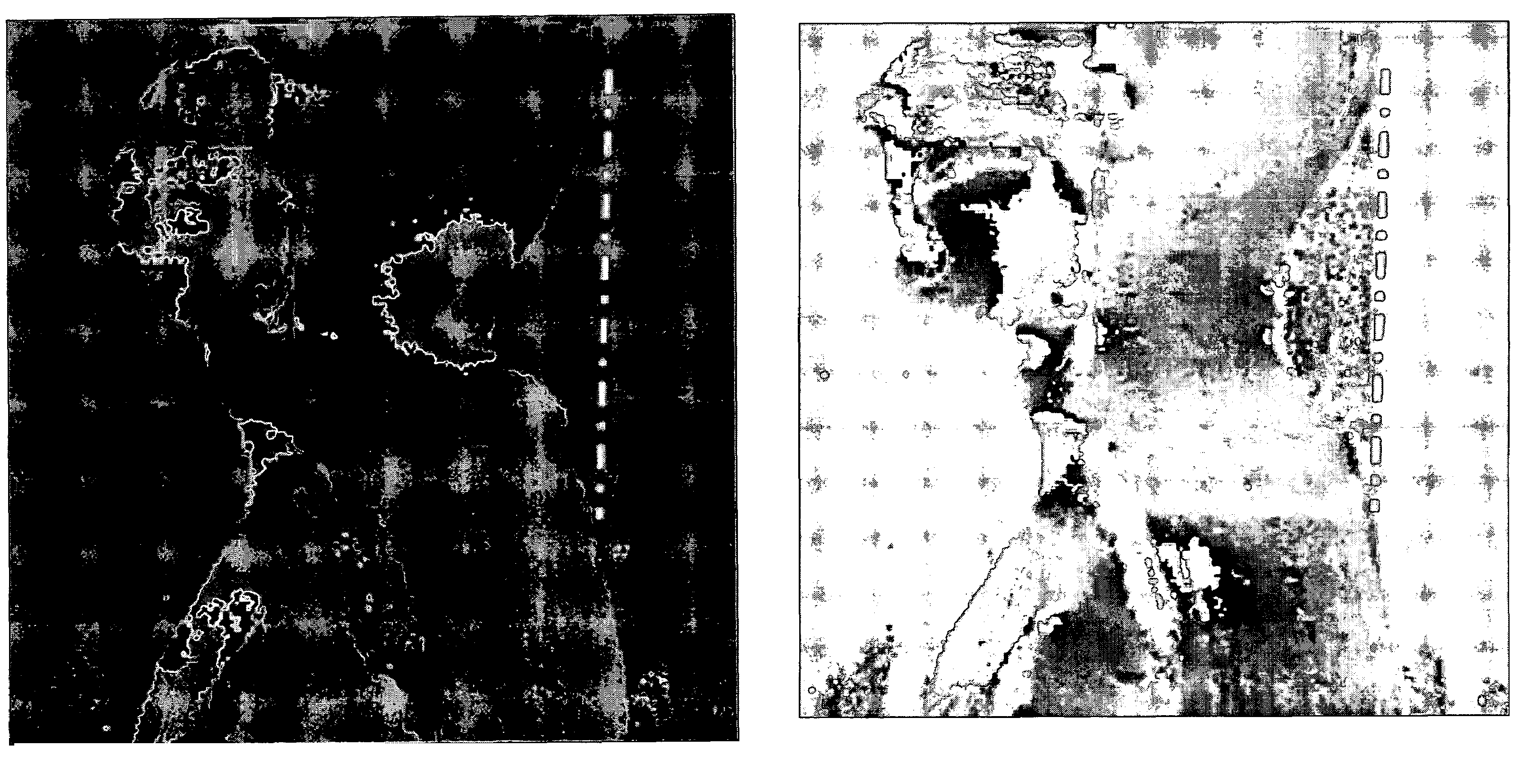
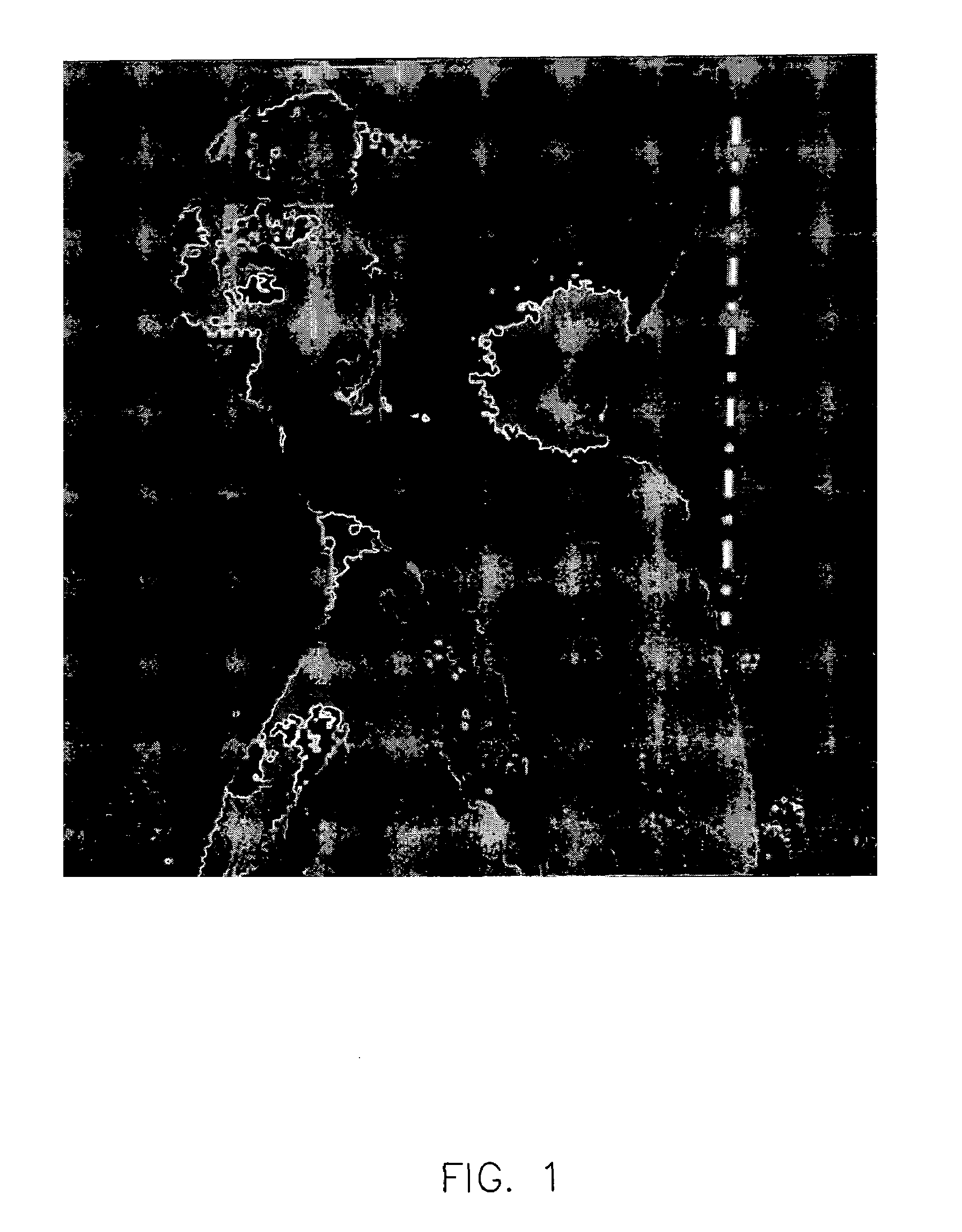
[0032]Now referring to the drawings in detail wherein like numbered elements refer to like elements throughout, FIG. 1 shows a map of magnetic field inhomogeneity. In a homogeneous field, the cross section of the person would be a uniform gray color. However, as shown, the shading varies from black to white due to the local inhomogeneity caused by the curvature of the neck. The dashed line from back of the torso to the back of the head is used to show the amount of inward curvature of the neck. Magnetic flux density is a vector quantity, defined by both a magnitude and a direction. The direction of the magnetic flux density in the magnet bore center is aligned parallel with the cylindrical bore magnet, usually annotated in images as the Superior-Inferior direction. From the torso to the head, the direction of the magnetic flux density lines are distorted due to the change in geometry in the neck region.

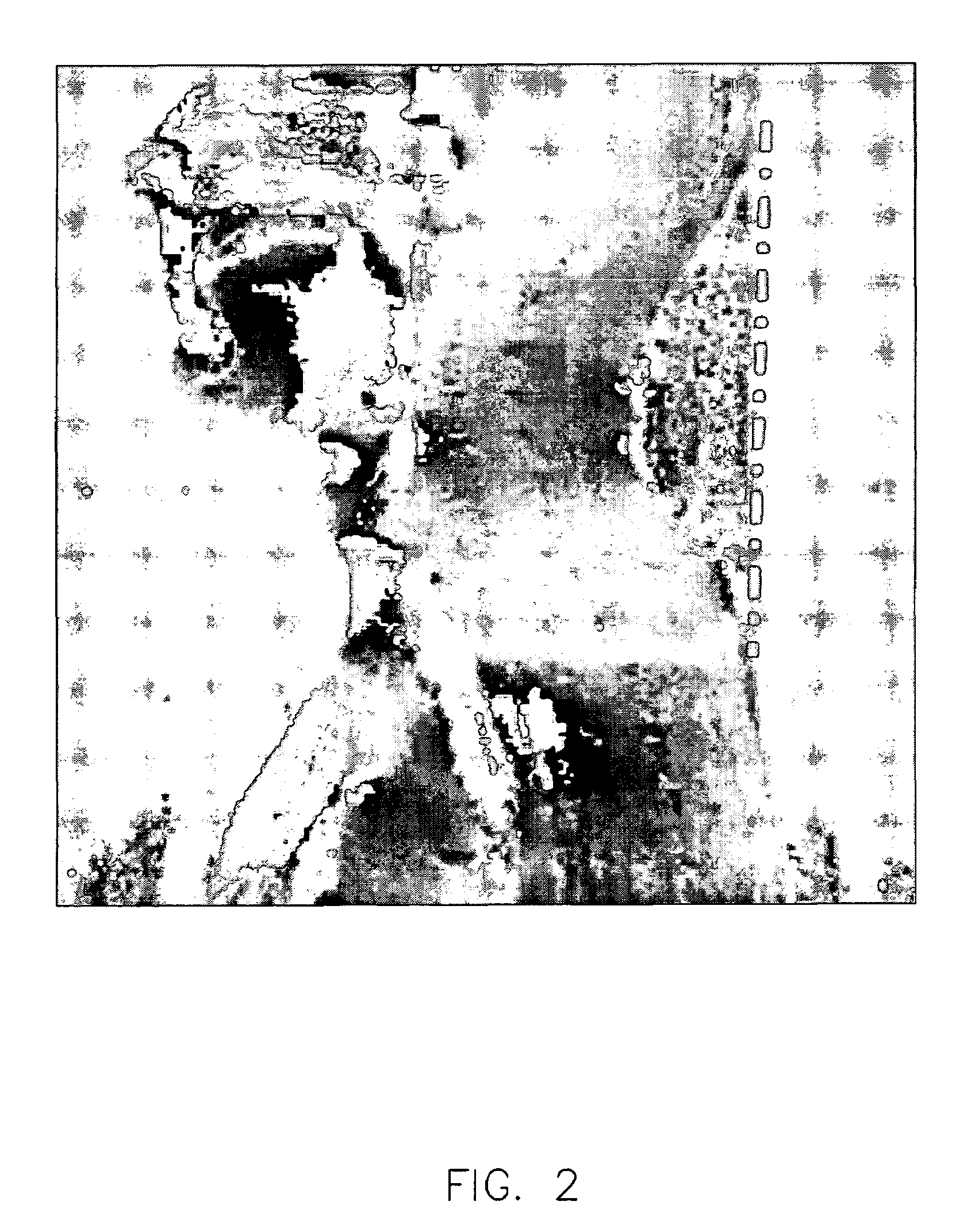
[0033]FIG. 2 shows a map of magnetic field inhomogeneity wherein the image has be...


PUM


Abstract

The present invention provides for a method for using a silicon dioxide (SiO2) material that is equally effective in improving magnetic field homogeneity during MR imaging. The present invention further provides for using silicon dioxide in its granular state. The granular silicon dioxide can then be poured inside a flexible container and the flexible container can be formed to fit any contour of the body.

Description

BACKGROUND OF THE INVENTION[0001]The present invention relates to a method for improving field homogeneity during magnetic resonance imaging when the homogeneity has been degraded by local susceptibility. More specifically, the invention relates to a method of placing a material with a magnetic susceptibility similar to body tissue in proximity to a part of a patient's body being imaged with a magnetic resonance imaging system and then imaging that body part, whereby field homogeneity is improved such that a homogeneity sensitive protocol provides an improved image of the body part.[0002]Magnetic resonance imaging, or MRI, is a method by which the location, size, and conformation of organs and other structures of the body may be determined. In the typical MRI system, a magnetic field is established across a body to align the spin axes of the nuclei of a particular chemical element, usually hydrogen, with the direction of the magnetic field. The aligned, spinning nuclei execute prece...

Claims


Application Information

Patent Timeline
Patent Type & Authority Patents(United States)
IPC IPC(8): G01R33/387G01R33/38
CPCG01R33/387
Inventor VAVREK, ROBERT M.BECERRA, RICARDOGUCLU, CEYLAN C.
Owner GE MEDICAL SYST GLOBAL TECH CO LLC
Learn More