Method for assaying protein nitrosylation
a protein nitrosylation and assay technology, applied in the direction of peptides, enzymology, material analysis by using resonance, etc., can solve the problems of complex photolytic-chemiluminescence technique used in these studies, s-nitrosylation cannot be elicited, and the inability to achieve widespread application
- Summary
- Abstract
- Description
- Claims
- Application Information
AI Technical Summary
Benefits of technology
Problems solved by technology
Method used
Image
Examples
example 1
Methods
Preparation of S-Nitroso-H-ras
[0031]GST-H-Ras was prepared in E. coli, bound to glutathione agarose, and eluted with 10 mM glutathione as described33. Glutathione was removed by passing the material through a MicroBioSpin-6 column (Biorad) equilibrated in HEN buffer (250 mM Hepes, pH 7.7, 1 mM EDTA, 0.1 mM neocuproine). GST-H-ras was incubated with 100 μM SNAP, 100 μM NAP, or the DMSO vehicle for 1 h at room temperature in the dark. The SNAP or NAP was removed by passing the material through a spin column as above three times.
[0032]Freshly isolated rat cerebella was homogenized in 50 volumes HEN buffer and then centrifuged at 800 g for 10 min at 4° C. The supernatant typically contained 0.8 μg protein per μl based on the BCA protein assay and was adjusted to 0.4% CHAPS. To 100 μl of the supernatant was added the appropriate concentration of the NO donor or control compound in a volume of 5 μl, and the mixture was incubated for one h at 25° C. Following th...
example 2
Assay Validation
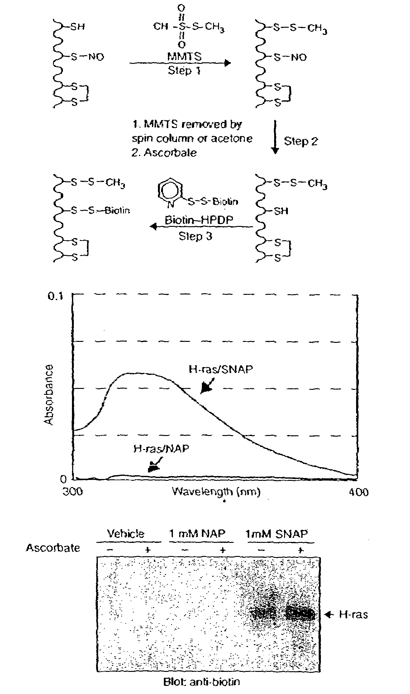
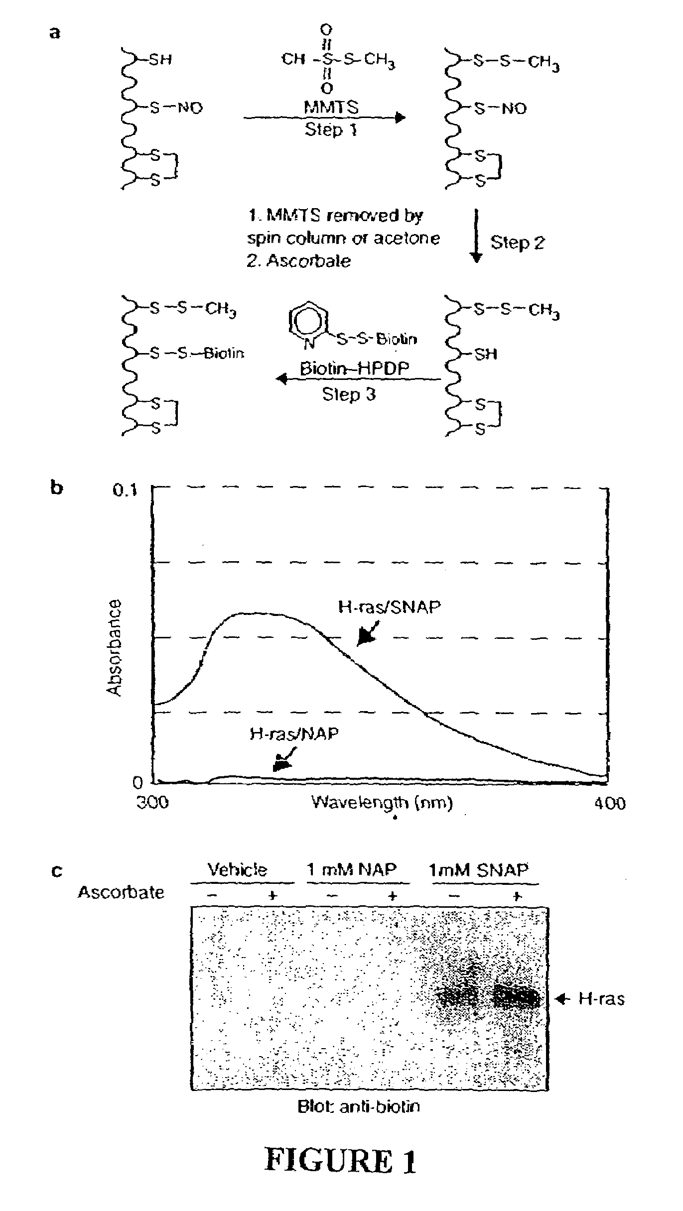
[0123]As an initial test of the system, we prepared S-nitroso-H-ras by incubation with NO donors. H-ras treated with S-nitrosoacetylpenicillamine (SNAP) is readily S-nitrosylated on a single cysteine5 which is detected spectrophotometrically by the characteristic absorbance of its nitrosothiol moiety (FIG. 1b). Reduced H-ras is prepared by incubating H-ras with the control compound N-acetylpenicillamine (NAP). In the S-nitrosylation assay, S-nitroso-H-ras is biotinylated (FIG. 1c), while H-ras, which lacks the nitrosothiol moiety, is unreactive. Ascorbate enhances the reactivity of S-nitroso-H-ras in the assay consistent with the idea that ascorbate accelerates the rate of nitrosothiol decomposition. Ascorbate does not appear to be essential for biotinylation in this assay, presumably because of spontaneous nitrosothiol decomposition. The specific biotinylation of the S-nitrosylated protein indicates that only S-nitrosylated proteins are capable of producing a signal...
example 3
Identification of Proteins Sensitive to Exogenous Nitrosylation
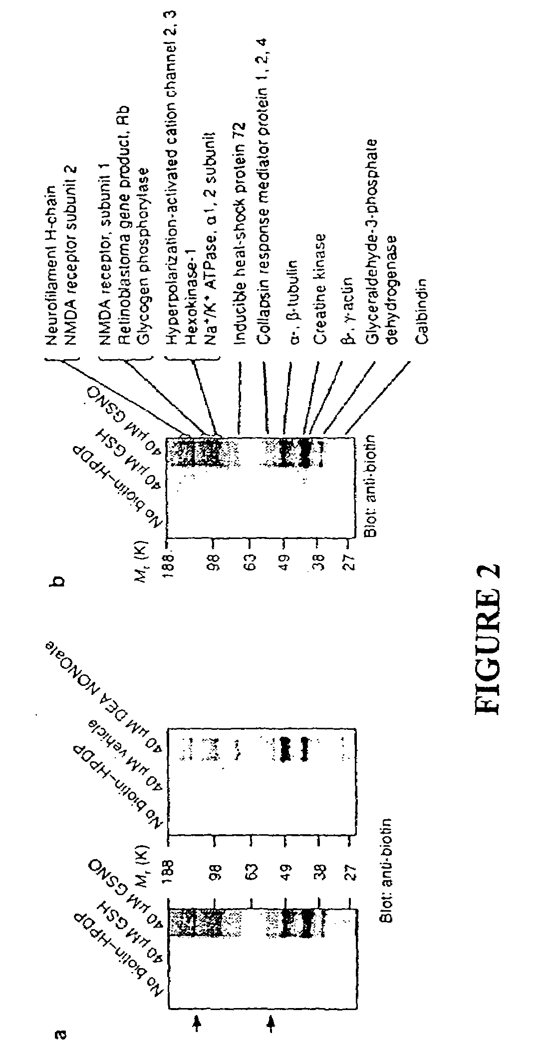
[0124]To identify tissue proteins that are uniquely sensitive to S-nitrosylation, we reacted brain lysates with NO donors of two different classes, 2-(N,N-diethylamino)-diazenolate-2-oxide (DEA-NONOate) and S-nitroso-glutathione (GSNO). GSNO is present in the brain at micromolar concentrations and might serve as an endogenous reservoir of NO groups16 and S-nitrosylate proteins by direct transnitrosation of thiols17. Following incubation with the NO donors, the tissue was passed through a spin column to remove the NO donors and then subjected to the S-nitrosylation assay. Biotinylated proteins were detected by immunoblotting with a biotin-specific antibody (FIG. 2a). A group of ˜15 protein bands are S-nitrosylated following treatment with either class of NO donor. In tissue preps subjected to the S-nitrosylation assay but in which the biotin-HPDP was omitted, faint reactivity of certain bands are visible, representing end...
PUM
| Property | Measurement | Unit |
|---|---|---|
| pH | aaaaa | aaaaa |
| pH | aaaaa | aaaaa |
| volume | aaaaa | aaaaa |
Abstract
Description
Claims
Application Information
 - R&D
- Intellectual Property
- Life Sciences
- Materials
- Tech Scout
- Unparalleled Data Quality
- Higher Quality Content
- 60% Fewer Hallucinations
Browse by: Latest US Patents, China's latest patents, Technical Efficacy Thesaurus, Application Domain, Technology Topic, Popular Technical Reports.
© 2025 PatSnap. All rights reserved.Legal|Privacy policy|Modern Slavery Act Transparency Statement|Sitemap|About US| Contact US: help@patsnap.com


