
Method and apparatus for monitoring blood condition and cardiopulmonary function

a technology for cardiopulmonary function and monitoring method, which is applied in the field of monitoring a patient's cardiopulmonary function and blood condition, can solve the problems of difficult and costly evaluation of a patient's cardiac output and respiratory efficiency, process cannot be performed as quickly as desirable, and analysis is costly

Inactive Publication Date: 2006-03-07
SENSICOR
View PDF61 Cites 86 Cited by
  • Summary
  • Abstract
  • Description
  • Claims
  • Application Information

AI Technical Summary

Benefits of technology

[0008]According to the present invention, electronically operated sensors are utilized to determine or evaluate certain characteristics of blood in certain major thoracic blood-containing structures, including particularly the pulmonary artery or the aorta of a mammal, particularly a human patient, and to obtain the desired information regarding those blood characteristics substantially instantaneously, without having to withdraw blood from the patient's body to analyze it. By measuring a characteristic such as oxygen saturation of hemoglobin of blood in certain blood vessels or portions of the heart the patient's pulmonary function can be evaluated. By measuring the level of oxygen saturation of the hemoglobin in mixed venous blood such as is found in the pulmonary artery, a reasonable estimate of cardiac function can be deduced.
[0009]By comparing the level of oxygen saturation of the hemoglobin in the mixed venous blood, such as is found in the pulmonary arteries, or in non-mixed venous blood such as is found in the superior or inferior vena cava, with the level of oxygen saturation in recently oxygenated blood, as may be found in the aorta or the left atrium, the efficiency of a patient's heart and lungs relative to the body's extraction of oxygen from the blood can be evaluated quickly and easily, so that a clinician can determine what, if any, intervention may be necessary for improvement of the patient's condition.
[0010]In addition, such an evaluation of the patient's condition on a repeated basis during and immediately after cardiac surgery can inform health care personnel whether a patient is satisfactorily enduring a surgical procedure and whether the patient's heart, lungs and other organs are performing as expected during recovery from surgery. Similarly, such repeated evaluation can inform health care experts as to whether devices such as an artificial heart or a ventricular assist device is providing the body with enough oxygenated blood. Such repeated evaluations could be used to signal such a support device, or a cardiac pacemaker, to increase or decrease its rate of operation in order to accommodate the variations in oxygen requirements of the body during exercise as compared with rest. By measuring and comparing the level of oxygen saturation of the hemoglobin of the blood in various parts of the heart or in various other blood vessels near the base of the heart various imperfections such as an inefficient part of lung, or an abnormal non-physiologic leak or “shunt” between the chambers of the heart, may be detected and surgical repair thereof may be evaluated.

Problems solved by technology

Using conventional techniques and available apparatus it is difficult and costly to evaluate a patient's cardiac output and respiratory efficiency, and the process cannot be performed as quickly as desirable.
Such analysis is costly, and it has usually not been practical to obtain such blood samples.
A measurement obtained using such a device can be used to evaluate major changes in arterial blood oxygenation, but does not provide enough information for determining a patient's cardiac output, since it does not provide enough information regarding oxygen extraction or utilization by tissues and thus is not a good enough tool for valid evaluation of a patient's cardiopulmonary function during and after cardiac surgery.
This is a relatively invasive procedure, however, and can be used for only a limited time, after which the sensors would become covered with protein deposits from the blood and would thereby lose their sensitivity.
There is no currently available implantable device that remains separate from and outside the flow of blood for measuring oxygen saturation in blood without blood samples having to be drawn for analysis.

Method used


Image

  • Method and apparatus for monitoring blood condition and cardiopulmonary function
  • Method and apparatus for monitoring blood condition and cardiopulmonary function
  • Method and apparatus for monitoring blood condition and cardiopulmonary function

Examples


Embodiment Construction

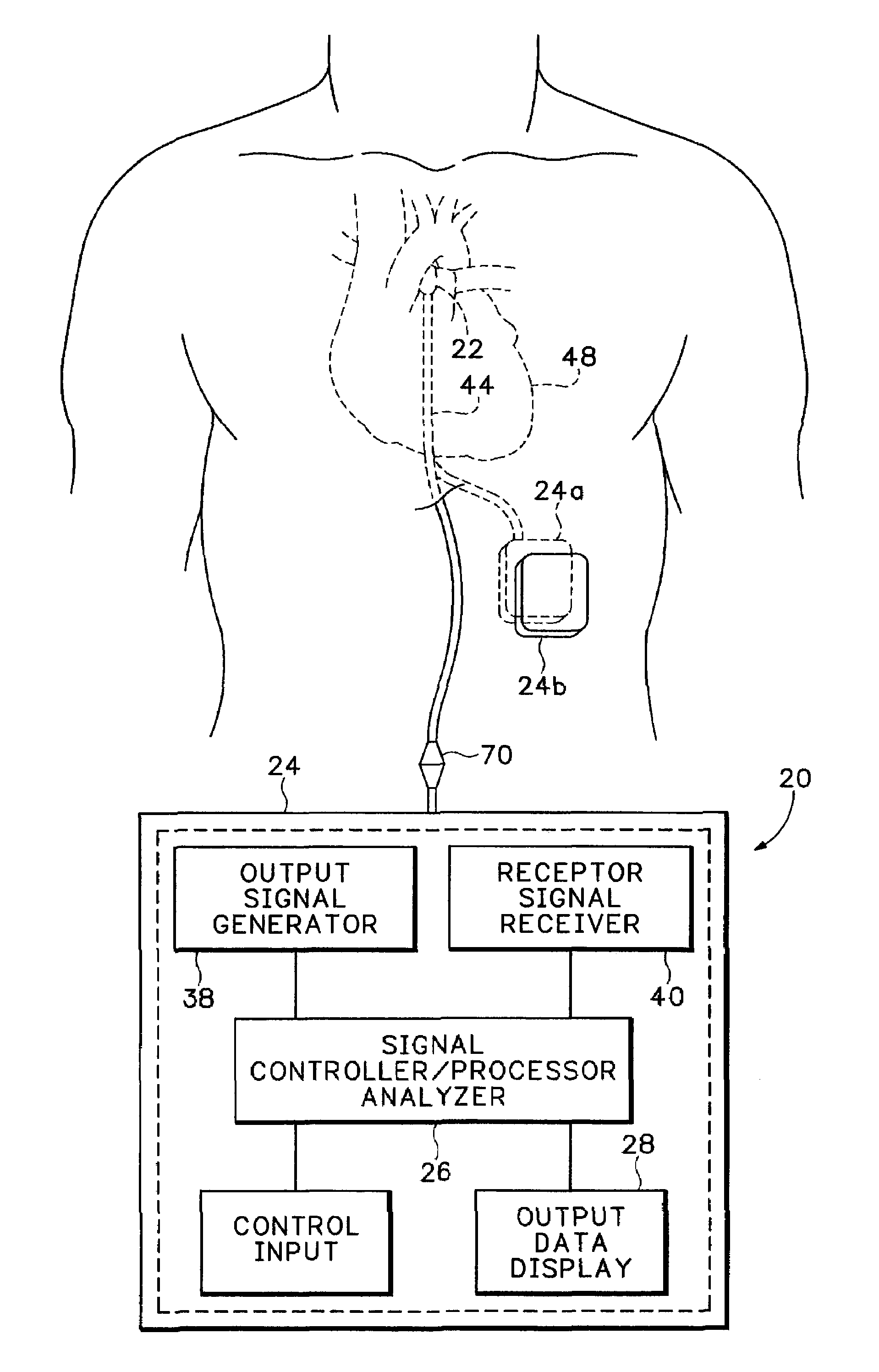
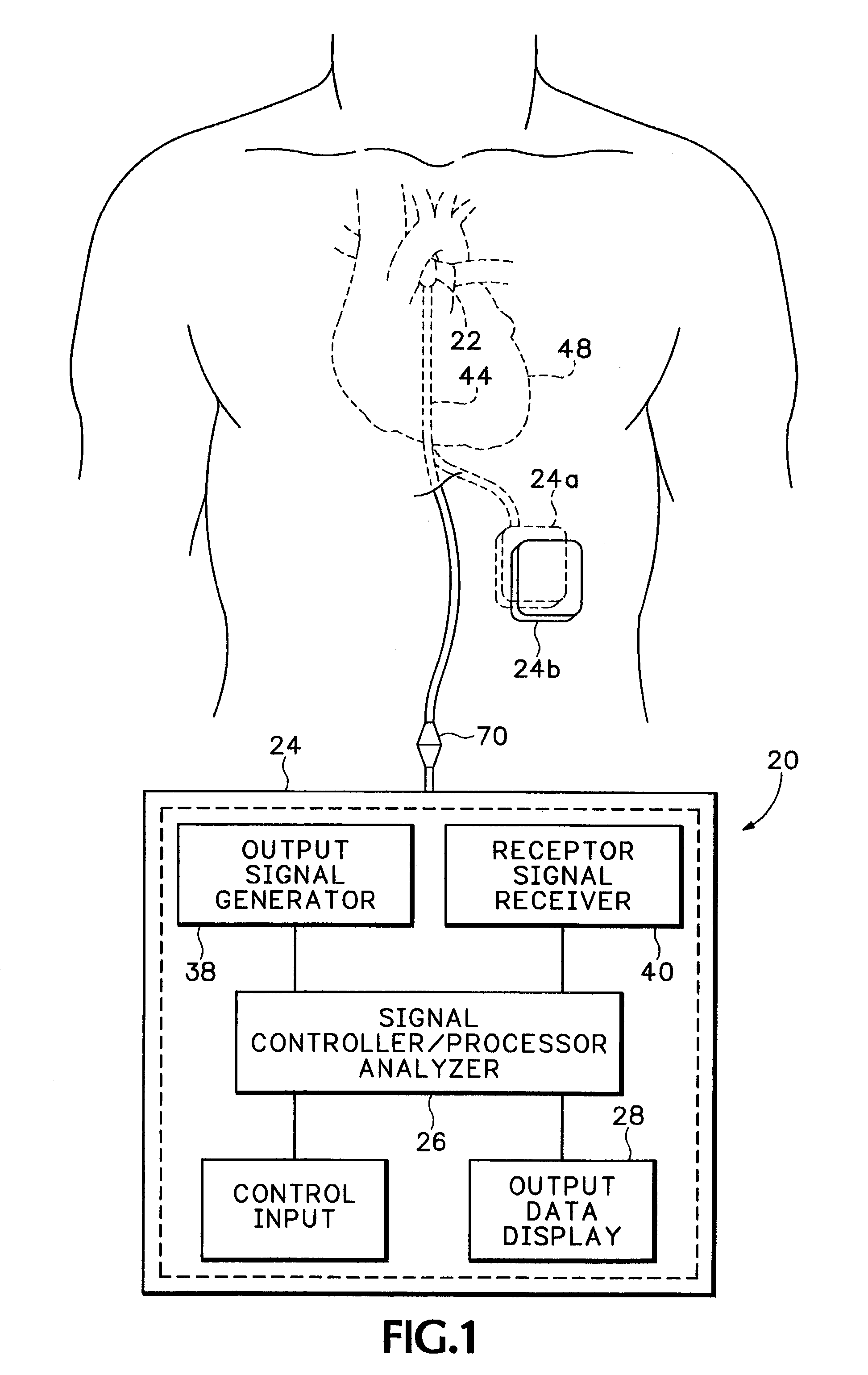
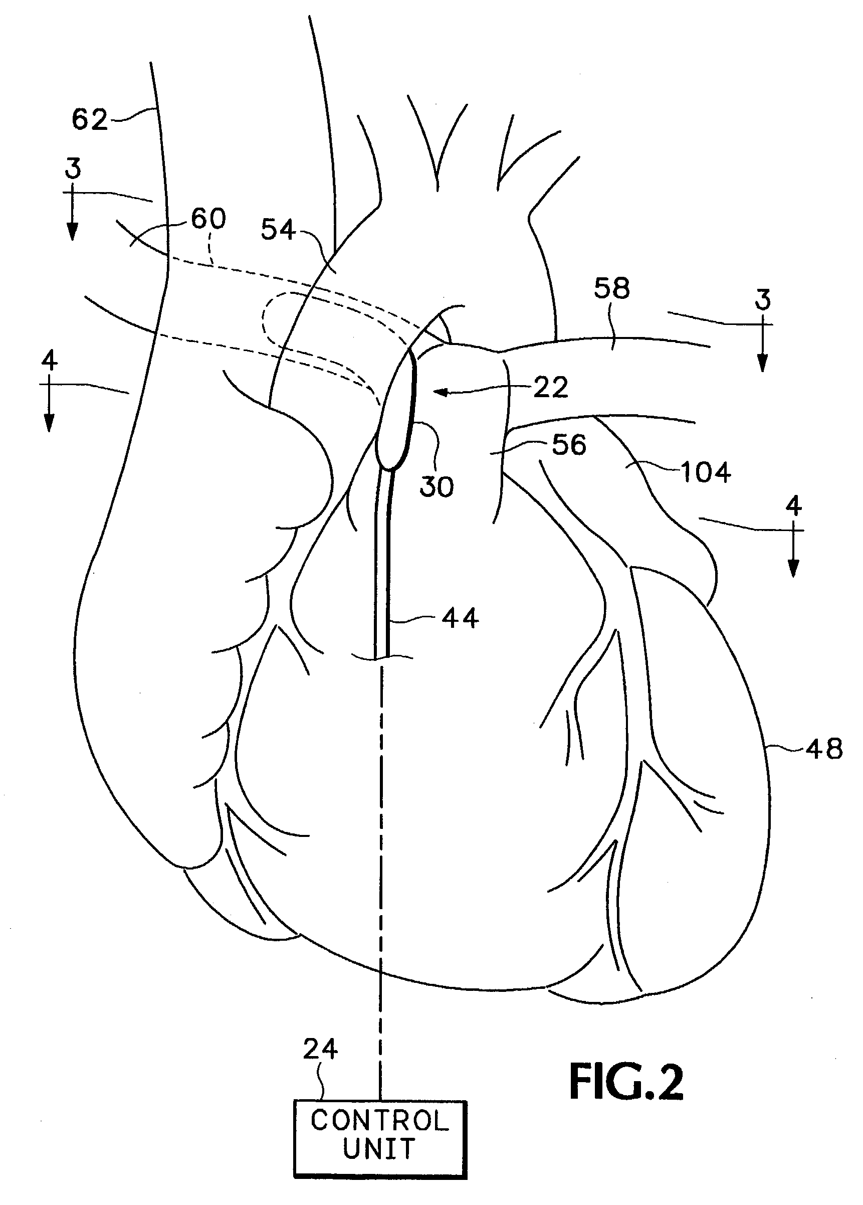
[0035]Referring now to FIGS. 1–7 of the drawings which form a part of the disclosure herein, a blood condition monitor 20 includes an implantable sensor section 22 and an electronics portion, or control unit 24, which may include an electronic controller and processor package 26 and an associated output data display section 28. The sensor section 22 of the blood condition monitor 20 includes a sensor carrier 30 and associated non-invasive sensors 32 and 34 used to quickly and conveniently determine the condition of a patient's blood without the need to withdraw blood samples from the patient.

[0036]The control unit 24 shown in simplified form in FIGS. 1–2 includes an electronic emitter signal generator portion 38, an electronic receptor signal receiver portion 40, and the output data display section 28. Preferably, the control unit 24 is provided as a self-contained unit incorporating suitable integrated circuit logic and data handling components to accept user instructions and provi...


PUM


Abstract

Sensors located on a sensor carrier are placed adjacent one or more of a surgical patient's major thoracic blood-containing structures such as the aorta or pulmonary artery, and characteristics of blood in the blood-containing structures are determined noninvasively by measuring transmission or reflection of light or other types of energy by the blood. Emitters and receptors included in the sensors are connected electrically with suitable electronic signal generating and processing components in a package remote from the sensor carrier.

Description

BACKGROUND OF THE INVENTION[0001]The present invention relates to monitoring a patient's cardiopulmonary function and blood condition, and particularly to a method and apparatus for monitoring the performance of a patient's heart and lungs during and following thoracic surgery.[0002]Cardiac output means the volume rate at which the heart pumps blood. The cardiac output is important to a clinician as an indication of how well a person's heart is able to function. Using conventional techniques and available apparatus it is difficult and costly to evaluate a patient's cardiac output and respiratory efficiency, and the process cannot be performed as quickly as desirable. Following surgery, and particularly heart surgery, it would be advantageous to be able to determine quickly and frequently how well a patient's heart and lungs are performing in delivering oxygenated blood to the patient's tissues in relation to how quickly the same tissues are removing the oxygen with which they are be...

Claims


Application Information

Patent Timeline
Patent Type & Authority Patents(United States)
IPC IPC(8): A61B5/00
CPCA61B5/14542A61B5/1459A61B2562/164A61B2562/043A61B2562/0233
Inventor FURNARY, ANTHONY P.LOWE, ROBERT I.
Owner SENSICOR
Learn More