Excisional devices having selective cutting and atraumatic configurations and methods of using same

a technology of selective cutting and surgical devices, applied in the field of distal tips, can solve the problems of time-consuming, multi-step process of mammography, and the threat of breast cancer, and achieve the effects of reducing the risk of breast cancer

Inactive Publication Date: 2006-04-18
ENCAPSULE MEDICAL
View PDF108 Cites 32 Cited by
  • Summary
  • Abstract
  • Description
  • Claims
  • Application Information

AI Technical Summary

Benefits of technology

[0027]The present invention, according to another embodiment thereof, is a surgical device for acting upon biological tissue, comprising a shaft defining a proximal end and a distal end; a work assembly adapted to act upon the tissue, the work assembly being disposed near the distal end of the shaft and including a sheath that may be configured to at least partially trail the distal end of the shaft as the device may be removed from the tissue, and a distal tip, the distal tip including a cutting surface that may be adapted to cut the tissue, the distal tip being configured such that the cutting surface does not damage the sheath as the device is removed from the tissue.

Problems solved by technology

Breast cancer is a major threat and concern to women.
The conventional method for diagnosing, localizing and excising nonpalpable lesions detected by mammography generally involves a time-consuming, multi-step process.
Obtaining good margins of normal tissue is extremely dependent upon the skill and experience of the surgeon, and often an excessively large amount of normal breast tissue is removed to ensure that the lesion is located within the specimen.
This increases the risk of post-operative complications, including bleeding and permanent breast deformity.
As 80% of breast biopsies today are benign, many women unnecessarily suffer from permanent scarring and deformity from such benign breast biopsies.
Fine needle aspiration is also very dependent upon the skill and experience of the operator and can result in a high non-diagnostic rate (up to about 83%), due to inadequate sample collection or preparation.
The stereotactic table requires awkward positioning of the patient and may be extremely uncomfortable.
The woman must lie prone during the entire procedure, which may be impossible for some patients.
This may be extremely difficult and uncomfortable for the patient if the lesion is very posterior near the chest wall or high towards the axilla.
Bleeding is generally not an issue with fine needle aspiration, but is a legitimate complication of the former two methods.
This causes increased post-procedural pain and delays healing.
However, documented instances of cancerous cells seeding locally into needle tracks exist.
The long-term consequences of neoplasm seeding into needle tracks are currently unknown, again because the techniques are relatively new.
The breast, however, may be torqued and distorted between the compression plates such that when the plates are removed after the biopsy, the large core track left behind may not be straight, but actually tortuous.
This can result in permanent breast deformity.
For the fine needle aspirate, core biopsy and vacuum-assisted devices, the incision is usually very small and the scar almost unappreciable.
Such a large incision often results in a non-aesthetically placed scar.
They are not, however, the final solution.
Also, the conventional biopsy devices do not consider margins in their excisions and if cancer is diagnosed, the patient must undergo a needle-localized breast lumpectomy to ensure that adequate margins are removed around the cancer.
However, while retracting a cut and isolated specimen from the surrounding mass of tissue, the cutting edge of the distal tip may damage the cutting and / or isolating assemblies, especially if the cut and isolated specimen trails the distal tip of the device as the device is retracted from the tissue.
Moreover, the distal cutting tip of known current excisional devices may prevent the excisional device from operating in close proximity to structures in the body that must not be damaged or cut by the excisional device.

Method used


Image

  • Excisional devices having selective cutting and atraumatic configurations and methods of using same
  • Excisional devices having selective cutting and atraumatic configurations and methods of using same
  • Excisional devices having selective cutting and atraumatic configurations and methods of using same

Examples


Embodiment Construction

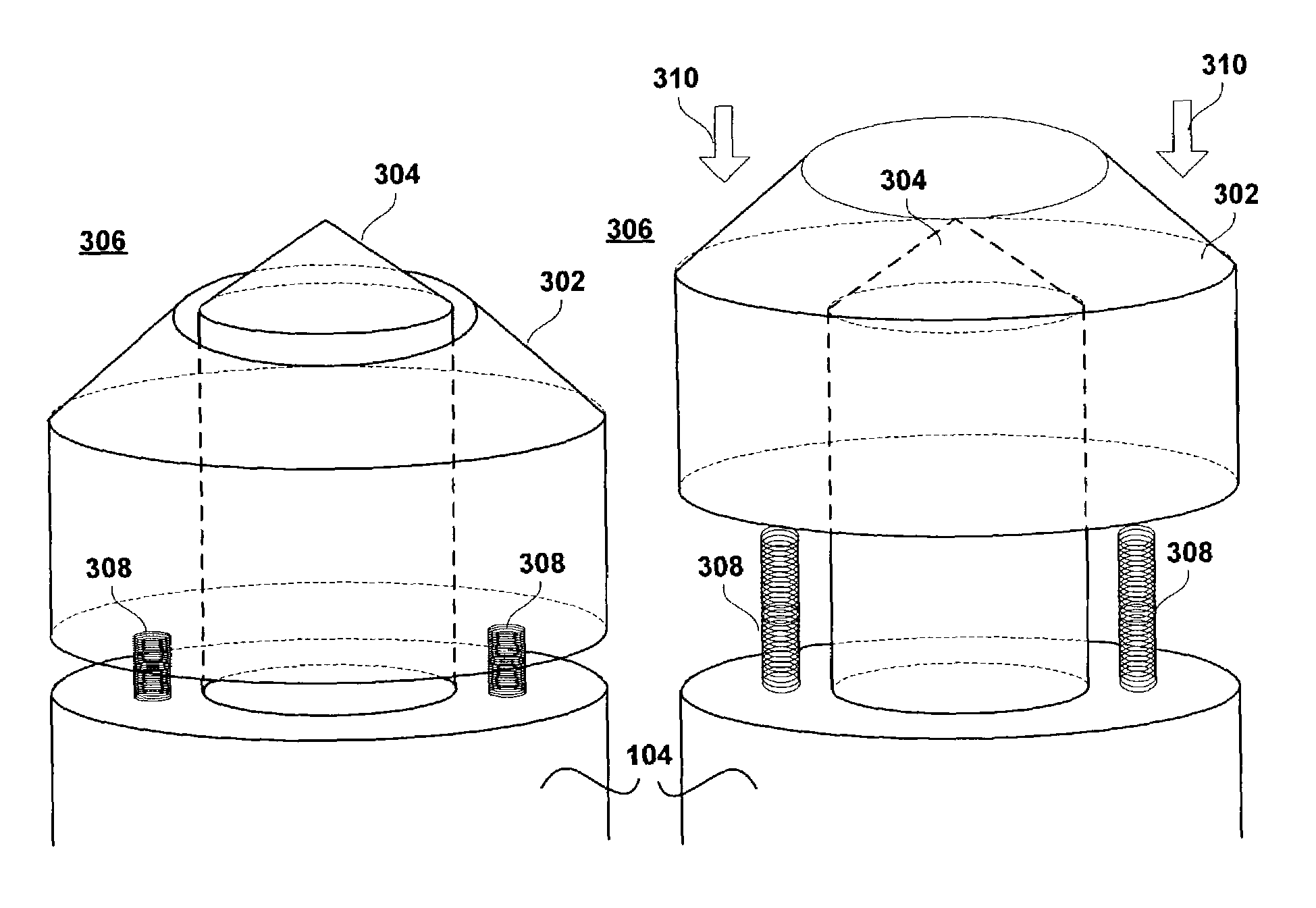
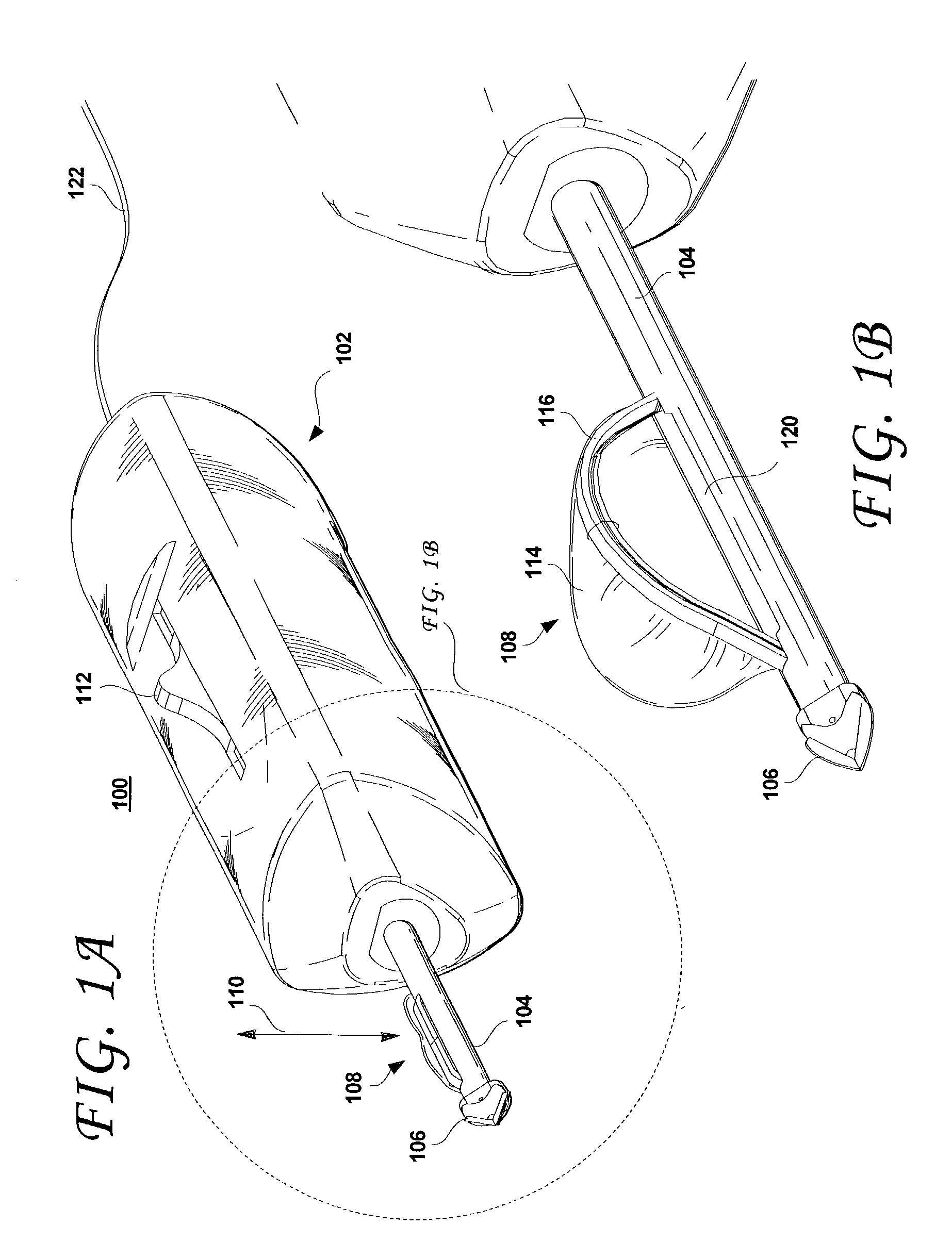
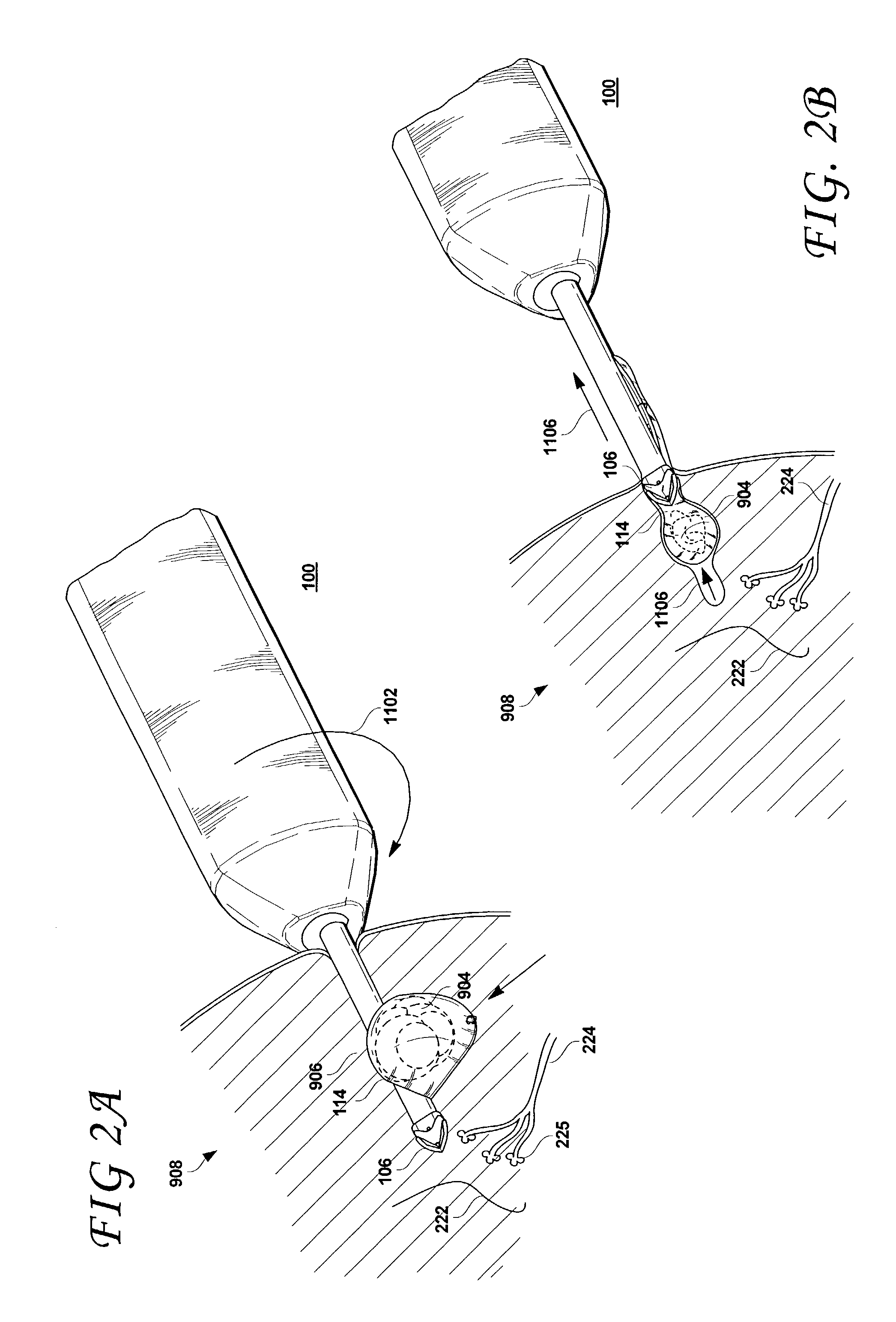
[0063]FIG. 1A shows an excisional biopsy device having a conventional distal tip. The excisional biopsy device shown in FIGS. 1A through 2B is disclosed in commonly assigned and co-pending US patent application entitled “Methods And Devices For Cutting And Collecting Soft Tissue”, Ser. No. 10 / 189,277 filed on Jul. 3, 2002, the disclosure of which is incorporated herewith by reference in its entirety. As shown, the excisional device 100 includes a proximal section 102 that may be configured to fit the physician's hand (or that may alternatively be configured for a stereotactic apparatus). Extending from the proximal section 102 is a shaft 104 that is terminated by a distal tip 106. The distal tip 106 is configured so as to easily penetrate a mass of tissue. The distal tip 106 may be configured to be energized by a radio frequency (RF) energy source, supplied via the electrical cord 122. However, the distal tip 106 need not be energized, as the cutting surface(s) of the distal tip 106...


PUM


Abstract

Surgical devices include a selectively cutting and atraumatic distal tip that is configured to assume a first configuration in which cutting surface or surfaces thereof are effective to cut tissue and a second configuration in which the cutting surface or surfaces thereof are ineffective to cut tissue.

Description

BACKGROUND OF THE INVENTION[0001]1. Field of the Invention[0002]The present invention pertains to the field of surgical devices. In particular, the present invention relates to distal tips for such devices and associated methods for treating soft tissue, such as breast tissue.[0003]2. Description of the Related Art[0004]Breast cancer is a major threat and concern to women. Early detection and treatment of suspicious or cancerous lesions in the breast has been shown to improve long-term survival of the patient. The trend is, therefore, to encourage women not only to perform monthly self-breast examination and obtain a yearly breast examination by a qualified physician, but also to undergo annual screening mammography commencing at age 40. Mammography is the only screening modality available today that can detect small, nonpalpable lesions. These nonpalpable lesions may appear as opaque densities relative to normal breast parenchyma and fat or as clusters of microcalcifications.[0005]...

Claims


Application Information

Patent Timeline
Patent Type & Authority Patents(United States)
IPC IPC(8): A61B10/00A61B10/02
CPCA61B10/0266
Inventor ANDERSON, SCOTT C.BROUNSTEIN, DANIEL M.CHERNOMORSKY, ARY S.CLIFFORD, MARK J.VETTER, JAMES W.
Owner ENCAPSULE MEDICAL
Why Eureka
Try Eureka