Eureka AIR delivers breakthrough ideas for toughest innovation challenges, trusted by R&D personnel around the world.

Attenuation of fibroblast proliferation

a fibroblast and proliferation technology, applied in the direction of lyase, peptide/protein ingredient, drug composition, etc., can solve the problems of inability to determine biological activity, no inhibitors which can selectively block synthesis or expression, and inhibit expression, so as to reduce the proliferative response of cells, reduce the secretion of collagen, and reduce the effect of growth factor receptors

Inactive Publication Date: 2006-06-06
BIOMARIN PHARMA INC
View PDF8 Cites 2 Cited by
  • Summary
  • Abstract
  • Description
  • Claims
  • Application Information

AI Technical Summary

Benefits of technology

[0011]Highly purified and specific glycosaminoglycan degrading enzymes, chondroitinase B and chondroitinase AC, are used to treat fibroproliferative diseases. The enzymatic removal of chondroitin sulfate B, and to a lesser extent, chondroitin sulfate A or C, from cell surfaces effectively decreases growth factor receptors on the cells and thereby decreases the cell proliferative response to such growth factors. In addition, removal of chondroitin sulfates reduces secretion of collagen, one of the major extracellular matrix components. Through the combined inhibition of fibroblast proliferation and collagen synthesis, treatment with chondroitinase B or chondroitinase AC decreases the size of fibrous tissue found in psoriasis, scleroderma, keloids and surgical adhesions.

Problems solved by technology

However both of these compounds inhibit the expression of all types of sulfated glycosaminoglycans.
There are currently no inhibitors which can selectively block the synthesis or expression of chondroitin sulfate A, B or C. However, specific glycosaminoglycan lyases which can remove either heparan or chondroitin sulfates A, B or C from cells are available.
Since chondroitinase ABC acts on more than one type of chondroitin sulfate, it is not possible to determine the biological activity of the individual types of chondroitin sulfates using this enzyme.

Method used


Image

  • Attenuation of fibroblast proliferation
  • Attenuation of fibroblast proliferation
  • Attenuation of fibroblast proliferation

Examples


example 1

Enzyme Substrate Specificity

[0043]Chondroitinase B (no EC number) and chondroitinase AC (EC 4.2.2.5) are recombinant proteins expressed in Flavobacterium heparinum (PCT / US95 / 08560 “Chondroitin Lyase Enzymes”). Specific activity and substrate specificity were determined for each enzyme, using a kinetic spectrophotometric assay, performed essentially as described in PCT / US95 / 0856. In these assays, enzyme concentrations were 0.25 IU / ml and substrate concentrations were 0.5 mg / ml (chondroitin sulfate B and chondroitin sulfate AC) or 0.75 mg / ml (heparan sulfate). The specific activities of the enzymes were: 97 IU / mg for Chondroitinase B and 221 IU / mg for chondroitinase AC.

[0044]The substrate specificity of ultra-purified Chondroitinase B and AC were determined by testing the ability of the enzymes to degrade chondroitin sulfate B, chondroitin sulfate A, chondroitin sulfate C, and heparan sulfate. As shown in Table 1, both enzymes were active towards the corresponding sulfated glycosamino...

example 2

Removal of Glycosaminoglycans from Cells

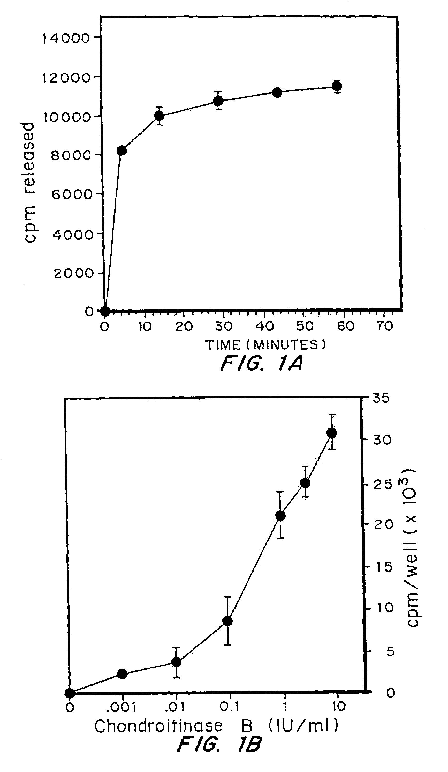
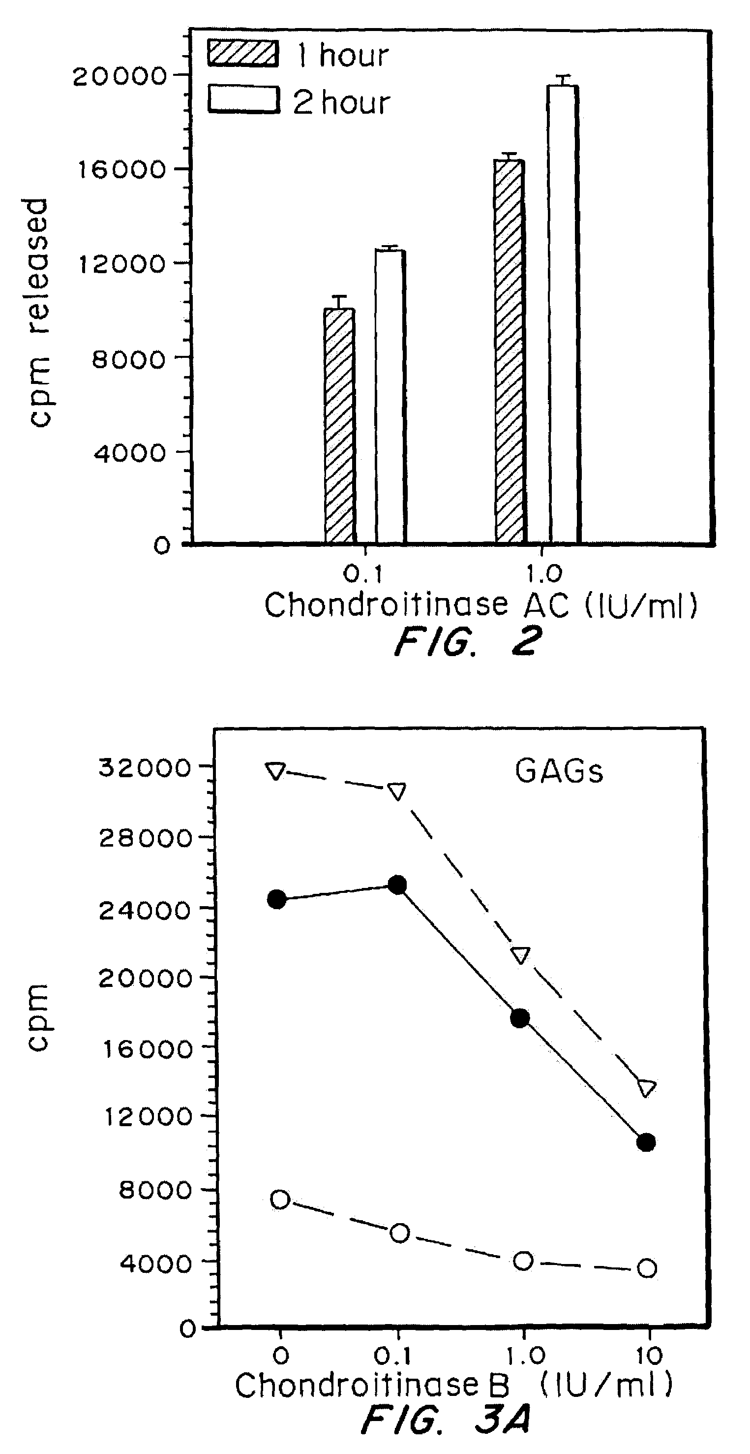
[0046]The effectiveness of the chondroitinases B and AC in removing sulfated glycosaminoglycans from cells was examined using cells with glycosaminoglycans labeled by incubation with 35S. Fibroblasts were plated at a density of 6×10⁴ cells / well in 24 well plates, in DMEM with 10% serum. After 24 hrs, medium was changed to Fisher's medium containing 10% serum and 25 microCi / ml of Na235SO4, and incubation continued for 2.5 days. The medium was removed and cells rinsed 2× with DMEM then treated with Chondroitinase B or AC as indicated. Medium was removed and radioactivity determined. The release of sulfated glycosaminoglycans from cells by enzyme was expressed as cpm / well of chondroitinase-treated cells minus cpm / well of untreated cells.

[0047]Cells were exposed to 1.0 IU / ml of Chondroitinase B, at 37° C. for variable lengths of time. As shown in FIG. 1A, maximal release of sulfated GAGs by chondroitinase B was achieved following an 1 hour exposur...

example 3

Effects on bFGF Binding

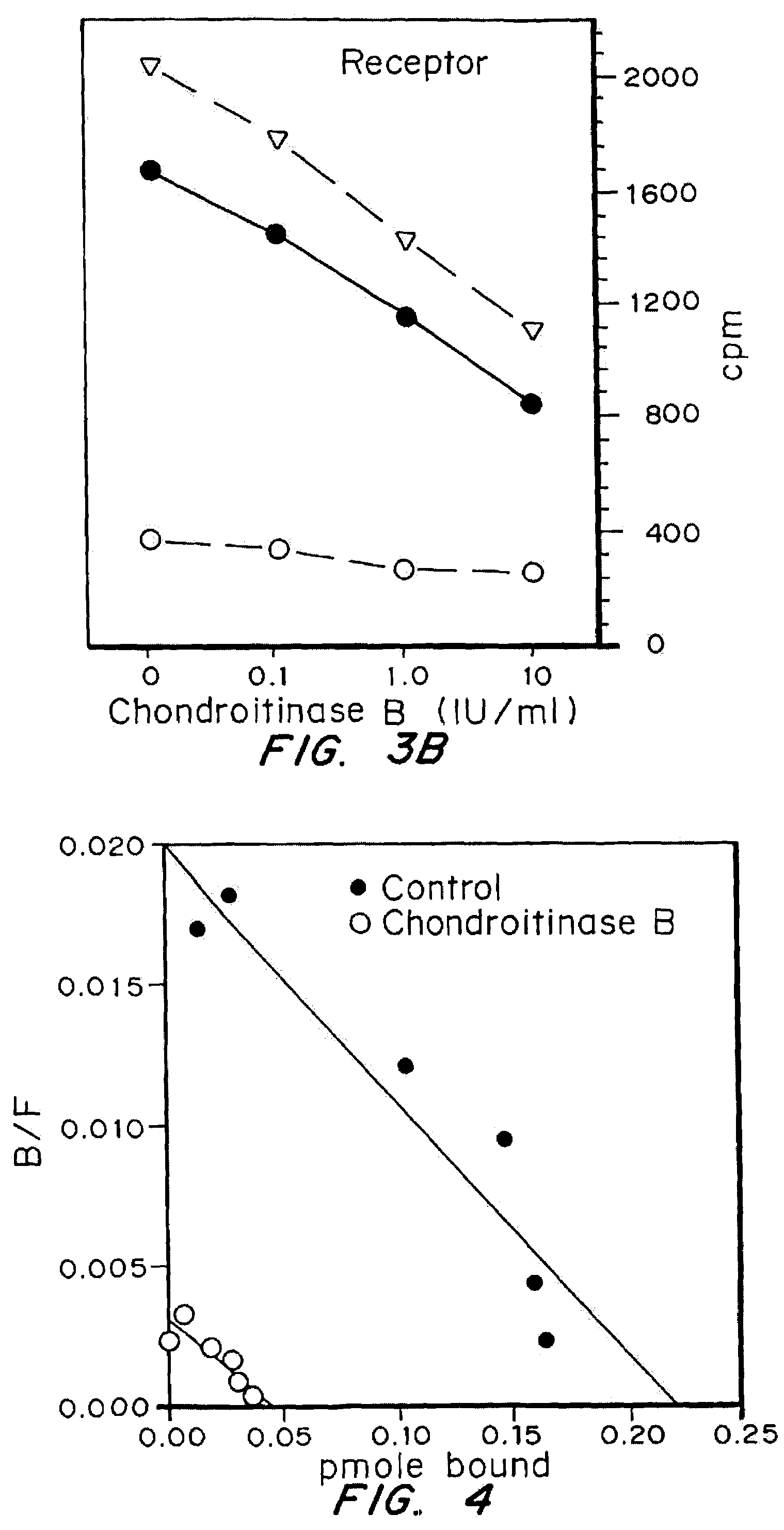
[0049]Iodinated bFGF was obtained from Dupont NEN, specific activity greater than 1200 Ci / mmol. Fibroblasts were plated in 48 well dishes and grown to confluence. Prior to binding assays, cells were treated with medium or enzyme as indicated for proliferation assays. Following enzyme treatments, cells were chilled and binding assays carried out at 4° C. Cells were incubated for one hour with 25 ng / ml of 1251-bFGF alone, or with the addition of 25 microg / ml of unlabelled bFGF in binding buffer (DMEM, 25 mM HEPES, 0.05% gelatin). Following incubation with bFGF, cells were washed 2× with ice cold binding buffer. Glycosaminoglycan-bound 1251-bFGF was removed with two rinses with wash buffer (2M NaCl in 20 mM HEPES, pH 7.4). Receptor bound 125I-bFGF was removed by washing 2× with wash buffer (pH 4.0) (Fannon and Nugent (1996) J. Biol. Chem. 271:17949–17956).

[0050]Numerous growth factors have been shown to bind to heparan sulfate proteoglycans on the cell surface. T...


PUM

PropertyMeasurementUnit
concentrationsaaaaaaaaaa
pHaaaaaaaaaa

Abstract

Highly purified and specific glycosaminoglycan degrading enzymes, chondroitinase B and chondroitinase AC, are used to treat fibroproliferative diseases. The enzymatic removal of chondroitin sulfate B(dermatan sulfate), and to a lesser extent, chondroitin sulfate A or C, from cell surfaces effectively decreases growth factor receptors on the cells and thereby decreases the cell proliferative response to such growth factors. In addition, removal of chondroitin sulfates reduces secretion of collagen, one of the major extracellular matrix components. Through the combined inhibition of fibroblast proliferation and collagen synthesis, treatment with chondroitinase B or chondroitinase AC decreases the size of fibrous tissue found in psoriasis, scleroderma, keloids, pulmonary fibrosis and surgical adhesions.

Description

[0001]This application claims priority to U.S. Ser. No. 60 / 168,518, filed Dec. 2, 1999.BACKGROUND OF THE INVENTION[0002]The present invention is a method and composition using chondroitinase B and chondroitinase AC, glycosaminoglycan degrading enzymes, to inhibit the formation of fibrotic tissue.[0003]Proteoglycans on the cell surface and in the extracellular matrix contain variable glycosaminoglycan chains, which include heparan sulfate and chondroitin sulfates A, B, or C. While some proteoglycans contain only one type of glycosaminoglycan, others contain a mixture of heparan and chondroitin sulfates (Jackson et. al., Physiol. Rev. 71:481–530,1991). Extracellular proteoglycans form a structural framework for cells and tissues, and together with cell-associated proteoglycans, have major functions in regulating cell adhesion, migration, and proliferation. The functions of proteoglycans and their component parts have been extensively studied, with much of the emphasis on the roles of ...

Claims


Application Information

Patent Timeline
Patent Type & Authority Patents(United States)
IPC IPC(8): C12N9/00A61K38/46A61K38/51A61P17/00A61P17/02A61P17/06A61P19/04A61P43/00
CPCC12Y402/02019A61K38/51A61P11/00A61P17/00A61P17/02A61P17/06A61P19/04A61P43/00
Inventor DENHOLM, ELIZABETH M.CAUCHON, ELIZABETHSILVER, PAUL J.
Owner BIOMARIN PHARMA INC
Why Eureka
Eureka Blog
Learn More