
Cardiac output measurement using dual oxygen sensors in right and left ventricles

a technology of right and left ventricles and oxygen sensors, applied in the field of cardiac monitoring, can solve the problems of inability to accurately detect current cardiac output, lack of mechanical ventricular synchrony, inability to adapt a pacing interval to changing cardiac output conditions, etc., and achieve the effect of continuous and accurate measurement, avoiding excessive fibrous growth and thrombosis, and simplifying deploymen

Inactive Publication Date: 2007-01-16
MEDTRONIC INC
View PDF53 Cites 91 Cited by
  • Summary
  • Abstract
  • Description
  • Claims
  • Application Information

AI Technical Summary

Benefits of technology

[0012]It is an object of the invention to provide a technique for measuring cardiac output accurately and continuously, and to improve the control of a pacing interval based on the measurement. It is a further object of the invention to provide a physician with cardiac output information useful in programming a pacing interval for an implantable medical device, and to enable adaptation of the pacing interval based on continuous measurements of cardiac output during operation of the device. In addition, it is an object of the invention to provide a measurement device that can be readily deployed with little risk to a patient, and thereafter provide accurate measurements of blood oxygen saturation for use in calculation of cardiac output.
[0013]In a typical embodiment described below, the invention may be applied to bi-ventricular pacing. In this application, the interval may be called the “V1–V2 interval,” which represents the time delay between delivery of pacing pulses to the ventricles. In some patients, simultaneous stimulation of the ventricles results in a lack of mechanical ventricular synchrony. The lack of synchrony may be caused by factors such as differences in placement of stimulating electrodes proximal to the ventricles or the differences in the conductive pathways of the ventricles. The lack of synchrony may cause the ventricles to begin ejection of blood at different times. For some patients, asynchronous blood ejection is inefficient and undesirable. The techniques of the invention may be useful in more accurately determining cardiac output, so that the ventricles can be brought into synchrony, resulting in improved hemodynamic performance.
[0016]The invention may also include a processor that computes a V1–V2 interval for bi-ventricular pacing such that pacing pulses, separated by this interval, cause the chambers to work in synchrony. In a typical application, the processor may set the interval to cause the right and left ventricles to commence blood ejection at substantially the same time in response to information including the measured cardiac output. In another application, the processor may set the interval to cause one ventricle to commence blood ejection prior to the other ventricle with a desired time offset, based on the cardiac output measured as described herein. The processor may further adjust the interval in response to changing conditions, such as a changes in heart rate, measured cardiac output, or other parameters. In addition, in some embodiments, the processor may drive a drug delivery device to deliver a drug to a patient in response to cardiac output measurements.
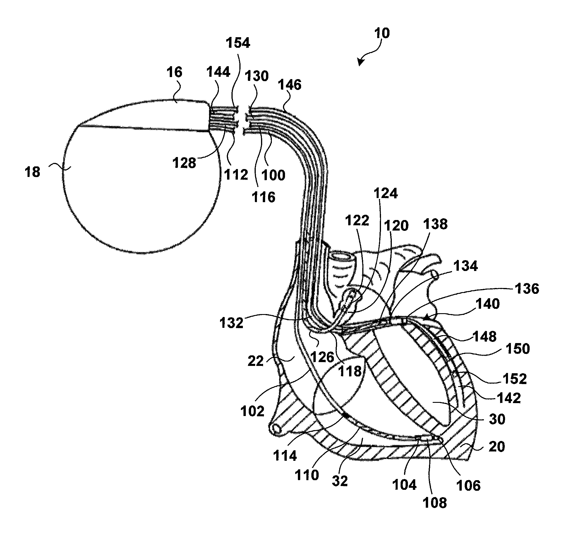
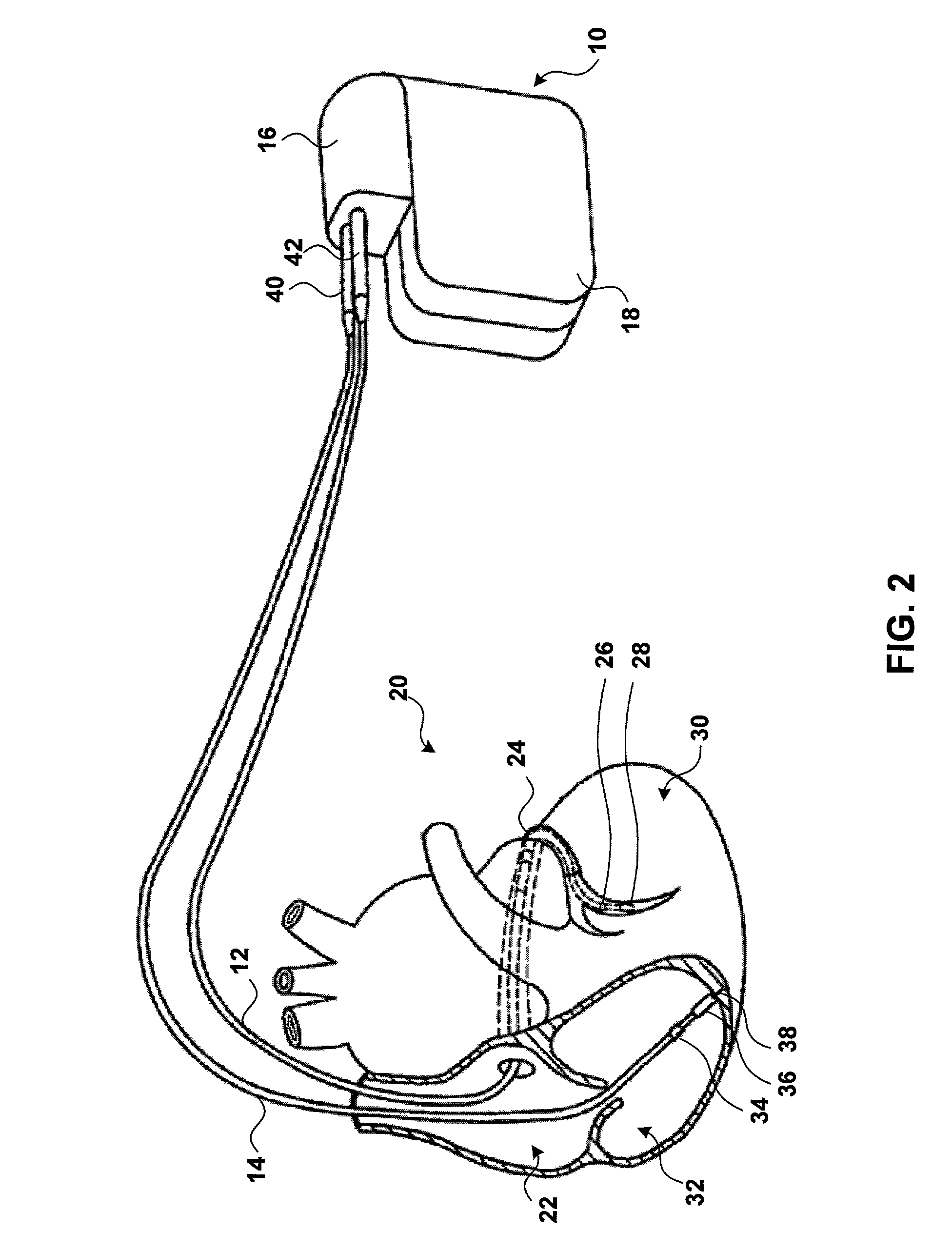
[0017]The invention may offer one or more advantages. Cardiac output can be measured continuously and accurately using dual oxygen saturation sensors, e.g., on a periodic basis over a series of cardiac cycles. In addition, a lead incorporating one or both of the sensors can be deployed within the ventricles to obtain a direct reading of cardiac output. When the sensors are provided in a single, trans-septal lead, deployment of the sensors can be accomplished in a single lead pass, simplifying deployment. Moreover, the trans-septal deployment may place one of the oxygen saturation sensors in close proximity with the left ventricle, but without significant protrusion of the sensor, thereby avoiding excessive formation of fibrous growth and thrombosis.
[0018]By selection of an interval that separates pacing pulses delivered to the ventricles using a continuous, more accurate measurement of cardiac output, the chambers of the heart may be synchronized for near-optimal cardiac performance. When the chambers are synchronized, in accordance with measured cardiac output, the patient may experience improved cardiac performance, such as improved stroke volume and cardiac output. Moreover, the chambers of the heart may be resynchronized for near-optimal cardiac performance in response to changing cardiac output conditions measured on a continuous basis.

Problems solved by technology

These problems include, for example, an inability to accurately detect current cardiac output conditions and thereby adapt a pacing interval to those conditions to promote hemodynamic efficiency, as well as an inability to adapt a pacing interval to changing cardiac output conditions.
In some patients, simultaneous stimulation of the ventricles results in a lack of mechanical ventricular synchrony.
The lack of synchrony may cause the ventricles to begin ejection of blood at different times. For some patients, asynchronous blood ejection is inefficient and undesirable.

Method used


Image

  • Cardiac output measurement using dual oxygen sensors in right and left ventricles
  • Cardiac output measurement using dual oxygen sensors in right and left ventricles
  • Cardiac output measurement using dual oxygen sensors in right and left ventricles

Examples


Embodiment Construction

[0034]In the following detailed description of the preferred embodiments, reference is made to the accompanying drawings that form a part hereof, and in which are shown by way of illustration specific embodiments in which the invention may be practiced. It is to be understood that other embodiments may be utilized and structural or logical changes may be made without departing from the scope of the present invention. The following detailed description, therefore, is not to be taken in a limiting sense, and the scope of the present invention is defined by the appended claims.

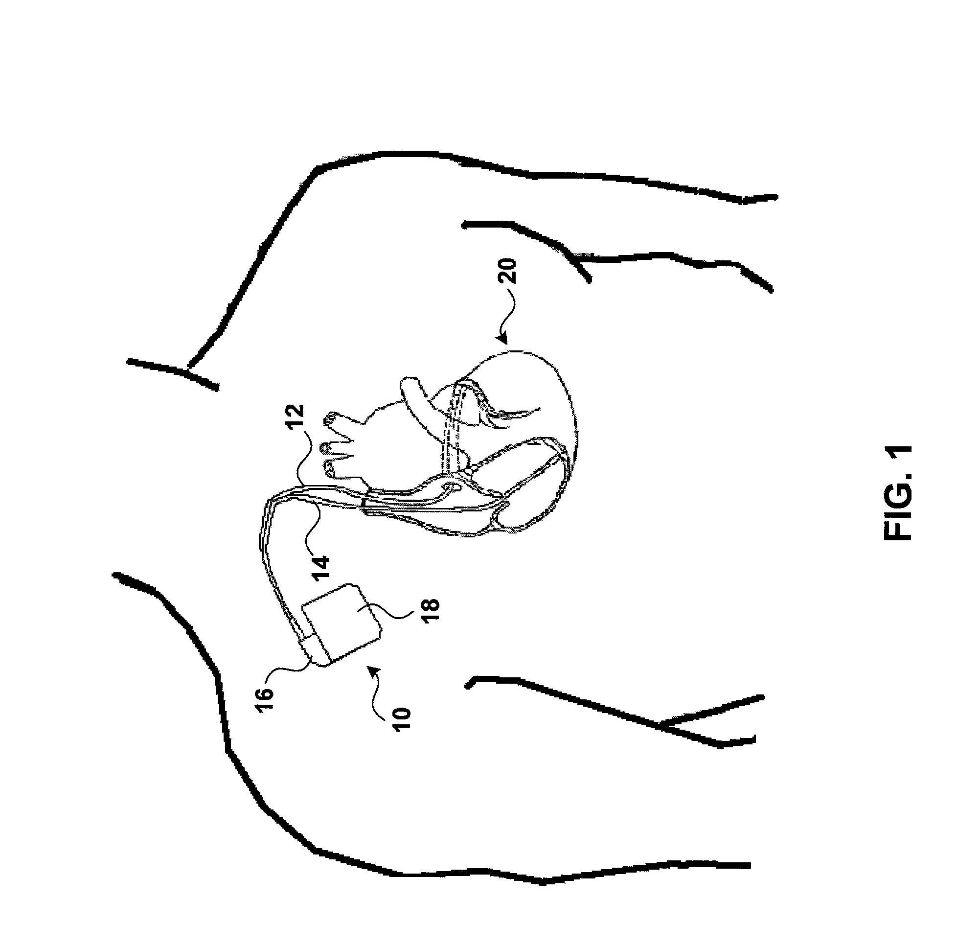
[0035]FIG. 1 is a simplified schematic view of one embodiment of implantable medical device (IMD) 10 of the present invention. In accordance with the invention, IMD 10 may be configured to measure cardiac output, respond to measured cardiac output, or both. In some embodiments, IMD 10 may incorporate one or more leads carrying sensors for sensing blood oxygen saturation levels within the left and right ventricles...


PUM


Abstract

A pacemaker provides multi-chamber pacing with a pacing interval that can be programmed and adapted in response to cardiac output measurements for a given patient. In a typical embodiment, the pacemaker may provide pacing stimuli to both ventricles of a heart. In addition, the invention may include a measurement device that incorporates first and second blood oxygen saturation sensors for deployment in the left and right ventricle. The oxygen saturation sensors provide a differential measurement that can be used to calculate cardiac output in accordance with the Fick method. The oxygen saturation sensors may be carried by a common trans-septal lead that positions one of the sensors proximate the right ventricle and the other sensor proximate the left ventricle. Alternatively, the oxygen saturation sensors may be deployed via separate leads. Whether single or dual leads are used to carry the oxygen saturation sensors, a respective lead may optionally carry electrodes for sensing, pacing, or both.

Description

FIELD OF THE INVENTION[0001]The invention relates to cardiac monitoring, and more particularly to cardiac output measurement.BACKGROUND[0002]Many patients that suffer from congestive heart failure (CHF) develop a wide QRS complex resulting from a delayed activation of one of the ventricles in the heart, and inter-and / or intraventricular electrical-mechanical dysynchrony. This ventricular “dysynchrony” may be caused by dilation of the heart, which disrupts the conductive pathways and interferes with depolarization sequences. Ventricular dysynchrony may worsen heart failure symptoms.[0003]In a classic case of ventricular dysynchrony, the patient's right ventricle activates first, and the left ventricle activates at a later time. The patient often experiences a reduction in cardiac output because the ventricles begin contraction at significantly different times. The timing imbalance may also cause the patient to experience paradoxical septal motion, mitral regurgitation or decreased ve...

Claims


Application Information

Patent Timeline
IPC IPC(8): A61N1/362A61B5/00A61B5/029A61N1/05
CPCA61N1/056A61B5/1459A61B5/029
Inventor STRUBLE, CHESTERMUHLENBERG, LAMBERTGRANDJEAN, PIERRE-ENDRO
Owner MEDTRONIC INC
Learn More