Cytotoxin-based biological containment

Inactive Publication Date: 2007-02-27
UNIV LIBRE DE BRUXELIES
22 Cites 20 Cited by

AI-Extracted Technical Summary

Problems solved by technology

The increasing application of recombinant DNA technology to engineer novel microorganism which are industrially useful have caused concerns in the general public over the potential risks involved.
These concerns are primarily related to the potential harm to humans and to undesirable and/or uncontrollable ecological consequences upon deliberate or unintentional release of such genetically engineered micro...

Method used

[0192]The medium copy number expression vector pBAD33 contains an arabinose inducible promoter (PBAD) with a multiple cloning site (mcs) and the araC gene (Guzman et al., 1995). The genetic structure of pBAD33 is shown in FIG. 5. Without arabinose added to the growth medium, the pBAD promoter is completely turned off. However, with arabinose, strong transcription is induced towards the cloning site. On top of this property, the pBAD promoter is repressible by the addition of glucose to the growth medium. Thus, by the addition of glucose, transcription from pBAD can be rapidly and efficiently turned off.
[0195]Plasmid pHA810 (+1 to +1122) is a pBR322 derivative that contains the relBP307 gene expressed from its own promoter. The strain MC1000/pHA810/pHA110 (relBP307) was subjected to a physiological growth experiment as descri...

Benefits of technology

[0015]These findings has opened up for an alternative, highly effective and versatile biological containment system as it is described in the following. Importantly, it has been discovered that such a system involves the significan...

Abstract

Method of conditionally controlling the survivability of a recombinant cell population and of containing such cells to an environment or containing replicons to a host cell is based on the use of proteic killer systems including the E. coli relBE locus and similar systems found in Gram-negative and Gram-positive bacteria and Archae. Such system are generally based on a cytotoxin polypeptide and an antitoxin or antidote polypeptide that in contrast to the cytotoxin is degradable by proteases. The recombinant cells are useful as vaccines, pollulant degrading organisms or as biogical pest control organisms e.g. expressing B. thuringiensis crystalline proteins.

Application Domain

Technology Topic

Image

  • Cytotoxin-based biological containment
  • Cytotoxin-based biological containment
  • Cytotoxin-based biological containment

Examples

  • Experimental program(16)

Example

Example 1
The Occurrence of relBE Operons in Bacteria and Archae
1.1. Nucleotide Sequence of the relBE Operon of E. coli K-12
[0162]The DNA sequence of the relBE operon from E. coli K-12 is shown in Table 1.1. In this Table the transcriptional start site of the relBE mRNA is indicated with two asterisks (heterogeneity). IR indicates inverted repeats in the promoter and terminator regions. Start codons and stop codons are shown in bold. The transcriptional termination point (ttp) of the relBE mRNA is also indicated with a vertical arrow. The DNA sequence is from Bech et al., 1985.
[0163]By visual inspection of the relBK-12 and relEK-12 genes there was found striking similarity with the so-called “proteic plasmid stabilization systems” as described by Jensen and Gerdes (1995). First, relEK-12 codes for a very basic protein (RelEK-12: pI=9.7) of 95 amino acids (aa), and relBK-12 codes for a very acidic protein (RelBK-12; pI=4.8) of 79 aa.
[0164]The sequences of proteins RelBK-12 and RelEK-12 are shown in Tables 1.5 and 1.6, respectively. These Tables show multiple sequence alignments of the RelB and RelE gene families. Conserved amino acids at a given position are shown with shading as follows: two amino acids are considered conserved if they both belong to one of the following groups: group 1: D and N; group 2: E and Q; group 3: S and T; group 4: K and R; group 5: F, Y and W; group 6: L, I, V and M. Light grey shading indicates 60–80% conservation, dark grey indicates 80–99% conservation and black indicates 100% conservation. Note in Table 1.6 the fully conserved glycine at position 69 (G in consensus line) and the fully conserved arginine at position 79 (R in consensus line). The entrez database accession numbers of the protein sequences are given in Tables 1.3 and 1.4.
[0165]The relBK-12 and relEK-12 genes are co-transcribed with a third gene, relF (also denoted orf-3 or hokC), which is homologous to the hok gene from plasmid R1 (Gerdes et al., 1986). The start site (i.e. the 5′-end) of the relBE mRNA was determined to be 31 nucleotides upstream of the relBK-12 AUG start-codon (Bech et al., 1985) and was confirmed (M. Gotfredsen and K. Gerdes, 1998). Inverted arrows in the relBE promoter region (Table 1.1) indicate putative binding sites for regulators of transcription (i.e. the RelBK-12 and RelEK-12 proteins themselves).
[0166]The properties described above suggested that RelE could be a cytotoxin and that RelB could be an antitoxin which counteracts the toxicity elicited by RelE.
[0167] TABLE 1.1 DNA sequence of the reIBE operon from E. coli K-12 (SEQ ID NO:9) 1 CTTAATTTCA GGCCCCATCG GATCACACAT GGAGAGTTTT TATGAATAAC 51 CCCGTCTGTC TTGATGAGTG CTTGATTGGC TTTAAAAGCT TGTTGACAGG 101 GGTAAACGTT CGGCAATAAT TTTCTGCCGC ATGCGGGTGT TGCATAAAAC 153 GTGTTACGTT CCTTTATCGA CAGGTCACGT CACCGCTCAC CCGCCGACGA 201 GAAAGCAACA CTGACATGCT AAAGCAAAAA ATAGATGAAT AAGTTGACTT 251 GTGCATATGT AGCCTGACCG TCACAAAGTA TATGGTGTCT GTACCAGTAA 301 GATGATGGCC GGACTCTTTA AAAACGAGCT GACCTGCACA ATACAGGATG 351 GACTTAGCAA TGGCTGCTCC TGGCACAAAG CGGACAGTGA TGACCGTTCT 401 TACGACTACT TTCTGACTTC CTTCGTGACT TGCCCTAAGC ATGTTGTAGT **→relBEF mRNA                   relB start 451 GCGATACTTG TAATGACATT TGTAATTACA AGAGGTGTAA GACATGGGTA ---- ---- --→IR←-- ------ -- 501 GCATTAACCT GCGTATTGAC GATGAACTTA AAGCGCGTTC TTACGCCGCG 551 CTTGAAAAAA TGGGTGTAAC TCCTTCTGAA GCGCTTCGTC CCATGCTCGA 601 GTATATCGCT GACAATGAAC GCTTGCCGTT CAAACAGACA CTCCTGAGTG 651 ATGAAGATGC TGAACTTGTG GAGATAGTGA AAGAACGGCT TCGTAATCCT End relB      Start relE 701 AAGCCAGTAC GTGTGACGCT GGATGAACTC TGATGGCGTA TTTTCTGGAT 751 TTTGACGAGC GGGCACTAAA GGAATGGCGA AAGCTGGGCT CGACGGTACG 801 TGAACAGTTG AAAAAGAAGC TGGTTGAAGT ACTTGAGTCA CCCCGGATTG 851 AAGCAAACAA GCTCCGTGGT ATGCCTGATT GTTACAAGAT TAAGCTCCGG 901 TCTTCAGGCT ATCGCCTTGT ATACCAGGTT ATAGACGAGA AAGTTGTCGT 951 TTTCGTGATT TCTGTTGGGA AAAGAGAACG CTCGGAAGTA TATAGCGAGG End relE 1001 CGGTCAAACG CATTCTCTGA ACCAAAGCAT GACATCTCTG TTTCGCACCG Start hokC (relF) 1051 AAGGTGACAC TTCTGCTTTG CGTTGACAGG AGAAGCAGGC TATGAAGCAG 1101 CAAAAGGCGA TGTTAATCGC CCTGATCGTC ATCTGTTTAA CCGTCATAGT 1151 GACGGCACTG GTAACGAGGA AAGACCTCTG GGAGGTACGA ATCCGAACCG End hokC 1201 ACCAGACGGA GGTCGCTGTC TTCACAGCTT ACGAACCTGA GGAGTAAGAG 1251 ACCCGGCGGG GGAGAAATCC CTCGCCACCT CTGATGTGGC AGGCATCCTC 1301 AACGCACCCG CACTTAACCC GCTTCGGCGG GTTTTTGTTT TTATTTTCAA ---- -- IR ---- - ttp 1351 CGCGTTTGAA GTTCTGGACG GTGCCGGAAT AGAATCAAAA ATACTTAAGT (data base accesion number X02405)
[0168] TABLE 1.3 relE homologues from Gram-positive and Gram-negative bacteria and Archae entrez Number Bacterial species accession genea) of aa MW (kD) p1 Gram-negative bacteria: E. coli K-12 132284 relEK-12 95 11.2 9.7 E. coli K-12 984581 relESOSb) 92 10.8 9.5 E. coli plasmid 516611 relEP307 95 11.2 9.9 P307 H. influenzae 1175293 relEHi 102 11.9 6.7 V. cholera 396846 relEVc 96 11.2 9.9 H. pylori 2314031 relEHp 88 10.4 7.9 Synechosystis 1653777 relESy 120 13.7 7.9 Gram-positive bacteria: B. thuringiensis 520407 relEBt 74 8.6 9.7 M. tuberculosis#1 2612811 relEMt1 87 10.2 11.0 M. tuberculosis#2 2695832 relEMt2 97 11.1 9.5 Archae: M. jannaschii#1 1498833 relEMj1 90 11.0 10.2 M. jannaschii#2 1499953 relEMj2(*) 88 10.6 10.0 M. jannaschii#3 1591583 relEMj3(*) 91 11.1 10.1 A. fulgidus#1 2648176 relEAf1 87 10.6 10.3 A. fulgidus#2 2649499 relEAf2 92 11.0 9.9 A. fulgidus#3 2649496 relEAf3 85 10.0 10.0 A. fulgidus#4 2649514 relEAf4 86 10.2 9.9 a)relE homologues marked with (*) are not located adjacent to a relB partner b)The relBESOS system of E. coli K-12 contains a LexA binding-site in the promoter region (Lewis et al:, 1994)
[0169] TABLE 1.4 relB homologues from Gram-positive and Gram-negative bacteria and Archae entrez Number Bacterial species accession genea) of aa MW (kD) p1 Gram-negative bacteria: E. coli K-12 132283 relBK-12 79 9.1 4.8 E. coli K-12 984582 relBSOSb) 86 9.4 5.2 E. coli K-12 984588 relBK-12.2(*) 97 11.2 5.5 E. coli plasmid 516610 relBP307 83 9.2 4.4 P307 S. typhimurium 731639 relBSt(*) 68 7.6 5.3 H. influenzae 1573712 relBHi 98 11.0 4.7 V. cholera 396847 relBVc 82 8.9 4.4 H. pylori 2314037 relBHp 95 11.4 9.8 Synechosystis 1653776 relBSy 86 9.9 4.7 Gram-positive bacteria: B. thuringiensis 520406 relBBt 85 10.1 4.5 M. tuberculosis#1 2612810 relBMt1 93 10.2 4.6 M. tuberculosis#2 2695833 relBMt2 89 9.8 5.1 Archae: M. jannaschii#1 1498832 relBMj1 82 9.6 4.5 A. fulgidus#1 2648190 relBAf1 65 7.8 4.8 A. fulgidus#2 2649516 relBAf2 62 7.4 4.3 A. fulgidus#3 2649510 relBAf3 72 8.5 4.5 A. fulgidus#4 269513 relBAf4 57 6.7 4.1 a)relB homologues marked with (*) are not located adjacent to a relE partner. b)The relBESOS system of E. coli K-12 contains a LexA binding-site in the promoter region (Lewis et al., 1994).
[0170] TABLE 1.5 Alignment of relE homologues from Gram-positive and Gram-negative bacteria and Archae (SEQ ID NOS: 10–26)
[0171] TABLE 1.6 Alignment of relB homologues from Gram-positive and Gram-negative bacteria and Archae (SEQ ID NOS: 27–43)
1.2. Nucleotide Sequence of the relBE Operon of Plasmid P307
[0172]By database searching it was found that the E. coli plasmid P307 codes for a gene system which exhibits both structural and sequence similarity with the E. coli relBE genes described above.
[0173]The DNA sequence of the relBEP307 genes is shown in Table 1.2. The transcriptional start site of the relBE mRNA is indicated with an asterisk, and the −10 and −35 sequence elements of the relBE promoter are underlined. The Shine & Dalgarno sequence of the relB and relE genes are doubly underlined. The DNA sequence is from Saul et al., 1989.
[0174]Again, relEP307 codes for a very basic protein of 95 aa (pI=9.9), and relBP307 codes for a very acidic protein of 83 aa (pI=4.4), see Tables 1.3 and 1.4. The protein sequences of RelEP307 and RelBP307 are also shown in Tables 1.5 and 1.6, respectively. The start site (i.e. the 5′-end) of the relBEP307 mRNA was determined to be located 27 nucleotides upstream of the relBP307 AUG start codon. Inverted arrows in the relBEP307 promoter region (Table 1.2) indicate putative binding sites for regulators of transcription (i.e. the RelBP307 and RelEP307 proteins).
[0175] Table 1.2 DNA sequence of the reIBE operon from the E. coli plasmid P307 (SEQ ID NO:44) 301 GAGTATCATA TTAGGATACG GGTGGGTGAC GCCCACCTCT GGCATAGAAC 351 GGACATTCAT TGATGCCATG CCAGAATGGA CGTTCAGGTT ATTCCGTCCA 401 GTTCTGCTGG CAACGCGAGA TCTCCCCTGG TATAGTGATG CCACAGCAAA 451 GCGCTCAAAC AGGGATAATA TGATGGAAAT CAAGGCTCAA CAGTTTTGTC 501 ACATCAACGG GGCGGCAAGT CCTTACTGAC AACGGACAAC AAGGTATGGG 551 CGGCGTGGCG GGTATCGGTT CCACGACTGA AAAGCATCAG GGGCGGGTGG 601 CGGAAGCGAT TTTTGCGAAC TGCGCGGAAC TGGATAACGA CCAGCTTAAC 651 GAGATCATCG AGTGGGTTCG CCTCTATCAG CGCTGAATGC CACTATCAGG 701 CTGCGCAAGC GGCCTTTTTT ACGCCCCTTG TTTAATTCCC GCACTACCTG −35 751 GACGTTCAGG TGATTCTGTC CATCTGTACA AAAAACAATA AAAGACTTGT −10          *→ relBEP307 mRNA 801 TAACAGGTCA TGTAAGGAGT ATCTTTGAGA CTGGTTAAAC AGTCTTGAAA SD     start relB 851 GGTGGCCTAT GCCTAACATT ATTCTCAGTG ATACAAGCGC CAGTGTCAGC 901 GAGCTGAAGA AAAACCCGAT GGCGACAGTC AGCGCCGGTG ATGGTTTCCC 951 GGTCGCTATC CTGAACCGTA ATCAGCCTGC TTTCTAGTGT GTACCCGCAG 1001 AGCTGTACGA AAAGATGCTT GATGCCCTAG ACGATCAGGA GTTGGTTAAA SD 1051 CTGGTAGCCG AACGCAGCAA CCAACGGCTG CATGATGTAG ATCTGGATAA end relB/start re1E 1101 ATACCTATGA GGTATCAGGT AAAATTCAGG GAAGATGCGC TGAAAGAGTG 1151 GCAAAAACTG GACAAGGCTA TTCAGCAACA GTTTGCGAAA AAGCTAAAAA 1201 AGTGCTGTGA CAATCCGCAT ATTCCTTCCG CAAAACTGCG TGGGATAAAG 1251 GACTGCTACA AAATAAAATT ACGTGCGTCA GGTTTTCGCC TGGTCTATCA 1301 GGTGATTGAC GAACAATTAA TTATCGCTGT TGTAGCTGTG GGTAAACGTG end relE 1351 AGCGCAGTGA CGTTTATAAT CTTGCCAGCG AAAGAATGAG ATAAAAGCAA 1401 TAAACACAGA AAGTTACTCT GGCGTTATGG GGTAATGCAA AGTATGAGTC 1451 GTAGACGGAA TTGCCTGGAT AATTCGCCGA TGGAAAGAGT CTTTCGCAGC 1501 CTTAAAAGTG AATGGCTTCC GAAAGGTGGT TATGGTGATT TTAGCCATGC (database accession number M26308)
1.3. Nucleotide Sequence and Proteins of a relBE Homologous Operon from Bacillus thuringiensis
[0176]Using BLAST database searching (Altschul et al., 1990) it was found that transposon Tn5401 from the Gram-positive organism B. thuringiensis contains, in one end or asymmetrically located, a two-component system which exhibits both structural and sequence similarity with the above described relBE systems from E. coli. This homology is surprising given that it has not previously been described that relBE-like genes are found in organisms other than E. coli.
[0177]The nucleotide sequence of the relBE operon from Tn5401 is shown in Table 1.7. In this Table the transcriptional start-site of the relBE mRNA is indicated with an asterisk (Baum, 1994). IR indicates inverted repeats in the relBEBt promoter region. Start codons and stop codons are shown in bold. The Shine & Dalgarno sequence of the relBBt gene is doubly underlined. The DNA sequence is from Baum et al., 1989.
[0178]The relEBt gene codes for a very basic protein of 74 aa (pI=10.6) and the relBBt gene codes for an acidic protein of 87 aa (pI=4.4). The protein sequences of RelEBt and RelBBt are aligned with the other RelE and RelB homologues in Tables 1.5 and 1.6, respectively. The modular, structural and physico-chemical similarities between the B. thuringiensis system and the E. coli systems suggested that the genes may exert similar functions in very different bacteria.
[0179] TABLE 1.7 DNA sequence of the reIBE operon from the Gram-positive organism B. thuringiensis (SEQ ID NO:45) 3701 CTCGTTTTTT CTGTTGGTAC AAACTTAATT GATTTTGAAT AATTTGTTTG 3751 TACCAGTCCT TTTTGCTTAG CCCAGTCAAA ATAACGTTTG ATTGAATTAA 3801 TGCGCCGGTT AATCGTAGAA GGTTTTAGTA ATCTTGTAAC TTGCATATGC 3851 CCTCGATATC GAGCAATAGT GCGAGCGGTA ACTTCTATTG GATGAAAAAG 3901 AGTATCCTCA GCATGTTTTC CCCACACATT TTCAAACCAA AATACAAAAT 3951 CTTTTAAATC ACTCGTATAT TCTTTTAGTG TTTTTGTATG CAAATCTCCT 4001 TCTTGAGATA AGCTACAAAT AAAATCGGAA ATCAAAGATG TTGCTTGTAT −35 4051 AGAAATTGTT TTAGTGGAAT GCATAAATAC CTCCTCTTTT ATTGACTTAC −10        *→ relBEBt mRNA 4101 ATTAGCGGAC ATGATATTTT AATCTTATCA ATTATGTTAG CGGACATCAA 4151 ACATTTATTT TCCCACACTT CATGTCCACT AATATTAATT AGTGGACATT --------- --→IR ←- --------- SD           Start relB 4201 TAAAACTATC TCGAAAGTAG GTGTAACACA TGGCTATTCG TAAAGATGAA 4251 TTGTATCGGT TAATTGATCA CCTGGATCAA CAAGATGAAA AAGCAGCATT 4301 TGACTTTTTA GAATTTCTTG TTCAACGGTC AAGAAGAAAA CCTAAAGAAT 4351 GGGAAAAAAT TGATATGGCA GATCCTGATC ATGAACCGCT GTCTACACAA 4401 GAGTTAGAAC AGTTAAACAG TGAAGAAGGA TATGTATCAG GGGAGGACGC End relB    Start relE 4451 AAAACGTGAA TTCGGACTAC AAATTGATTT ACCATAAGTC CGCGGTGAAA 4501 TTTATTGCAA AGCAAGAAAA AGGGATTCAA AAAAGAATTG CAGAAGGATT 4551 GAAGGGACTT CTTAAGATTC CTCCTGAAGG AGATATTAAA AGTATGAAAG 4601 GTTACACAGA ACTATATCGA TTACGGATTG GAACCTTTCG AATTTTATTT 4651 GAAATAAATC ATGATGAGAA AGTCATATAC ATACAAGCAA TTGGAAATCG End relE 4701 TGGTGACATC TATAAATAAG GCAAACATGC ATTTTTATAA GAAAGGTCTT 4751 CTGAATCGAA GAACCTTCCT TTTTTGTGTG CCAATAATGT CCGCTAATGC 4801 TTGTTGCGTG ATTCTGTTCC ATTGCTACAC ATACCCC (database accession number U03554)
1.4. The Archaeon Methanococcus jannaschii Encodes a relBE Homologous System
[0180]Again using database searching it was found that the completely sequenced genome of the methanogenic archaeon Methanococcus jannaschii codes for three relE homologous genes, one of which are located just downstream of relB homologous genes. This finding was surprising since, in many respects, archaeal organisms are more similar to eukaryotes than to bacteria (e.g. in their macromolecular synthesis apparatuses).
[0181]The DNA sequence of the relBEMj1 system is shown in Table 1.8. In this Table start codons and stop codons are shown in bold. The DNA sequence is from Bult et al., 1996.
[0182]Gene relEMj1 codes for a very basic polypeptide of 90 aa (pI=11.0) and gene relBMj1 codes for an acidic polypeptide of 82 aa (pI=4.4). The aa sequences of the RelEMj1 and RelBMj1 proteins are aligned with the other RelBE homologues in Tables 1.5 and 1.6, respectively. Thus, these basic similarities suggested that the relBEMj1 system may carry out similar or related functions in bacteria and archae. The properties of the second and third relE homologues of M. jannaschii are also given in Table 1.3. These comparisons show that M. jannaschii codes for one complete relBE homologous gene system and for two relE homologues without an adjacent relB partner.
[0183] TABLE 1.8 DNA sequence of a reIBE homologous gene system from the archaeon Methanococcus jannaschii (SEQ ID NO:46) 751 CCGATACCGT TGCTGGAGAC ATAGCTGGAG CTTTGAAGGC GGAGAAGCTT 801 ATTTTAATAA CAGATGTTGA TGGAATAATG GATGATATAA ATAATCCAGA 851 GACGTTGCAT AGAAAATTAA CAGCTTCAGA ACTAAAAGAA ATGATAGAAG 901 ATGGAAGAAT AAAGGGAGGG ATGATTCCAA AGGCTGAAAG TGCCTTATAT 951 GCCTTAGAGC ATGGAGTTAA GAGCGTTCAT ATAATAAATG GAAAGATTCC 1001 TCATGCTTTG TTGTTGGAGA TATTTACAGA GGAGGGTATT GGGACGATGA 1051 TAACAAGAGA TTAAAGTTTT TATATTATAA ACTACTTAAG AATTAAAATA Start relBMj1 1101 AGACAAATAA GGGGATAACT ATGCTCAATA TAAACAAAGA GATAGCACAA 1151 ATAGAAACTG AATTGAATGA ATTGAAAAAA TTGAGAGATG AAATCTCTGA 1201 AAGGATTGAA AAATTAGAAA TAAAGTTATT AAAATTGAAA GCATTAGGTA 1251 TTCCAGAGGA GGAATTTGAA GAGGATTATG AAGAAATTAT AGAAGATGTT 1301 AAAAAATCTC TGGATAAAAA AGAGACTGTG CCAGCAGAAG AGGCTTTGAA End relBMj1/start relEMj1 1351 AGAATTGGGA TTATTATGAA GTTTAACGTT GAGATACATA AAAGAGTCTT 1401 AAAAGATTTA AAGGATTTGC CTCCCTCAAA CTTAAAGAAG TTTAAAGAAC 1451 TAATAGAAAC ATTAAAAACC AATCCCATTC CAAAAGAAAA ATTTGATATT 1501 AAAAGATTAA AAGGCAGTGA TGAGGTTTAT AGAGTTAGAA TTGGAAAATT 1551 TAGAGTTCAA TATGTTGTTT TATGGGATGA TAGAATAATA ATAATTAGAA End relEMj1 1601 AGATAAGTAG AAGAGAAGGA GCTTATAAAA ATCCCTAAGC TATTAAAAAT 1651 TCTAATGGCT ACATTTTTAT ATCTCTTTTC TTAATTCAAA TAGAAAAAAC 1701 AGATTCGGCT GATACCATGA TTATTCTTTT AGATTTAAAT GGAACAATAG (database accession number U67464)
1.5. relBE Homologous Genes are Ubiquitous in Prokaryotes
[0184]Further relBE homologous two-component systems were discovered. The corresponding RelB and RelE homologous proteins are aligned in Tables 1.5 and 1.6, respectively. It appears that relE homologous genes are present in a wide variety of Gram-negative bacteria (E. coli, H. influenzae, V. cholera, H. pylori and Synechosystis), in Gram-positive bacteria (B. thuringiensis and M. tuberculosis) and in Archae (M. jannaschii and A. fulgidus). Most strikingly, the archaeon A. fulgidus contains four complete relBE homologous gene systems.
[0185]A number of features become evident from the alignments of the proteins (Tables 1.5 and 1.6) and from the properties listed in Tables 1.3 and 1.4. First, all RelE homologues are basic with pH's around 8–10 whereas the RelB homologues are acidic with pI's about 4–5. Secondly, the RelE proteins are in general slightly larger (90–120 aa) than the RelB homologues (70–80 aa). Thirdly, the start codons of the relE genes are juxtaposed or even overlap with the stop codons of the linked relB partner, thus indicating translational coupling of relE to relB. These properties suggest that the proteins could exert similar functions in very different organisms.

Example

Example 2
Demonstration of Translation of the relBK-12 and relEK-12 Genes
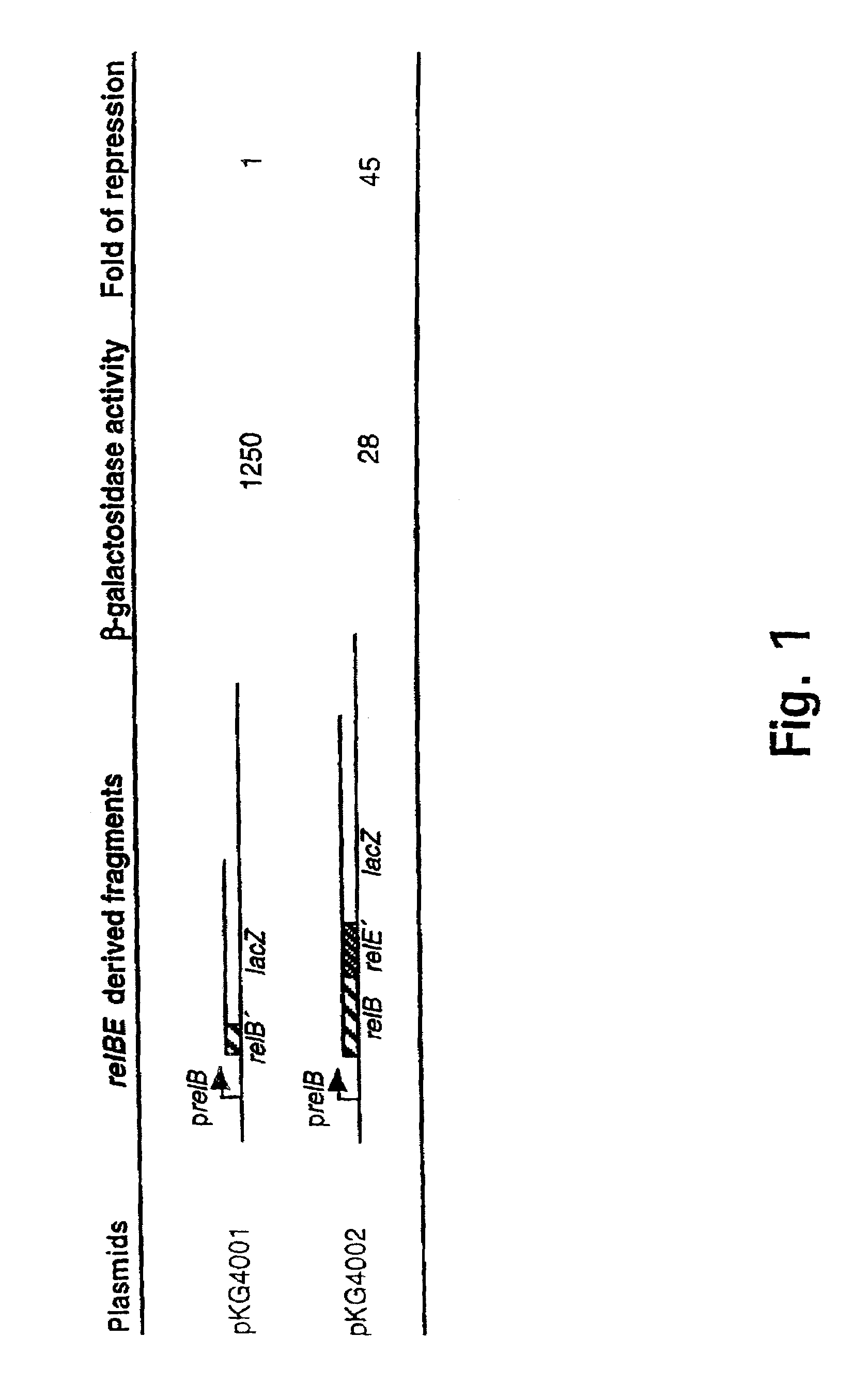
[0186]Using the low copy-number lacZ fusion vector pOU253 (Table 0.1) in frame gene fusions between relBK-12 and relEK-12 and the lacZ gene were constructed (see Materials and methods). Thus plasmid pKG4001 (+388 to +596) carries a fusion between relBK-12 and lacZ, and pKG4002 (+388 to +921) carries a fusion between relEK-12 and lacZ. The structure of the relevant parts of these reporter plasmids are shown in FIG. 1. When present in strain MC1000, both plasmids expressed significant amounts of—galactosidase fusion proteins, indicating that genes relBK-12 and relEK-12 are translated (FIG. 1). The relEK-12-lacZ fusion (pKG4002) expressed significantly lower amounts of β-galactosidase than the relBK-12-lacZ fusion, mainly because pKG4002 encodes an intact relBK-12 gene which produces the RelBK-12 autorepressor which inhibits transcription from the relB promoter.

Example

Example 3
Demonstration of Translation of the relBP307 and relEP307 Genes
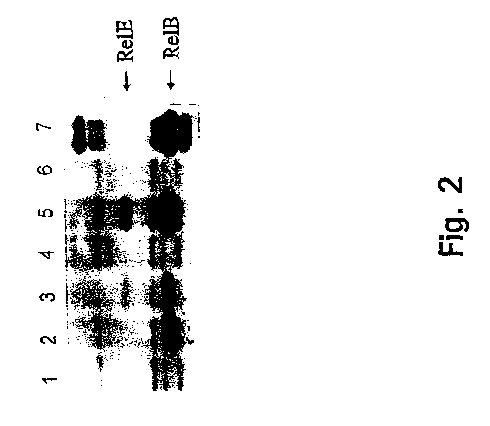
[0187]To detect authentic RelBP307 and RelEP307 proteins, in vitro translation reactions were carried out using high copy number pUC-plasmids carrying genes relEP307 (pHA403), relBP307 (pHA402) or both genes (pHA100) (for construction of these plasmids, see Materials and Methods and Table 0.1). Proteins produced in the in vitro translation reactions were labelled with 35S-Methionine and separated by SDS-page. FIG. 2 shows the direct visualization of RelBP307 and RelEP307, thus providing evidence that the corresponding genes are translated.

PUM

PropertyMeasurementUnit
Volume1.994E-15L
Volume1.995E-15L
Volume1.978E-15L
tensileMPa
Particle sizePa
strength10

Description & Claims & Application Information

We can also present the details of the Description, Claims and Application information to help users get a comprehensive understanding of the technical details of the patent, such as background art, summary of invention, brief description of drawings, description of embodiments, and other original content. On the other hand, users can also determine the specific scope of protection of the technology through the list of claims; as well as understand the changes in the life cycle of the technology with the presentation of the patent timeline. Login to view more.

Similar technology patents

Cell differentiation inducer

InactiveUS20080188489A1Highly effectiveDrugs usedBiocideOrganic chemistryParasitic animalSolid Carcinoma
Owner:BAYER INTELLECTUAL PROPERTY GMBH

Classification and recommendation of technical efficacy words

  • Highly effective
  • Low frequency

Vitreoretinal silicone tamponades made by supercritical fluid extraction

InactiveUS7276619B2EconomicalHighly effectiveSilicon organic compoundsSilicon compound active ingredientsSupercritical fluid extractionOligomer
Owner:BARCLAYS BANK PLC AS SUCCESSOR AGENT

Bispecific, bivalent Anti-vegf/Anti-ang-2 antibodies

ActiveUS20110236388A1Valuable propertyHighly effectiveAnimal cellsHybrid immunoglobulinsEndothelial Growth FactorsAntibody
Owner:F HOFFMANN LA ROCHE & CO AG

Controlled synthesis of highly monodispersed gold nanoparticles

InactiveUS20070125196A1High monodispersityHighly effectiveTransportation and packagingMetal-working apparatusSodium AcrylateNanometre
Owner:THE RES FOUND OF STATE UNIV OF NEW YORK

Method and System for Managing Vehicle Travel

ActiveUS20110246246A1Low monetary costLow frequencyAnalogue computers for vehiclesReservationsProgram planningTravel mode
Owner:THE CRAWFORD GROUP

Computer program product for performing digital-to-analog conversion

InactiveUS20050040979A1Low frequencyElectric signal transmission systemsDifferential modulationGreek letter sigmaDigital input
Owner:AVAGO TECH WIRELESS IP SINGAPORE PTE
Why Eureka
Try Eureka