Tissue material and process for bioprosthesis

a bioprosthesis and tissue material technology, applied in the field of tissue material and process for bioprosthesis, can solve the problems of increasing the risk of serious hemorrhage for users, the longest durability of available replacement heart valves for mechanical valves, and the danger of continuous use of anticoagulants, so as to improve the mechanical properties of bioprosthesis and the effect of increasing the elasticity of anisotropic materials

Active Publication Date: 2007-03-13
CLEMSON UNIV RES FOUND
View PDF43 Cites 150 Cited by
  • Summary
  • Abstract
  • Description
  • Claims
  • Application Information

AI Technical Summary

Benefits of technology

[0011]In one embodiment, the bioprostheses of the invention can include an anisotropic tissue, for example, an anisotropic tissue exhibiting greater stiffness in a first direction and greater elasticity in a second direction.
[0016]In one embodiment, wherein the tissue used in the bioprosthesis is an anisotropic material, the tissue portion that is excised from the flat tissue can be oriented so as to improve the mechanical properties of the bioprosthesis. For example, in one embodiment, the excised portion can have the general shape of a heart valve leaflet. In this particular embodiment, the direction of greater elasticity of the anisotropic material can be substantially equivalent to the center-most radius of the leaflet as cut from the sheet.

Problems solved by technology

Presently, mechanical valves have the longest durability of available replacement heart valves.
Unfortunately, continuous use of anticoagulants can be dangerous, as it greatly increases the user's risk of serious hemorrhage.
In addition, a mechanical valve can often be audible to the recipient and may fail without warning, which can result in serious consequences, even death.
However, the availability of human valves for transplantation continues to decline as a percentage of cardiac surgeries performed each year.
One disadvantage of current xenograft materials is poor durability.
As a consequence of the increased stiffness, the periodic opening and closing of the valve leads to material fatigue of the bioprosthetic replacement tissue.
In addition, the recipient's heart will be required to work harder to overcome the stiffness of the bioprosthetic valve as compared to the exertion required for the original valve to function.
Additionally, fatigue and mechanical degradation of the xenograft valve is associated with increased calcification of the valve.
The calcification causes additional stiffening which further degrades the physical and biological integrity of the valve.
Unfortunately, due to the heterogenic nature of the pericardium, the pericardium source material used to form the BHV leaflets can have notable variations in physical properties, even when harvested from the same pericardial sac.

Method used


Image

  • Tissue material and process for bioprosthesis
  • Tissue material and process for bioprosthesis
  • Tissue material and process for bioprosthesis

Examples


example 1

Tissue Collection and Characterization

[0054]Porcine inferior vena cava was collected from a slaughterhouse and placed in ice-cold saline. The vena cava was manually cleaned of adherent tissues, followed by opening the veins using a longitudinal incision. After rinsing the vena cava material in fresh saline, the tissue was fixed for 7 days at room temperature in 0.6% glutaraldehyde (GA) prepared in 50 mM HEPES buffered saline at pH 7.4. The fixative was changed with fresh GA solution following an initial 24-hour fixation interval.

[0055]Glutaraldehyde fixed vena cava was cut with a template to obtain samples approximately 40 mm long and 5 mm wide. Six samples were obtained and examined in both a longitudinal direction (parallel to the axis of the blood flow) and in a circumferential direction (perpendicular to blood flow). Stress and strain tests were performed at room temperature on a Vitrodyne V-1000 mechanical tester (Lifeco Inc, Burlington, Vt.) equipped with a 10-pound load cell....

example 2

Thermal Denaturation (Td) Profiles Assessed by Differential Scanning Calorimetry (DSC)

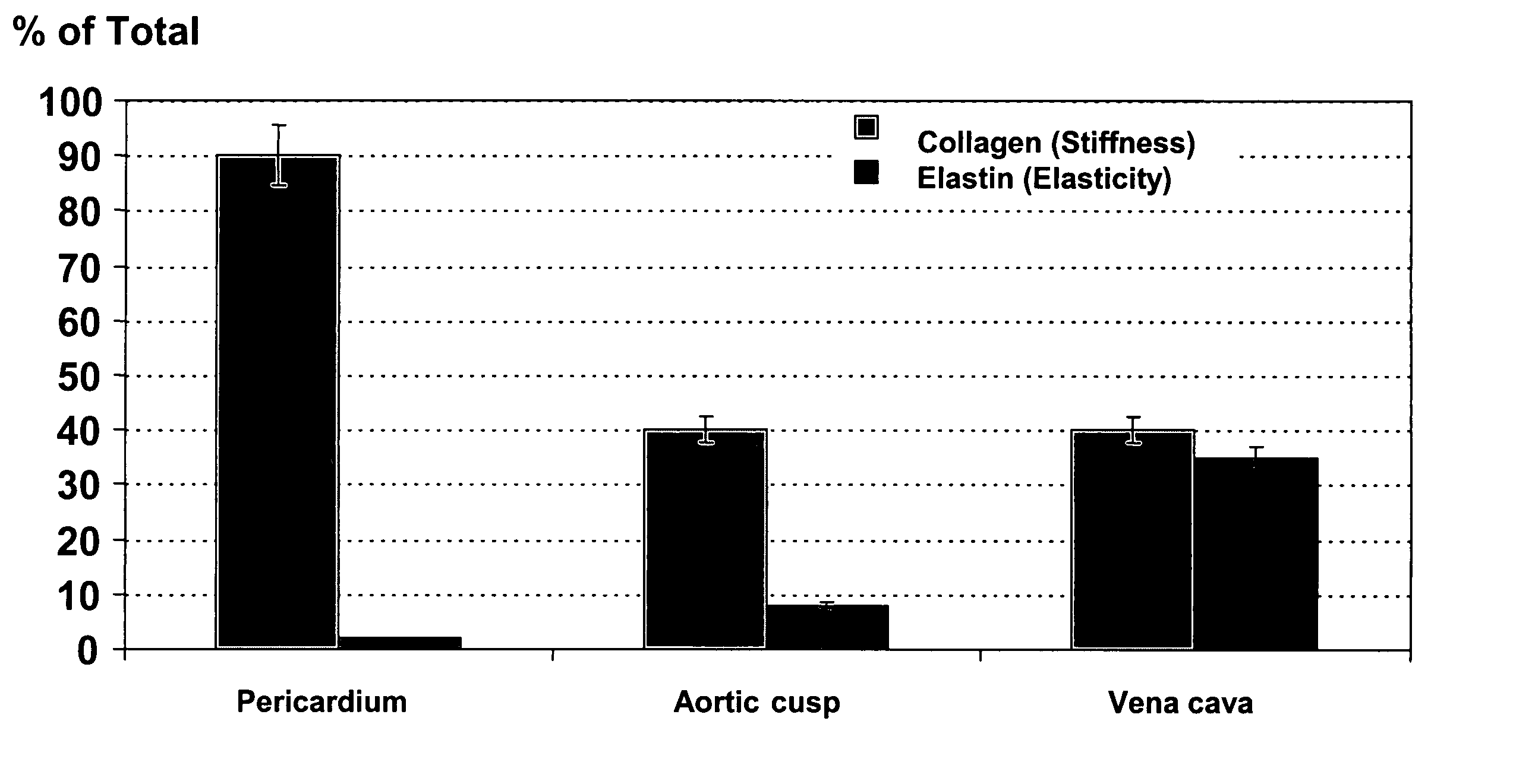
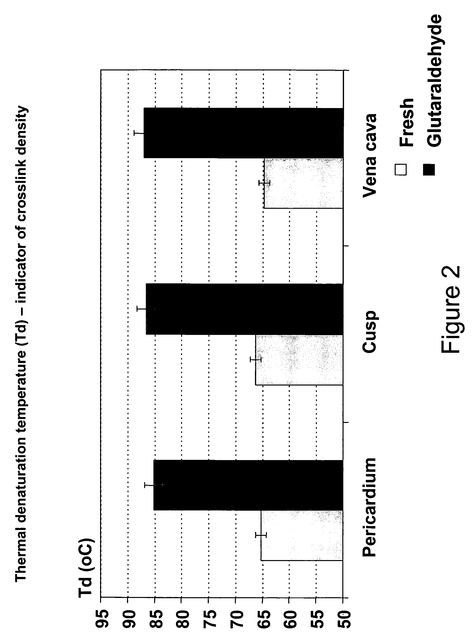
[0056]Td indicates the amount of energy absorbed by a sample. In the case of connective tissues, Td represents the temperature at which native collagen molecules unravel. This process leads to protein denaturation and is recorded as a peak maximum for three different tissues types in FIG. 2. Fresh, native tissues exhibit a Td of around 65° C., while chemically cross-linked tissues require a larger amount of heat to denature, and therefore their Td increases proportionally to the number of cross-links.

[0057]Porcine vena cava tissue (native and GA fixed, as described above) was rinsed in saline and 2 mm2 samples were cut and hermetically sealed in Differential Scanning Calorimetry (DSC) aluminum pans. Samples were heated at a rate of 10° C. / min, from 25° C. to 110° C. and the temperature of thermal denaturation (Td) for each sample was recorded on a Perkin Elmer DSC 7 machine.

[0058]Fresh vena cava ex...

example 3

Comparison of Elastic Properties of Various Tissues

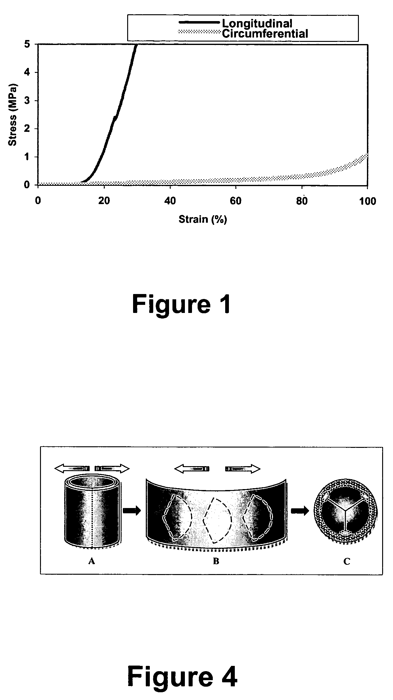
[0061]Porcine vena cava tissue, porcine aortic cusps, and bovine pericardial tissue were prepared and subjected to stress / strain tests as disclosed above in Example 1. FIG. 3A sets forth stress / strain data obtained for both GA-fixed and fresh porcine aortic cusps in both the radial and circumferential directions. The circumferential direction of an individual cusp runs parallel to the aortic wall, and the radial direction runs from the center of the cusp to the edge of the aorta. FIG. 3B sets forth the data obtained for GA-fixed pericardium, and FIG. 3C sets forth the data obtained for GA-fixed porcine vena cava in both the longitudinal (with blood flow) direction and circumferential (perpendicular to blood flow) direction.

[0062]As seen in reference to FIG. 3C, GA-fixed vena cava is anisotropic, exhibiting greater elasticity in the circumferential direction than in the longitudinal direction. Further, in comparison to pericardial ti...


PUM


Abstract

A biomaterial useful for bioprostheses such as bioprosthetic heart valves is provided in which the fixed tissue has improved elastic properties. The high elastin-containing biomaterial is further characterized by having anisotropic properties wherein the biological material has a greater stiffness in one direction and a greater elasticity in a cross direction. For instance, the biological material has an elastin content of about 30% by weight. In one embodiment, the biological material is vena cava tissue.

Description

CROSS REFERENCE TO RELATED APPLICATION[0001]This application claims benefit to U.S. Provisional Application Ser. No. 60 / 429,191 filed Nov. 26, 2002.FEDERALLY SPONSORED RESEARCH AND DEVELOPMENT[0002]The United Stated Government may have rights in this invention pursuant to Grant No. HL 61652 between Clemson University and the National Institutes of Health.BACKGROUND OF THE INVENTION[0003]Prosthetic heart valves are used to replace damaged or diseased heart valves. Prosthetic heart valves may be used to replace a heart's natural valves including aortic, mitral, and pulmonary valves. The predominant types of prosthetic heart valves are either mechanical valves or bioprosthetic valves. Bioprosthetic valves include allograft valves, which include biomaterial supplied from human cadavers; autologous valves, which include biomaterial supplied from the individual receiving the valve; and xenograft valves, which include biomaterial obtained from non-human biological sources including pigs, c...

Claims


Application Information

Patent Timeline
Patent Type & Authority Patents(United States)
IPC IPC(8): A61F2/24A61B
CPCA61F2/2412A61F2/2418Y10S623/909
Inventor SIMIONESCU, DAN T.VYAVAHARE, NARENDRA
Owner CLEMSON UNIV RES FOUND
Why Eureka
Try Eureka