Multiple mode flat panel X-ray imaging system

a x-ray imaging and multi-mode technology, applied in tomography, instruments, applications, etc., can solve the problems of increasing the aforementioned time delay, and reducing the spatial resolution of the image,

Active Publication Date: 2007-06-12
VAREX IMAGING CORP
View PDF8 Cites 154 Cited by
  • Summary
  • Abstract
  • Description
  • Claims
  • Application Information

AI Technical Summary

Problems solved by technology

However, developing exposed X-ray film typically takes a minimum of ninety seconds which can be too long in emergency situations.
Further, the relatively low dynamic range of X-ray film can result in under- or over-exposed images and, therefore, necessitate additional exposures which increase the aforementioned time delay as well as the X-ray dosage received by the patient.
The image intensifier tube used in fluoroscopy has a greater exposure latitude than X-ray film, but also has a more limited active detection area and lower spatial resolution.
However, the image intensifier tube is typically heavy, bulky and expensive, and can introduce image distortion which can only be partially removed during post processing.
However, after exposure to X-rays, the photostimulable phosphor plate must be scanned with a laser system which is large and expensive, and the readout process is just as slow as the development of film.

Method used


Image

  • Multiple mode flat panel X-ray imaging system
  • Multiple mode flat panel X-ray imaging system
  • Multiple mode flat panel X-ray imaging system

Examples


Embodiment Construction

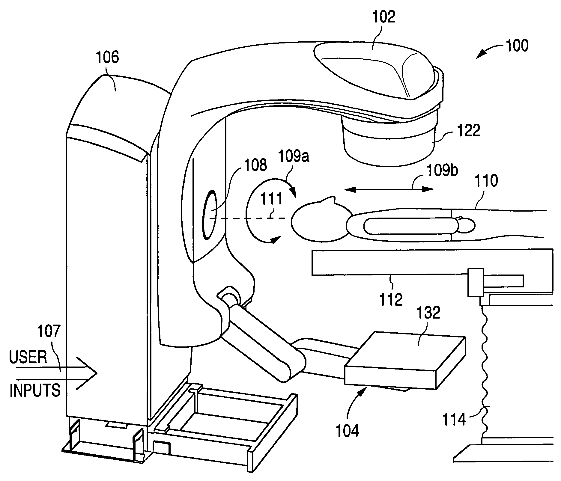
[0021]The following detailed description is of example embodiments of the presently claimed invention with references to the accompanying drawings. Such description is intended to be illustrative and not limiting with respect to the scope of the present invention. Such embodiments are described in sufficient detail to enable one of ordinary skill in the art to practice the subject invention, and it will be understood that other embodiments may be practiced with some variations without departing from the spirit or scope of the subject invention.

[0022]Throughout the present disclosure, absent a clear indication to the contrary from the context, it will be understood that individual circuit elements as described may be singular or plural in number. For example, the terms “circuit” and “circuitry” may include either a single component or a plurality of components, which are either active and / or passive and are connected or otherwise coupled together (e.g., as one or more integrated circ...


PUM

PropertyMeasurementUnit
cone beam computed tomographyaaaaaaaaaa
rotational velocitiesaaaaaaaaaa
signal-to-noise ratioaaaaaaaaaa

Abstract

A flat panel X-ray imaging system capable of acquiring images at multiple frame rates with multiple fields of view.

Description

BACKGROUND OF THE INVENTION[0001]1. Field of the Invention[0002]The present invention relates to X-ray radiation imaging systems, and in particular, to flat panel X-ray radiation imaging systems using solid state flat detector panels and capable of operating in multiple detection modes.[0003]2. Description of the Related Art[0004]The use of X-ray radiation has become a valuable and widespread tool in medical diagnoses and treatments. In film radiography, a burst of X-rays, after passing through the body, is recorded on high resolution X-ray film. In fluoroscopy, an image intensifier tube converts X-ray radiation to a video signal for viewing and recording interior body activity as a video image.[0005]Film radiography is commonly used due to its good spatial resolution, high signal-to-noise ratio (SNR), large detection area and low cost. However, developing exposed X-ray film typically takes a minimum of ninety seconds which can be too long in emergency situations. Further, the relat...

Claims


Application Information

Patent Timeline
Patent Type & Authority Patents(United States)
IPC IPC(8): G01N23/04H05G1/64
CPCA61B6/4233A61B6/4441A61B6/488A61B6/5235G01N23/04A61B6/032A61B6/4085
Inventor LEVY, RICHARD M.
Owner VAREX IMAGING CORP
Why Eureka
Try Eureka