Imaging and targeting tumors using sickle cells

a technology of sickle cell and tumor, applied in the field of delivery vehicles, can solve the problems of limited radiation therapy effectiveness, no routine technique available to target tumor tissue, and no routine clinical tool specifically applicable to imaging tumor hypoxia

Inactive Publication Date: 2007-07-24
HENRY FORD HEALTH SYST +1
View PDF4 Cites 13 Cited by
  • Summary
  • Abstract
  • Description
  • Claims
  • Application Information

AI Technical Summary

Benefits of technology

[0018]FIG. 5 are images relating to the Nuclear Magnetic Resonance Blood Oxygen Level Dependent or BOLD imaging, where a large 9L rat tumor implanted in the leg was imaged using Blood Oxygen Level Dependent, BOLD imaging where Panel A is a T2* image of a large 9L rat leg tumor before the intravenous infusion of 1 ml of human sickle red blood cells, SRBCs, Panel B are T2* images obtained before the infusion of sRBCs, Panel C illustrates the five largest regions of interest, each with 14 pixels or more, and were within the tumor core, not around the periphery, which accounted for approximately 2% of the tumor pixels (suggesting 2% hypoxia) and the number ...

Problems solved by technology

Low oxygen levels within regions of a tumor or cancer limits the effectiveness of radiation therapy, since cells maintained in a hypoxic environment are resistant to radiation damage.
Presently, there is no routine technique available to target tumor tissue.
More importantly, no routine clinical tool is available specifically applicable towards imaging t...

Method used


Image

  • Imaging and targeting tumors using sickle cells
  • Imaging and targeting tumors using sickle cells
  • Imaging and targeting tumors using sickle cells

Examples


examples

[0040]In the following examples, red blood cells from patients with sickle cell anemia are shown to preferentially aggregate in tumor regions using cells labeled with fluorescence, radioactivity, or loaded with Gd-DTPA. In all cases, control cells from normal patients do not show statistically significant preferential aggregation in tumor tissue.

[0041]Various techniques are utilized to demonstrate the preferential aggregation of sickle red blood cells in tumor regions. For sickle red blood cells labeled with fluorescence, laser scanning confocal microscopy is used. For sickle red blood cells labeled with radioactivity, a nuclear medicine imaging or an autoradiography technique is utilized. Finally, sickle red blood cells loaded with Gd-DTPA are analyzed using nuclear magnetic resonance imaging. Additionally, through the use of functional magnetic resonance imaging, sickle red blood cells are shown to aggregate in tumor regions, particularly the hypoxic regions of the tumors.

General ...

example one

Nuclear Medicine Imaging

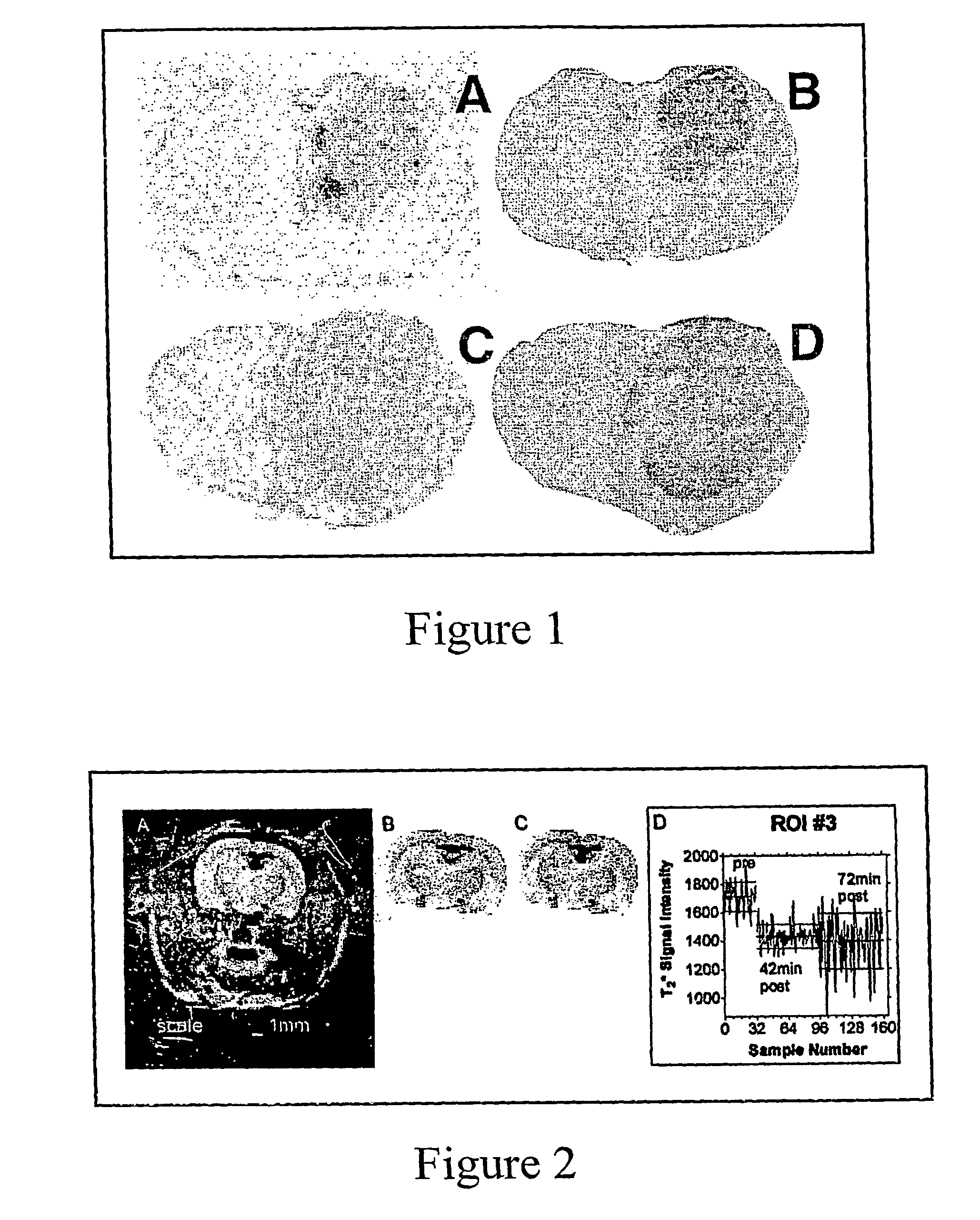
[0047]Radiolabeled sickle cells can be imaged relatively non-invasively using nuclear medicine imaging. FIG. 3 illustrates an example of the resulting image from a clinical scintillation scanner following the intravenous administration of human sickle cells to a rat with a large 9L tumor implanted in its right leg. Radioactivity increases sharply over the first several minutes and continues to increase more slowly over the next hour. The highest activity is found in regions of the rat corresponding to the tumor and some normal tissues, for example, the spleen or liver. Regions of interest are drawn around the liver, spleen, bladder, normal leg muscle, and tumor in order to monitor changes in radioactivity as a function of time. The results are shown in FIG. 3.

[0048]FIG. 3b demonstrates preferential accumulation of sickle cells in tumor immediately. Furthermore, the temporal data shown in FIG. 3b suggests that further accumulation in the tumor over the next ho...

example two

Laser Scanning Confocal Microscopy

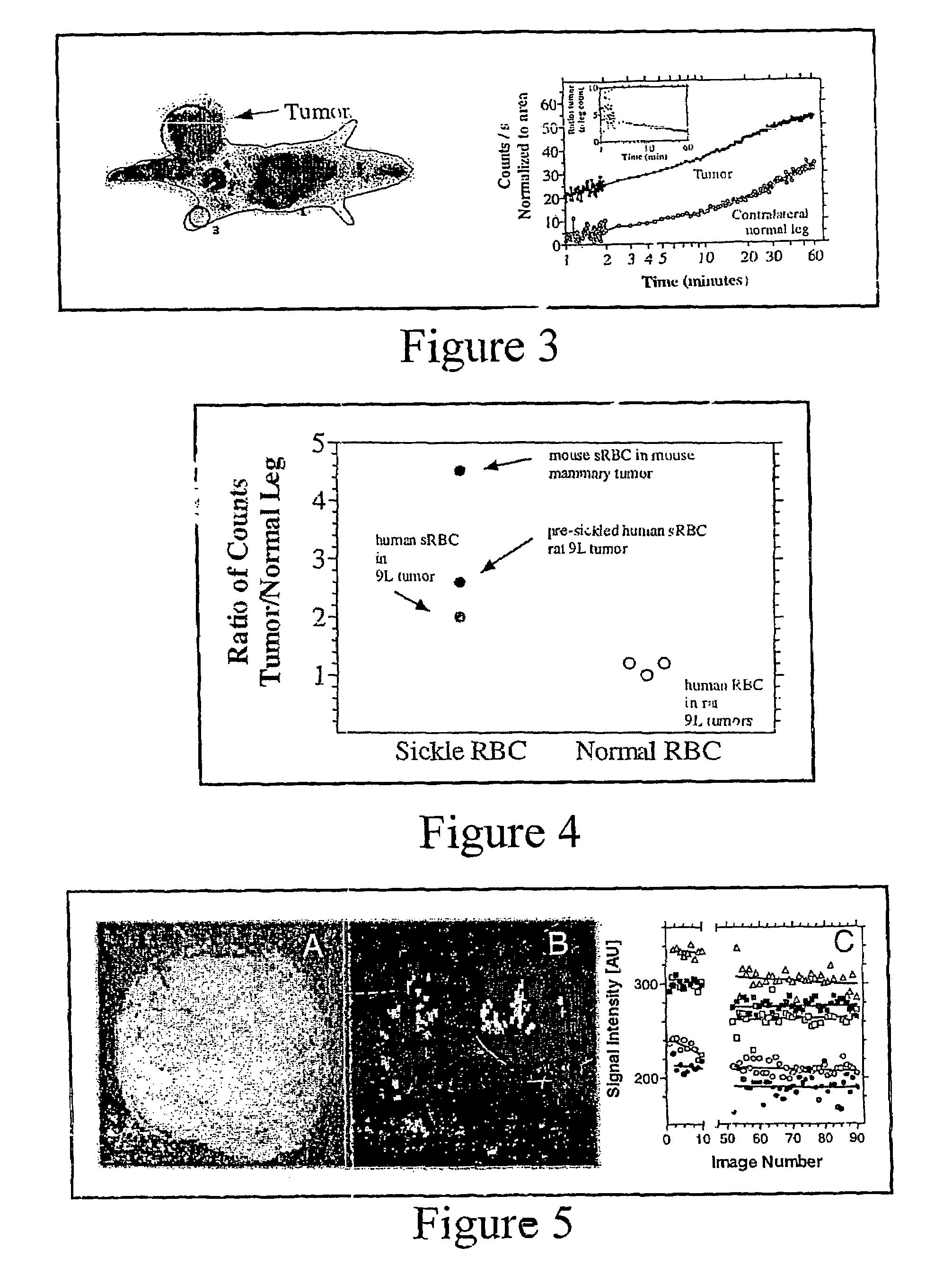
[0052]Preferential aggregation of sickle cells to tumor is observed in rats when sRBC (0.5 ml packed) are labeled with fluorescence (Oregon Green 488 conjugated to a streptavidin-biotin complex that binds to RBC surface proteins (biotinoyl-amino-hexanoic acid, sulfosuccinimidyl ester; Molecular Probes, Inc., Eugene Oreg.) and delivered through the femoral vein one hour before sacrifice. Vasculature is visualized by injecting another fluorescence compound, Evans Blue, which conjugates to albumin in vivo. Evans Blue (2 mls per kg of a 2% w / v solution) is delivered within 10 seconds of sacrificing the rats. Of note is that the distribution of sRBCs within the tumor is not uniform. Some regions, particularly the periphery, sequestered extremely high activity, indicating sickle red blood cells preferentially aggregate to particular regions in a tumor (number of studies, n=9). Congested sRBCs are even visible on bright field (FIG. 6). Normal RBCs did not ...


PUM

PropertyMeasurementUnit
Therapeuticaaaaaaaaaa

Abstract

According to the present invention, there is provided a delivery vehicle including sickle red blood cells carrying a moiety. The moiety can be any type of diagnostic or therapeutic agent. The present invention further provides for a method of diagnosing systemic hypoxia, acidosis, or hypertonicity by administering sickle red blood cells to a patient and detecting the location of the sickle red blood cells. Additionally, the present invention provides for a method of therapeutic treatment of systemic hypoxia, acidosis, or hypertonicity by administering sickle red blood cells to a patient. Further, the present invention provides for a delivery vehicle that specifically localizes or concentrates at systemic hypoxia, acidosis, or hypertonicity areas. The delivery vehicle is used in diagnosing and therapeutically treating these areas of hypoxia, acidosis, or hypertonicity also. The present invention further provides for a method of making the delivery vehicle described herein.

Description

CROSS-REFERENCE TO RELATED APPLICATION[0001]This patent application is a National Phase Concerning a Filing Under 35 U.S.C. 371, claiming the benefit of priority of PCT / US01 / 19846, filed Jun. 22, 2001, which claims the benefit of priority of U.S. Provisional Application Ser. No. 60 / 213,516 filed Jun. 22, 2000, all of which are incorporated herein by reference.BACKGROUND OF THE INVENTION[0002]1. Field of the Invention[0003]This invention generally relates to the field of delivery vehicles for use in diagnostic and therapeutic applications, and specifically towards diagnostic and therapeutic applications involving hypoxic areas of a patient.[0004]2. Description of Related Art[0005]Hypoxic tissue areas are defined as areas having low oxygen content. Generally, the hypoxic tissue area is a result of numerous factors including, but not limited to, a reduction of the oxygen-carrying capacity of blood as a result of a decrease in the total hemoglobin or an alteration of the hemoglobin cons...

Claims


Application Information

Patent Timeline
IPC IPC(8): G01N33/555A61K51/00A61K47/48A61K49/00A61K49/18A61K51/12
CPCA61K47/48776A61K49/0002A61K49/1896A61K51/1203A61K47/6901
Inventor KIM, JAE HOBROWN, STEPHEN LAWRENCESWERDLOW, PAUL S.
Owner HENRY FORD HEALTH SYST
Why Eureka
Try Eureka