Methods and apparatus for characterization of tissue samples

a tissue sample and tissue technology, applied in the field of tissue classification, can solve the problems of indeterminate diagnosis, insufficient colposcopic technique, insufficient direct visual observation alone, etc., and achieve the effect of increasing diagnostic sensitivity and specificity and increasing diagnostic accuracy

Inactive Publication Date: 2007-12-18
LUMA IMAGING CORP
View PDF378 Cites 41 Cited by
  • Summary
  • Abstract
  • Description
  • Claims
  • Application Information

AI Technical Summary

Benefits of technology

[0010]The invention provides increased diagnostic sensitivity and specificity by combining a plurality of statistical classification techniques to determine tissue-class probabilities for a tissue sample. Furthermore, in one embodiment, the invention comprises combining one or more statistical classification techniques with one or more non-statistical approaches in order to determine a condition of a tissue sample.
[0011]The invention provides increased diagnostic accuracy by applying two or more statistical classification techniques to data fro

Problems solved by technology

However, direct visual observation alone may be inadequate for proper identification of an abnormal tissue sample, particularly in the early stages of disease.
Colposcopic techniques are not perfect.
Colposcopic images may contain complex and confusing patterns and may be affected by glare, shadow, or the presence of blood or other obstruct

Method used


Image

  • Methods and apparatus for characterization of tissue samples
  • Methods and apparatus for characterization of tissue samples
  • Methods and apparatus for characterization of tissue samples

Examples


Embodiment Construction

[0167]

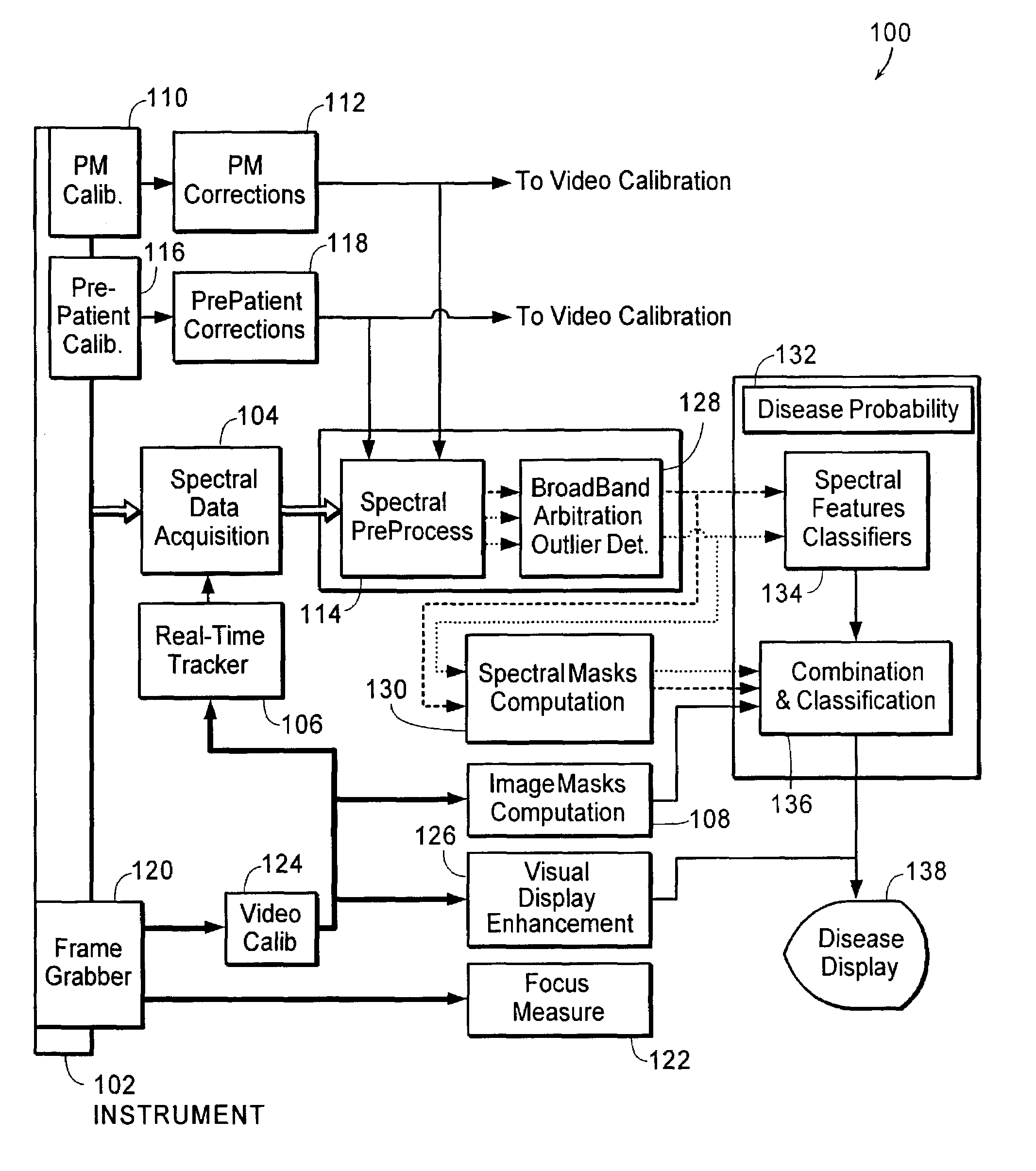
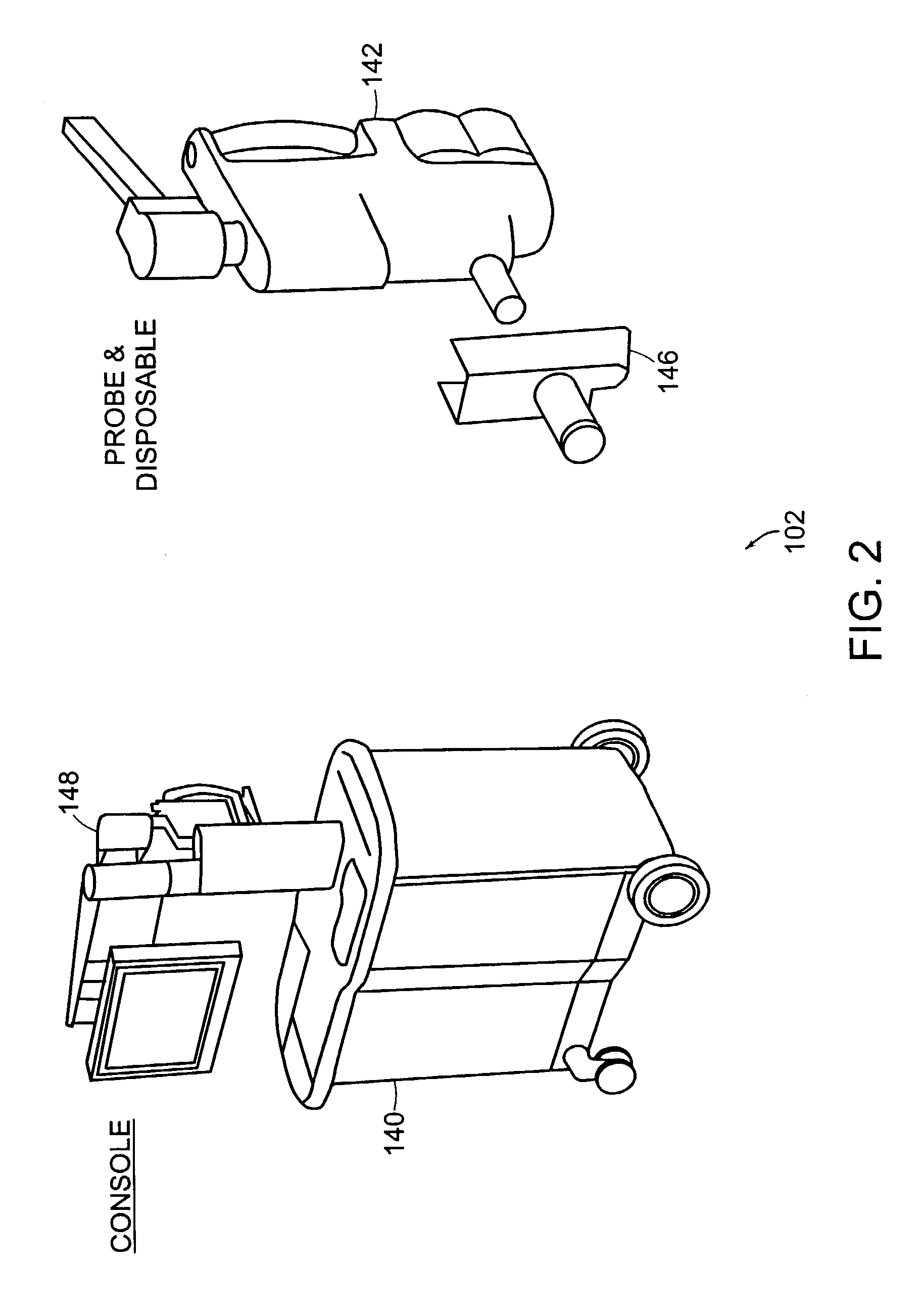
Table of ContentsPageSystem overview32Instrument38Spectral calibration51Patient scan procedure99Video calibration and focusing102Determining optimal data acquisition window114Motion tracking131Broadband reflectance arbitration and low-signal masking158Classification system overview180Spectral masking186Image masking197Glarevid203[ROI]vid208[ST]vid209Osvid217Bloodvid222Mucusvid226[SP]vid231[VW]vid242[FL]vid256Classifiers265Combining spectral and image data276Image enhancement285Diagnostic display291

[0168]The Table of Contents above is provided as a general organizational guide to the Description of the Illustrative Embodiment. Entries in the Table do not serve to limit support for any given element of the invention to a particular section of the Description.

System 100 Overview

[0169]The invention provides systems and methods for obtaining spectral data and image data from a tissue sample, for processing the data, and for using the data to diagnose the tissue sample. As used here...


PUM


Abstract

The invention provides methods for determining the probability that a given region of a tissue sample contains tissue of a given category, such as CIN 1 (cervical intraepithelial neoplasia, grade 1), CIN 2/3 (cervical intraepithelial neoplasia grades 2 and/or 3), normal squamous, normal columnar, and metaplasia, for example. The invention provides increased diagnostic accuracy by combining a plurality of statistical classification techniques. Furthermore, in one embodiment, the invention comprises combining one or more statistical techniques with one or more non-statistical classification techniques.

Description

RELATED APPLICATIONS[0001]This application is related to the following commonly-owned applications: entitled, “Methods and Apparatus for Characterization of Tissue Samples”; entitled, “Methods and Apparatus for Displaying Diagnostic Data”; entitled, “Methods and Apparatus for Visually Enhancing Images”; entitled, “Methods and Apparatus for Processing Image Data for Use in Tissue Characterization”; entitled, “Methods and Apparatus for Processing Spectral Data for Use in Tissue Characterization”; entitled, “Methods and Apparatus for Evaluating Image Focus”; and “Methods and Apparatus for Calibrating Spectral Data,” all of which are filed on even date herewith.FIELD OF THE INVENTION[0002]This invention relates generally to the classification of tissue. More particularly, in certain embodiments, the invention relates to the combination of statistical and / or non-statistical tissue classification schemes.BACKGROUND OF THE INVENTION[0003]It is common in the field of medicine to perform vis...

Claims


Application Information

Patent Timeline
IPC IPC(8): G01N21/64A61B5/00G01N21/31
CPCA61B5/0059A61B5/7203G01N21/31A61B5/7264A61B5/725A61B5/7267A61B5/7257G16H50/20
Inventor COSTA, PETER J.SUM, STEPHEN T.FLEWELLING, ROSS F.SCHOMACKER, KEVIN T.SCHOTT, JEAN-PIERREDRAAYER, BRET F.
Owner LUMA IMAGING CORP
Why Eureka
Try Eureka