Magnetic resonance spectroscopy to classify tissue

Inactive Publication Date: 2008-02-26
SYDNEY UNIV OF
View PDF1 Cites 16 Cited by
  • Summary
  • Abstract
  • Description
  • Claims
  • Application Information

AI Technical Summary

Benefits of technology

[0008]Magnetic resonance spectroscopy (MRS) is a modality with a proven record in the diagnosis of minimally invasive malignant lesions (5-11). MR spectra of small samples of tissue or even cell suspensions enable the reliable determination of whether the tissue of origin is malignant or benign. Often MRS is able to detect malignancy before morphological manifestations are visible by light microscopy (8).

Problems solved by technology
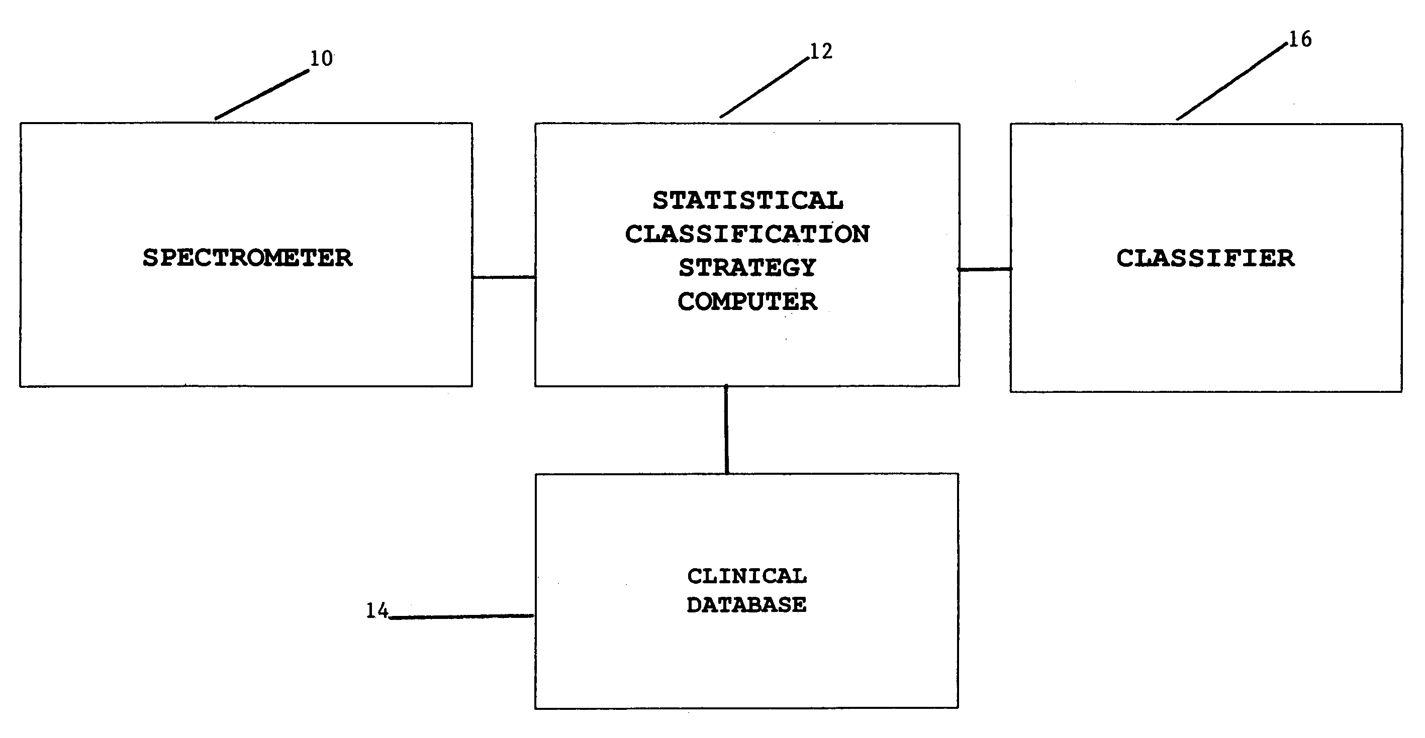
Twenty percent of the spectra were discarded because low aspirate cellularity yielded inadequate SNR.

Method used


Image

  • Magnetic resonance spectroscopy to classify tissue
  • Magnetic resonance spectroscopy to classify tissue
  • Magnetic resonance spectroscopy to classify tissue

Examples


Embodiment Construction
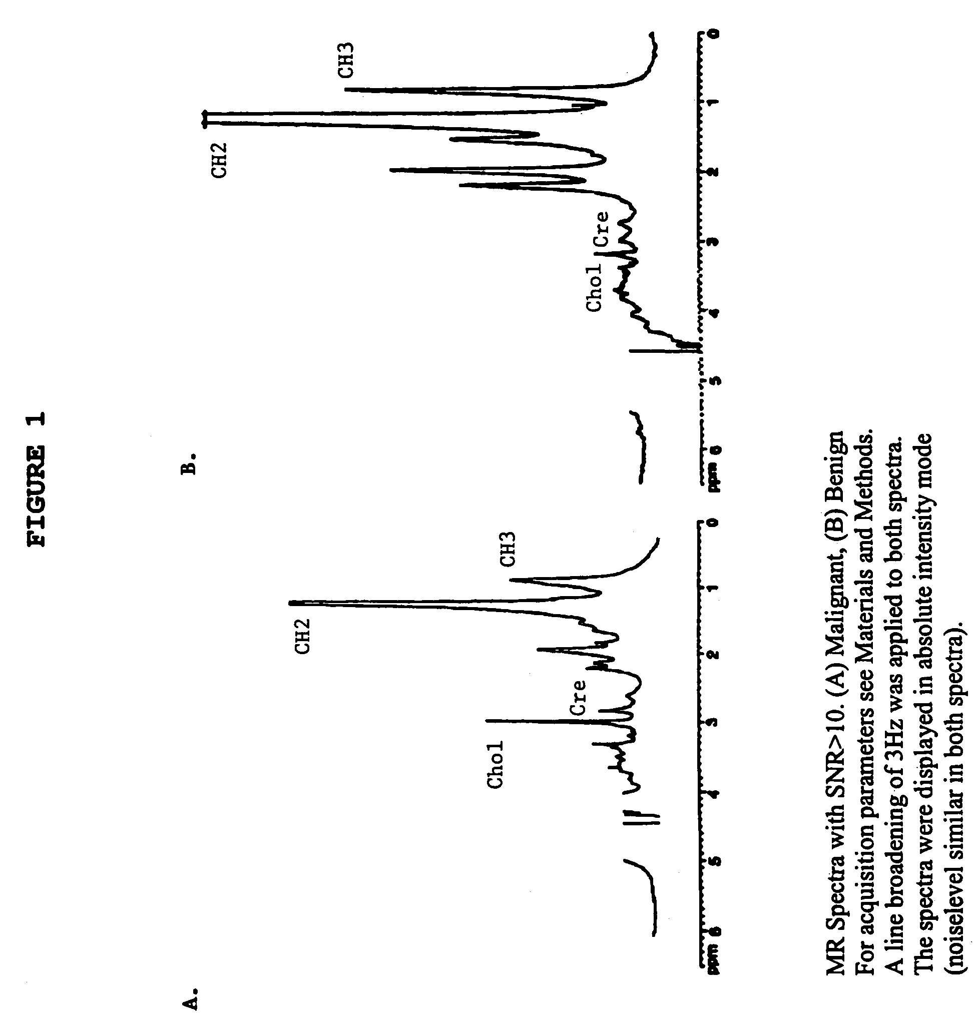
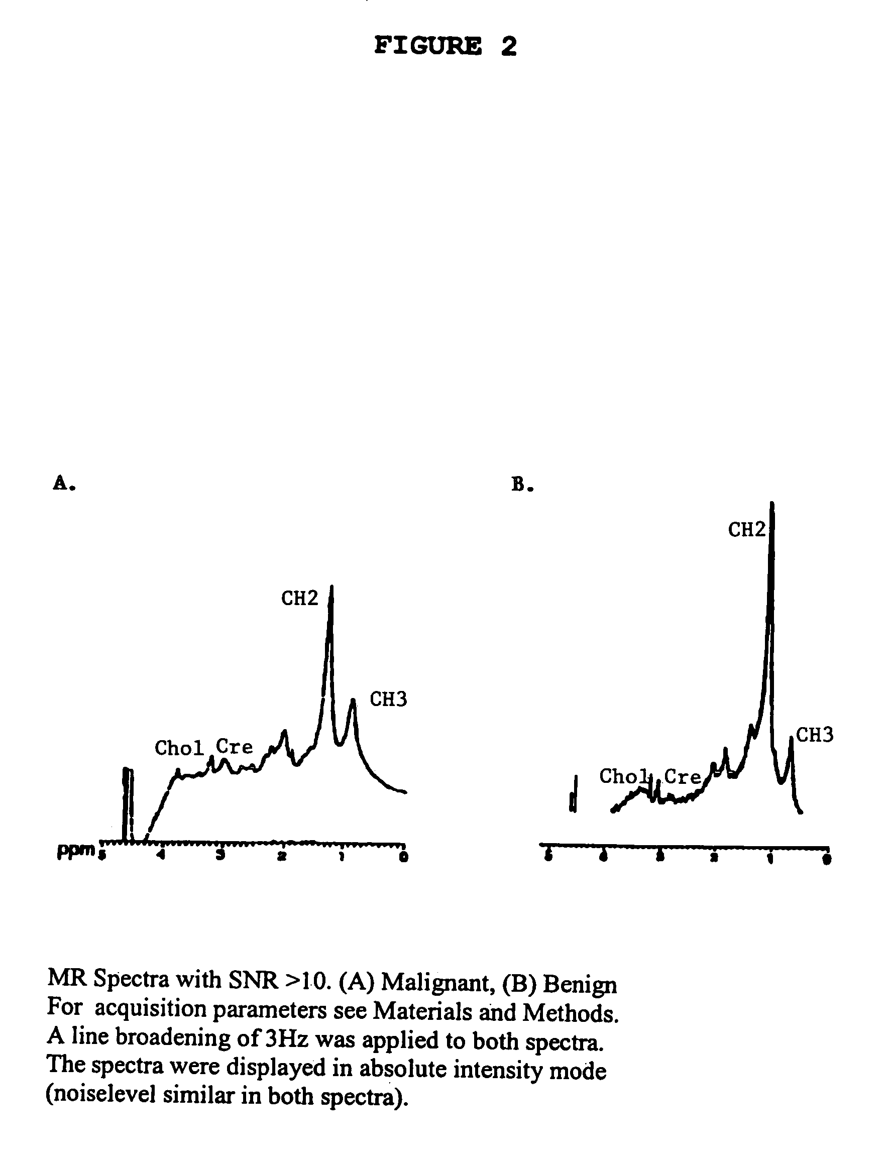
Methods

Preparation of Patients:

[0039]Intra-operative FNAB were taken from 139 patients undergoing breast surgery for malignant and benign conditions (Table 1) by three surgeons in separate hospitals. In order to provide a sufficiently large data set for SCS an additional 27 patients joined the study (see Table 1). Impalpable breast lesions that had been localised by carbon track or hook wire were included except if the lesion was not palpable at excision or when the pathology specimen could have been compromised. All samples were taken during surgery under direct vision after the lesion had been identified and incised sufficiently widely to ensure that the FNAB and tissue specimens represented the same lesion and were thus comparable. The lesion was identified and incised in-vivo via the margin with the greatest apparent depth of normal tissue between it and the lesion to ensure the pathologist could report upon the lesion according to a standard protocol. Malignant and suspicious l...


PUM

PropertyMeasurementUnit
volumeaaaaaaaaaa
temperatureaaaaaaaaaa
chemical shiftsaaaaaaaaaa

Abstract

Robust classification methods analyze magnetic resonance spectroscopy (MRS) data (spectra) of fine needle aspirates taken from breast tumors. The resultant data when compared with the histopathology and clinical criteria provide computerized classification-based diagnosis and prognosis with a very high degree of accuracy and reliability. Diagnostic correlation performed between the spectra and standard synoptic pathology findings contain detail regarding the pathology (malignant versus benign), vascular invasion by the primary cancer and lymph node involvement of the excised axillary lymph nodes. The classification strategy consisted of three stages: pre-processing of MR magnitude spectra to identify optimal spectral regions, cross-validated Linear Discriminant Analysis, and classification aggregation via Computerised Consensus Diagnosis. Malignant tissue was distinguished from benign lesions with an overall accuracy of 93%. From the same spectrum, lymph node involvement was predicted with an accuracy of 95% and tumor vascularisation with an overall accuracy of 92%.

Description

CROSS REFERENCE TO RELATED APPLICATIONS[0001]This application claims priority on, and incorporates by reference, U.S. Provisional Application Ser. No. 60 / 160,029 filed Oct. 18, 1999, now abandoned, and this application is a continuation of U.S. patent application Ser. No. 09 / 691,776 filed Oct. 18, 2000, now U.S. Pat. No. 6,835,572.STATEMENT REGARDING FEDERALLY SPONSORED RESEARCH DEVELOPMENT[0002]The work described herein was supported by U.S. Army Grant number DAMD 17-96-1-6077 and NH & MRC 950215 and NH & MRC 973769.BACKGROUND OF THE INVENTION[0003]1. Technical Field of the Invention[0004]The present invention relates to the use of magnetic resonance spectroscopy, and more particularly to such use for determining pathology, vascularization and nodel involvement of a biopsy of breast tissue.[0005]2. Description of the Related Art[0006]Within this application several publications are referenced by arabic numerals within parentheses. Full citations for these and other references may b...

Claims


Application Information

Patent Timeline
Patent Type & Authority Patents(United States)
IPC IPC(8): G01N33/48A61B5/05G06F19/00G01N24/08G01R33/46G01R33/465
CPCG01N24/08G01R33/4625G01R33/20G06F19/366G01R33/465Y10T436/24G16H10/40A61P43/00
Inventor MOUNTFORD, CAROLYN E.RUSSELL, PETERSMITH, IAN C. P.SOMORJAI, RAJMUND L.
Owner SYDNEY UNIV OF
Why Eureka
Try Eureka