Method and apparatus for real time quantitative three-dimensional image reconstruction of a moving organ and intra-body navigation

a three-dimensional image and moving organ technology, applied in the field of three-dimensional medical imaging and navigation, can solve problems such as prone to rupture and provoke fatal vessel obstruction

Inactive Publication Date: 2008-03-11
ST JUDE MEDICAL INT HLDG SARL
View PDF60 Cites 400 Cited by
  • Summary
  • Abstract
  • Description
  • Claims
  • Application Information

AI Technical Summary

Problems solved by technology

Intra-vascular plaques may be prone to rupture and may provoke fatal vessel obstruction.

Method used


Image

  • Method and apparatus for real time quantitative three-dimensional image reconstruction of a moving organ and intra-body navigation
  • Method and apparatus for real time quantitative three-dimensional image reconstruction of a moving organ and intra-body navigation
  • Method and apparatus for real time quantitative three-dimensional image reconstruction of a moving organ and intra-body navigation

Examples


Embodiment Construction

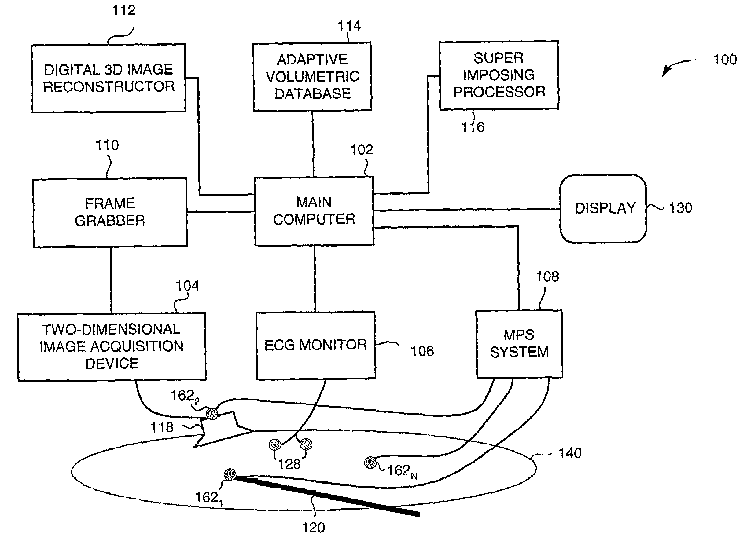
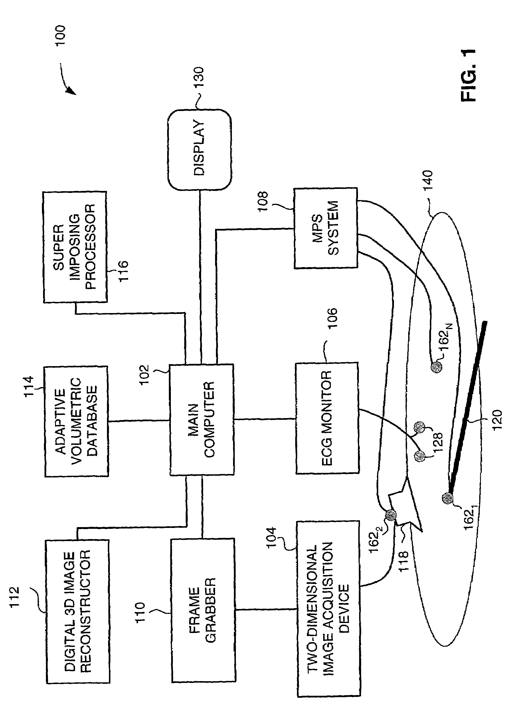
[0092]The present invention overcomes the disadvantages of the prior art by providing methods and systems for constructing and displaying three-dimensional images of moving organs, synchronously with the movement of these organs and synchronously with an invasive tool, such as a catheter. According to a preferred embodiment, the three-dimensional images and the presentation of the invasive tool, all reside within a single coordinate system, and no registration of a plurality of coordinate systems is required.

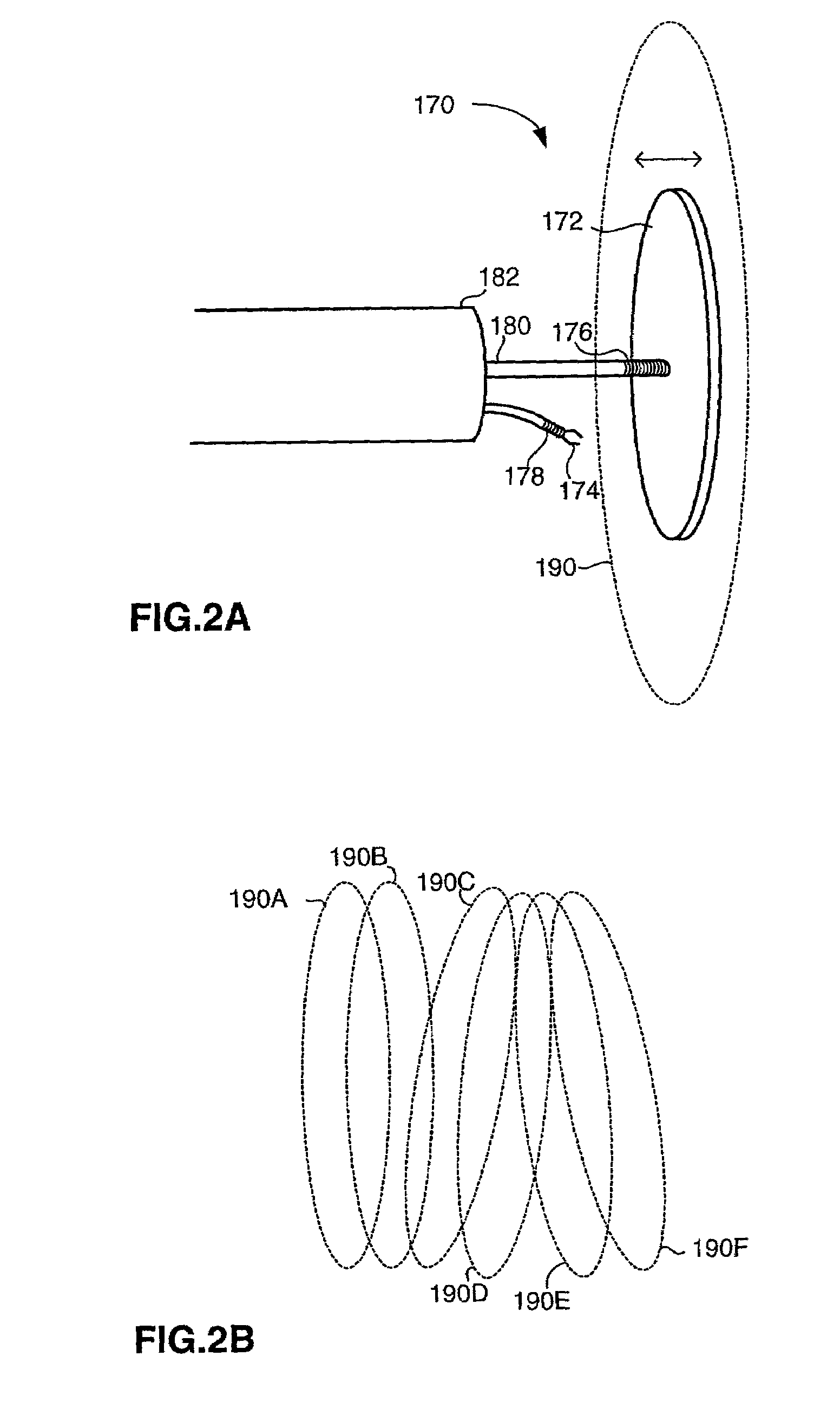
[0093]According to one aspect of the invention there is provided a pseudo real time imaging system, for minimal invasive surgery. This system includes a two-dimensional image acquisition system, a medical positioning system (MPS) which is basically a location and orientation detection system, a specific organ monitor and an image processing system. The location and orientation detection system includes at least three sensors. The first sensor is mounted on the image detector of ...


PUM


Abstract

Medical imaging and navigation system including a processor, a medical positioning system (MPS), a two-dimensional imaging system and an inspected organ monitor interface, the MPS including an imaging MPS sensor, the two-dimensional imaging system including an image detector, the processor being coupled to a display unit and to a database, the MPS being coupled to the processor, the imaging MPS sensor being firmly attached to the image detector, the two-dimensional imaging system being coupled to the processor, the image detector being firmly attached to an imaging catheter.

Description

CROSS REFERENCE INFORMATION[0001]This application is a Continuation-in-Part of application Ser. No. 09 / 782,528, filed Feb. 13, 2001, which is a Continuation-in-Part of application Ser. No 09 / 314,474, filed May 18, 1999 now U.S. Pat. N. 6,233,476.FIELD OF THE INVENTION[0002]The present invention relates to medical diagnostic and surgery systems and methods in general, and to methods and systems for three-dimensional medical imaging and navigation, in particular.BACKGROUND OF THE INVENTION[0003]Methods and systems for acquiring and presenting two-dimensional and three-dimensional images are known in the art. Three-dimensional imaging enhances modern diagnostics, therapy and surgery procedures.[0004]A two-dimensional imaging system processes and represents two-dimensional internal body slices in static or in dynamic form on a display. A conventional two-dimensional ultrasound imaging system includes an ultrasound transducer, an image capturing module and an image-processing unit.[0005]...

Claims


Application Information

Patent Timeline
Patent Type & Authority Patents(United States)
IPC IPC(8): A61B5/00A61B1/00G01B7/00A61B5/055A61B5/06A61B5/352A61B6/00A61B6/03A61B8/08A61B8/12A61B19/00A61G13/00A61M25/00G01B7/30G01B17/00G01B17/02G01B17/06G01T1/00G01T1/161
CPCA61B5/0066A61B5/7445A61B5/06A61B6/5247A61B8/0833A61B8/0841A61B8/12A61B8/4254A61B8/463A61B8/5238A61B19/52A61B19/5244A61B5/0456A61B5/7285A61B6/503A61B6/504A61B6/541A61B8/0891A61B8/483A61B19/20A61B19/50A61B19/5212A61B19/56A61B2019/505A61B2019/507A61B2019/5251A61B2019/5272A61B2019/5276A61B2019/5278A61B2019/528A61B2019/5291A61B2019/5295A61B2019/5458A61B2019/566A61B5/015A61B8/4227A61B90/10A61B90/36A61B2034/2072A61B2090/3782A61B2090/367A61B2034/256A61B34/20A61B34/25A61B90/361A61B2090/3784A61B34/10A61B2034/105A61B2034/107A61B2034/2051A61B2090/378A61B2090/365A61B2090/3958A61B5/062A61B5/352
Inventor STROMMER, GERA M.EICHLER, UZISCHWARTZ, LIAT
Owner ST JUDE MEDICAL INT HLDG SARL
Why Eureka
Try Eureka