
PD-1-lacking mouse and use thereof

a mouse and pd-1 technology, applied in the field of programmed cell death1 receptors, can solve the problems of no available treatment method, no known treatment method, and threaten life, and achieve the effect of easy development of lupus

Inactive Publication Date: 2008-08-19
ONO PHARMA CO LTD +1
View PDF1 Cites 44 Cited by
  • Summary
  • Abstract
  • Description
  • Claims
  • Application Information

AI Technical Summary

Benefits of technology

The present invention relates to a mouse with a deficiency in the PD-1 receptor, which is involved in regulating the immune system. The mouse is useful for screening methods for identifying medicines that can treat autoimmune diseases, particularly those caused by the production of autoantibodies. The mouse also has a specific protein that is associated with dilated cardiomyopathy, a type of autoimmune disease that causes heart failure. The invention provides a diagnostic method for detecting the protein and a potential genetic marker for the disease. The mouse model also suggests a possibility that dilated cardiomyopathy may be caused by an autoimmune response targeting cardiac muscle.

Problems solved by technology

In both cases, the disease is progress, and threatens the life, and there is no available treatment method excluding cardiac transplant now.
However, it really has been not known to what influence the difference of the genetic background influences the loss of PD-1, in case that PD-1 is lacked in other strain mice.

Method used


Image

  • PD-1-lacking mouse and use thereof
  • PD-1-lacking mouse and use thereof
  • PD-1-lacking mouse and use thereof

Examples


example 1

Making of PD-1 Deficient BALB / c Mice

[0036]PD-1 receptor deficient BALB / c mice written were made by backcrossing PD-1 receptor deficient C57BL / 6(B6) mice with BALB / c mice tenth generation, according to method described in International Immunology. Vol. 10(10), 1563-1572(1998), with embryo stroma (ES) cell lines from BALB / c mice. Obtained mice were bred in facilities of the sterility.

example 2

Cause of Death of PD-1 Deficient BALB / c Mice

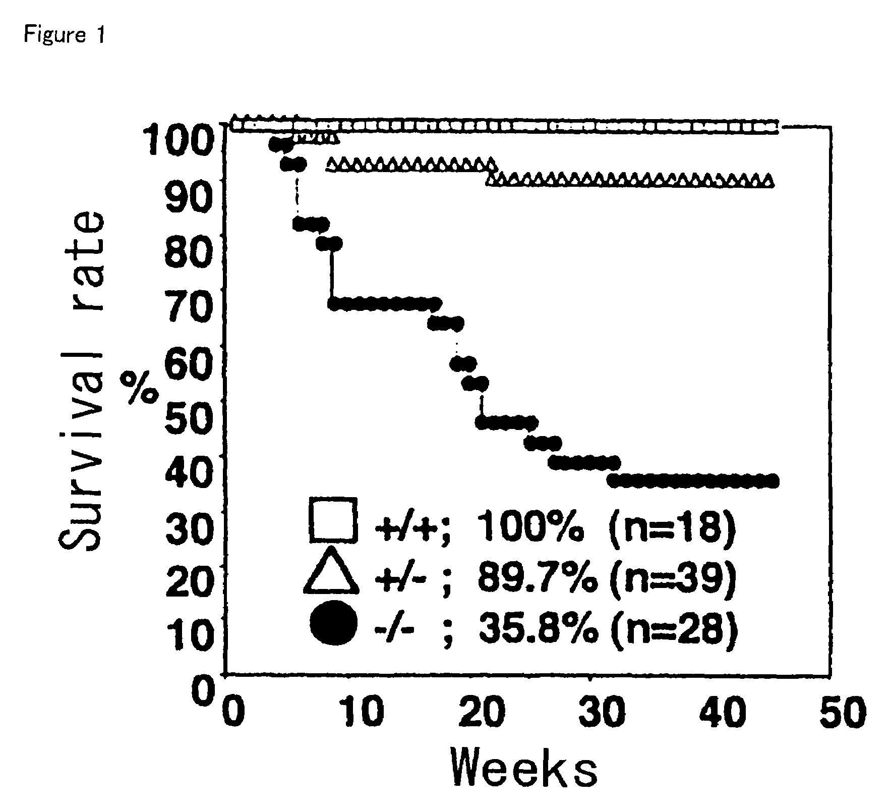
[0037]Breeding PD-1 deficient BALB / c mice (PD-1(− / −)) obtained in example 1, they started to die as early as 5 weeks of age, as black circles in FIG. 1 show. Then, by 30 weeks, two thirds of mice died. On the other hand, in normal BALB / c (PD-1(+ / +)) mice (void squares in FIG. 1), which are not deficient of PD-1, those deaths were not observed. FIG. 1 shows with the result of breeding of PD-1 deficient (PD-1(+ / −)) B6 mice.

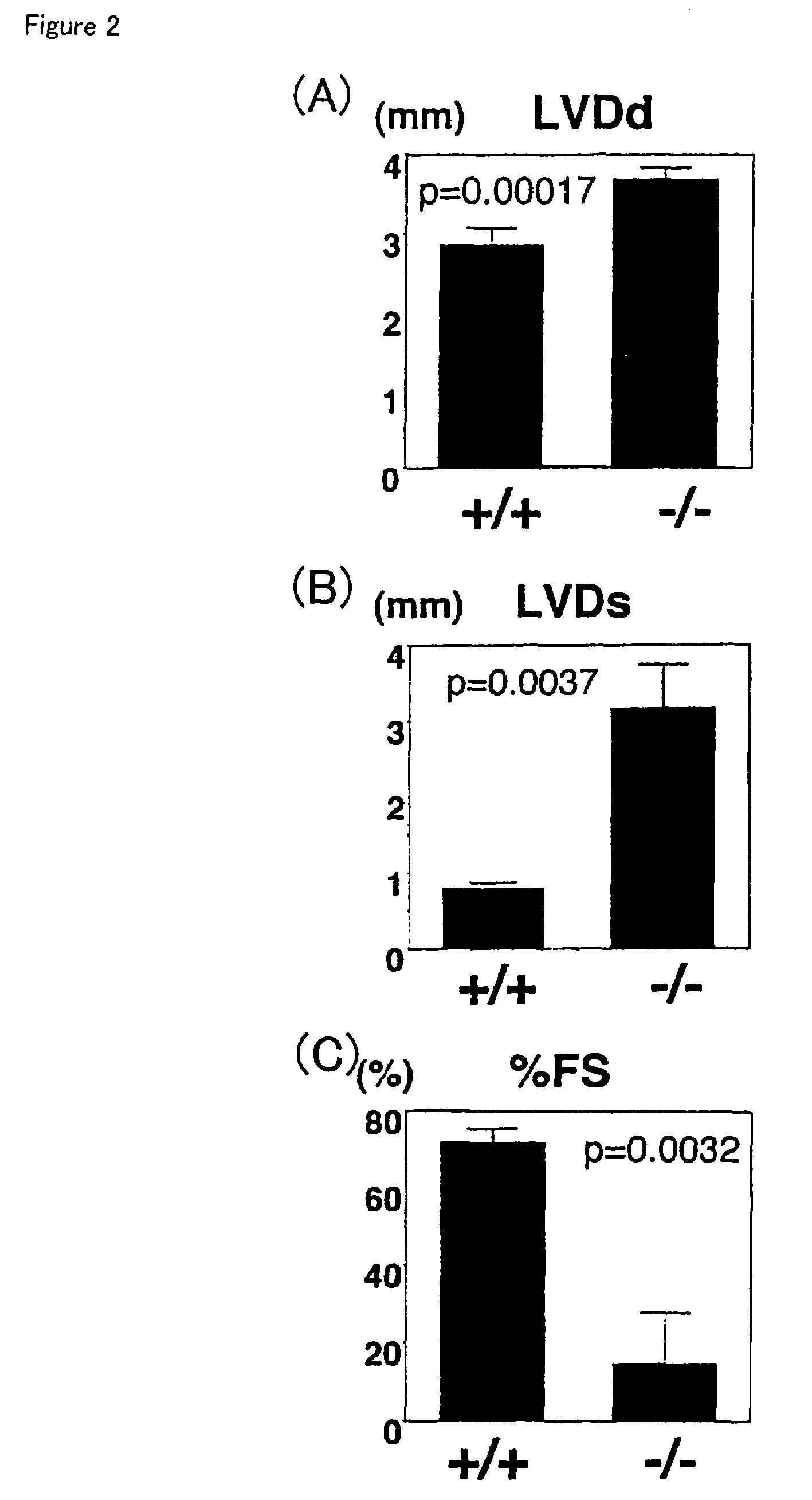
[0038]PD-1(− / −) mice show the projection of the eyeball several days before death, and as a result of the autopsy, hearts in all mice were enlarged to anomalous. Moreover, tumefaction of livers were shown, and suggested that the cause of death was congestive heart failure.

example 3

Histological Examination

[0039]Histological examinations of dead PD-1 deficient BALB / c mice were done. Right ventricular walls of PD-1(− / −) mice were thinner than those of normal mice, and both ventricles were more enlarged to about two fold in diameter.

[0040]Though a sporadic fibrous react was observed including cellular interstitial fibrosis accompanied with scarring plasticand, ventricular walls appeared grossly normal. Electron microscopic examination revealed that throughout ventricular walls the scattered and disrupted myofilaments and the degeneration of cardiomyocytes with irregularly shaped mitochondria disarrayed. In atriums of large majority of mice, the thrombus seemed to depend on various sizes and almost huge congestion was observed.


PUM

PropertyMeasurementUnit
molecular weightaaaaaaaaaa
pHaaaaaaaaaa
hydrophobicaaaaaaaaaa

Abstract

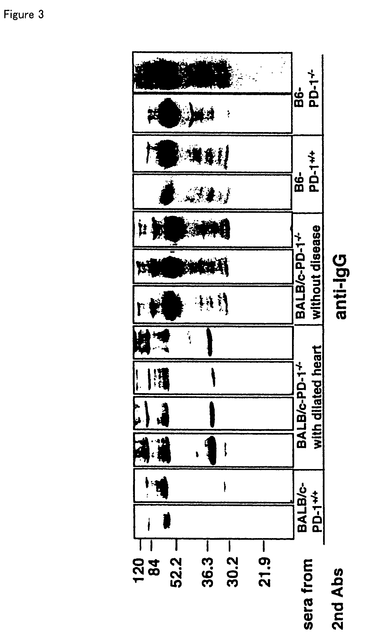
The invention relates to BALB / c mice that are deficient in the programmed cell death-1 receptor (PD-1), a screening method for autoimmune disease medicine using these mice, an IgG self-reactive antibody that the mice produce, a protein that specifically reacts to the antibody and is produced in heart, and a diagnostic method for dilated cardiomyopathy using the protein. Because PD-1 deficient BALB / c mice spontaneously develop autoimmune disease, and specifically dilated cardiomyopathy, they are useful for screening for medicines against these diseases.

Description

TECHNICAL FIELD OF THE INVENTION[0001]The present invention relates to programmed cell death-1 receptor (hereafter, it is abbreviated as PD-1)-deficient BALB / c mice and the use. More particularly, it relates to PD-1 receptor-deficient BALB / c mice, a screening method of medicines against autoimmune disease by use of the mice, IgG self-reactivity antibody of which these mice produce specifically, a protein specifically reacted to the antibody and produced in heart and an diagnostic method in dilated cardiomyopathy by use of the protein.BACKGROUND ART[0002]Programmed cell death controlled embryologically or physiologically can be observed in all most tissues of various animals. Such programmed cell death is generally called, “Programmed cell death” or “Premeditated programmed cell death”, and distinguished from “unexpected cell death” which could be caused by a pathologic mechanism.[0003]First, PD-1 has been found in mice as a receptor that cells are related to process to premeditated ...

Claims


Application Information

Patent Timeline
Patent Type & Authority Patents(United States)
IPC IPC(8): A01K67/00A01K67/027A01K67/033C07K14/47C07K14/705C07K16/18C12N15/12C12N15/85
CPCA01K67/0276C12N15/8509C07K14/47C07K14/70503C07K16/18A01K2217/075A01K2227/105A01K2267/0325A01K2267/0375A01K67/027
Inventor HONJO, TASUKUNISHIMURA, HIROYUKI
Owner ONO PHARMA CO LTD
Learn More