Method and medical imaging system for compensating for patient motion

a medical imaging and patient technology, applied in the field of patient motion compensation, can solve the problems of inability to accurately compensate for motion, complicated image processing methods that require extensive processing power, and difficult to implement in real time, so as to improve image results

Inactive Publication Date: 2008-09-02
SIEMENS HEALTHCARE GMBH
View PDF12 Cites 81 Cited by
  • Summary
  • Abstract
  • Description
  • Claims
  • Application Information

AI Technical Summary

Benefits of technology

[0012]Using this prior art as a starting point, an object of the present invention is to specify a method as well as an associated imaging system to compensate for the motion of patients when recording a series of images in medical imaging, with which the motion of the patient can be compensated for while the images are being recorded without time-consuming user interaction, with the method being able to be implemented in real time. The method and the associated imaging system should in particular improve image results in a digital subtraction angiography and roadmapping with the lowest possible outlay as regards the operator's time.
[0017]The present method, by contrast with most previously known methods of motion correction, manages without interaction with the operator. The proposed method only requires a position sensor to be accommodated on the patient where necessary. Consequently no further user interaction is required for motion compensation. The principle of previous methods for retrospective image processing dictates that they only operate approximately. It is barely possible to correct large movements using these methods, and small movements can only be approximately corrected. The proposed method operates with high precision even for large movements, so that the need to record a mask image more than once is avoided. The saves time, contrast means and reduces the applied X-ray dose in the case of X-ray image recording. The present method also makes it possible in particular cases to dispense with sedating or anesthetizing the patient merely for the purpose of minimizing motion artifacts.
[0020]To compensate for the detected deviations it is further possible to geometrically modify the image content of the recorded images. This relates in particular to a rotation of the image content at right angles to the image plane as well as to the translation in the two degrees of translation freedom of the two-dimensional recorded image. Furthermore scaling of the image is possible. Depending on the type of patient motion a combination of the two compensation techniques can also be advantageous, i.e. the modification of the geo-metrical circumstances of the imaging system and the geometrical adaptation of the image content.
[0021]Naturally, with the present method, after the at least approximate compensation of the movement, retrospective image processing methods can also be used in order to further improve the image results. The approximate compensation by modifying the geometrical circumstances of the imaging system can be used in this case to compensate for rough movements, while remaining small errors can be rectified by retrospective image processing.

Problems solved by technology

Retrospective image processing can however only approximately compensate for motion.
Furthermore the complicated image processing methods demand extensive processing power and are thus difficult to implement in real time.
Manual methods for image processing (pixel shifting) need user interaction and can demand significant amounts of time.

Method used


Image

  • Method and medical imaging system for compensating for patient motion
  • Method and medical imaging system for compensating for patient motion
  • Method and medical imaging system for compensating for patient motion

Examples


Embodiment Construction

[0028]The present method is described below with reference to an X-ray angiography system for applications in neuroradiology. The method can naturally also be used in other fields in which digital subtraction angiography and / or roadmapping are employed. The present method can also be used with other medical imaging techniques involving having to record a series of images and relate them to one another.

[0029]The embodiments below are typically restricted to the case of correcting the patient's head movements. Since the head can be approximately regarded as a rigid body, the motion can be restricted to the 6 degrees of freedom of the translation and rotation of a rigid body in three-dimensional space.

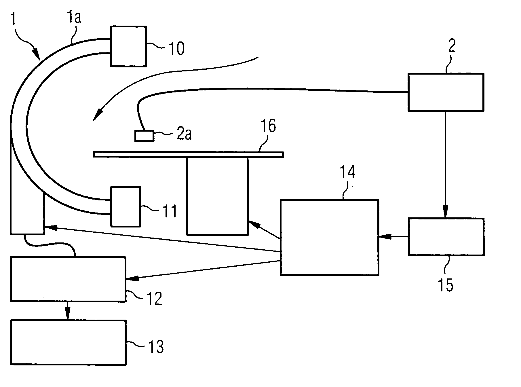
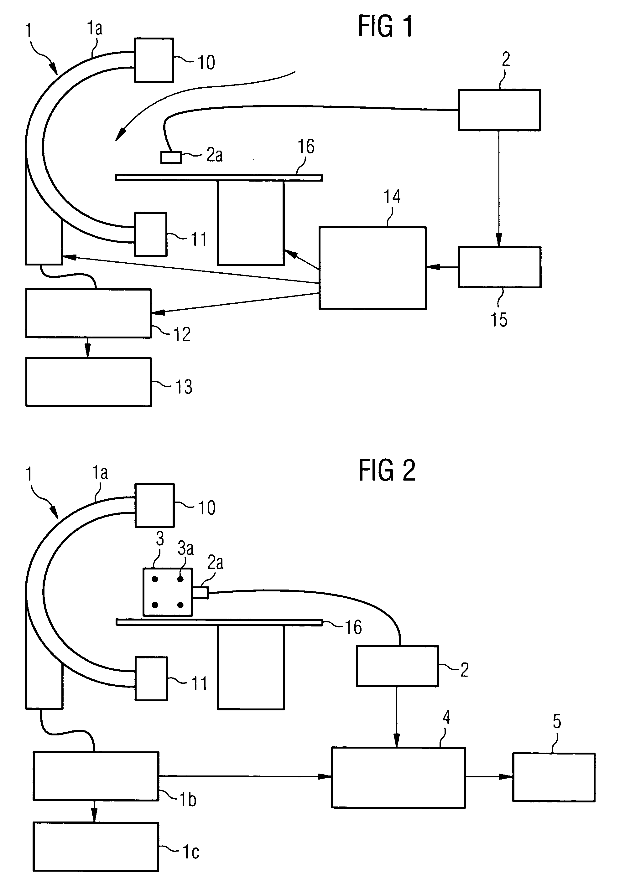
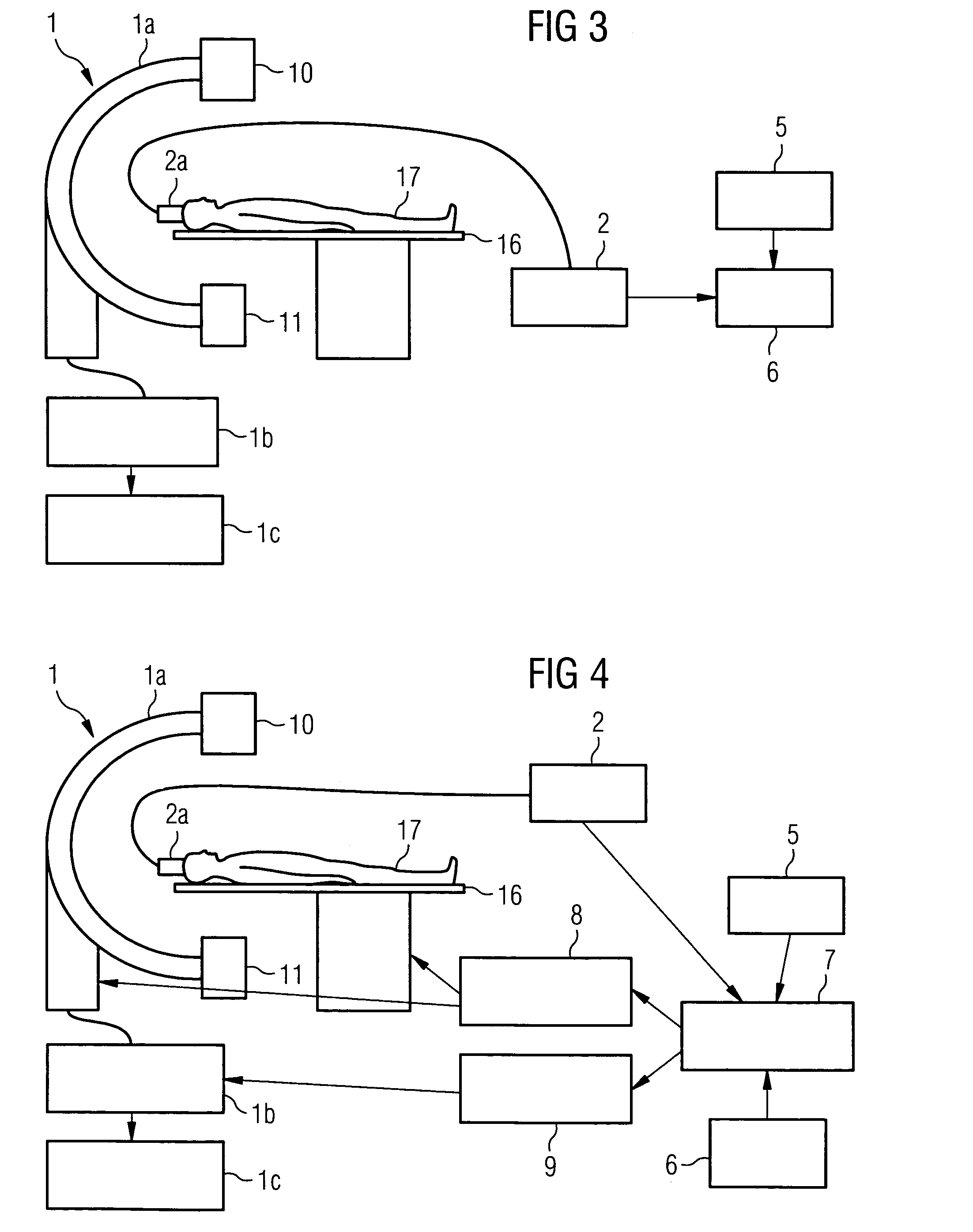
[0030]An X-ray angiography system 1 for neuro-radiology, which is shown schematically in FIG. 1, is used to record the images. The X-ray angiography system 1 includes a C-arm 1a which can be rotated around two axes, to which an X-ray tube 10 and a detector 11 arranged opposite the X-ray t...


PUM


Abstract

The present invention relates to a method and also an imaging system to compensate for patient motion when recording a series of images in medical imaging, in which a number of images of an area under examination of a patient (17) are recorded at intervals with an imaging system (1) and are related to one another. With the method a localization system (2) is used as the series of images are being recorded to permanently or at a time close to the recording of the individual images, record a momentary spatial location of the area under examination in a reference system permanently linked to the imaging system (1), a first spatial location of the area under examination recorded close to the time of recording of a first image is stored, and a deviation of the images recorded momentarily in each case of the first spatial location is determined and by changing the geometrical circumstances of the imaging system (1) at a time to close the recording of the spatial location and / or through geometrical adaptation of an image content of an image just recorded, is at least approximately commentated for, so that the images show the area under examination in the same position and orientation. The method does not require any time-consuming interaction with the operator and is also suitable for compensating for larger movements of the patient.

Description

CROSS REFERENCE TO RELATED APPLICATIONS[0001]This application claims priority to the German application No. 10 2004 004 604.2, filed Jan. 29, 2004 which is incorporated by reference herein in its entirety.FIELD OF INVENTION[0002]The present invention relates to a method to compensate for patient movements when recording a series of medical images in which a number of images of an area under examination of the patient are taken at intervals with an imaging system and are compared to one another, as is the case for example in Digital Subtraction Angiography (DSA) or roadmapping. The invention further relates to a medical imaging system with radiation source, detector, patient support, image processing unit and image display unit which is embodied to execute the method.BACKGROUND OF INVENTION[0003]In a main area of application of the present method, the area of digital subtraction angiography, blood vessels of the human body are recorded with an imaging system, in this case an X-ray sy...

Claims


Application Information

Patent Timeline
Patent Type & Authority Patents(United States)
IPC IPC(8): H05G1/60H05G1/64A61B5/113A61B6/00A61B6/04
CPCA61B5/721A61B6/584A61B6/504A61B6/527A61B6/481A61B5/1135A61B6/0457A61B6/0487
Inventor BOESE, JANRAHN, NORBERT
Owner SIEMENS HEALTHCARE GMBH
Why Eureka
Try Eureka