

Method for discrimination of metaplasias from neoplastic or preneoplastic lesions
a metaplasia and preneoplastic technology, applied in the field of metaplasia discrimination from neoplastic or preneoplastic lesions, can solve problems such as loss of genes, and achieve the effect of high risk
- Summary
- Abstract
- Description
- Claims
- Application Information
AI Technical Summary
Benefits of technology
Problems solved by technology
Method used
Image

Examples
example 1
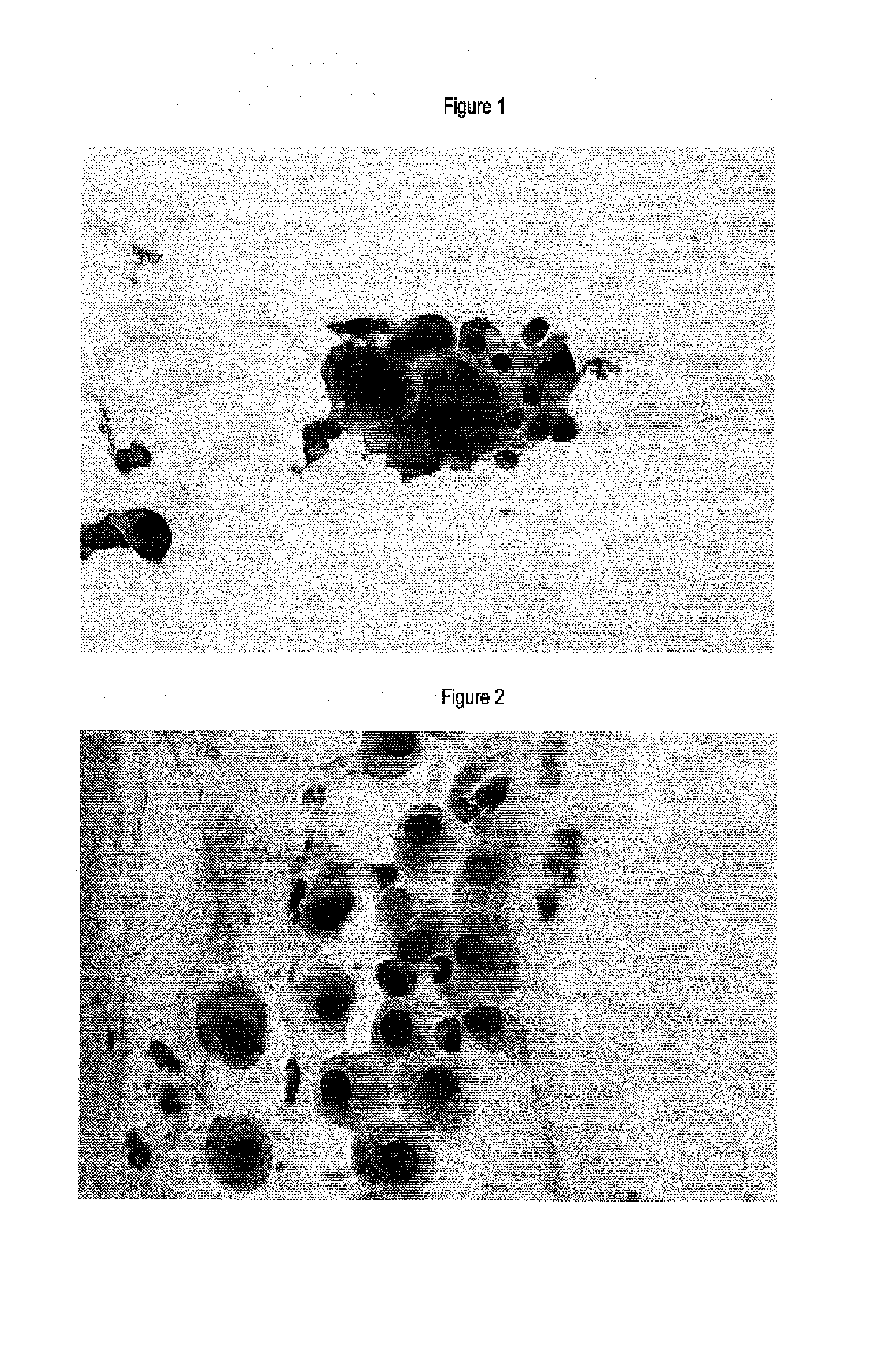
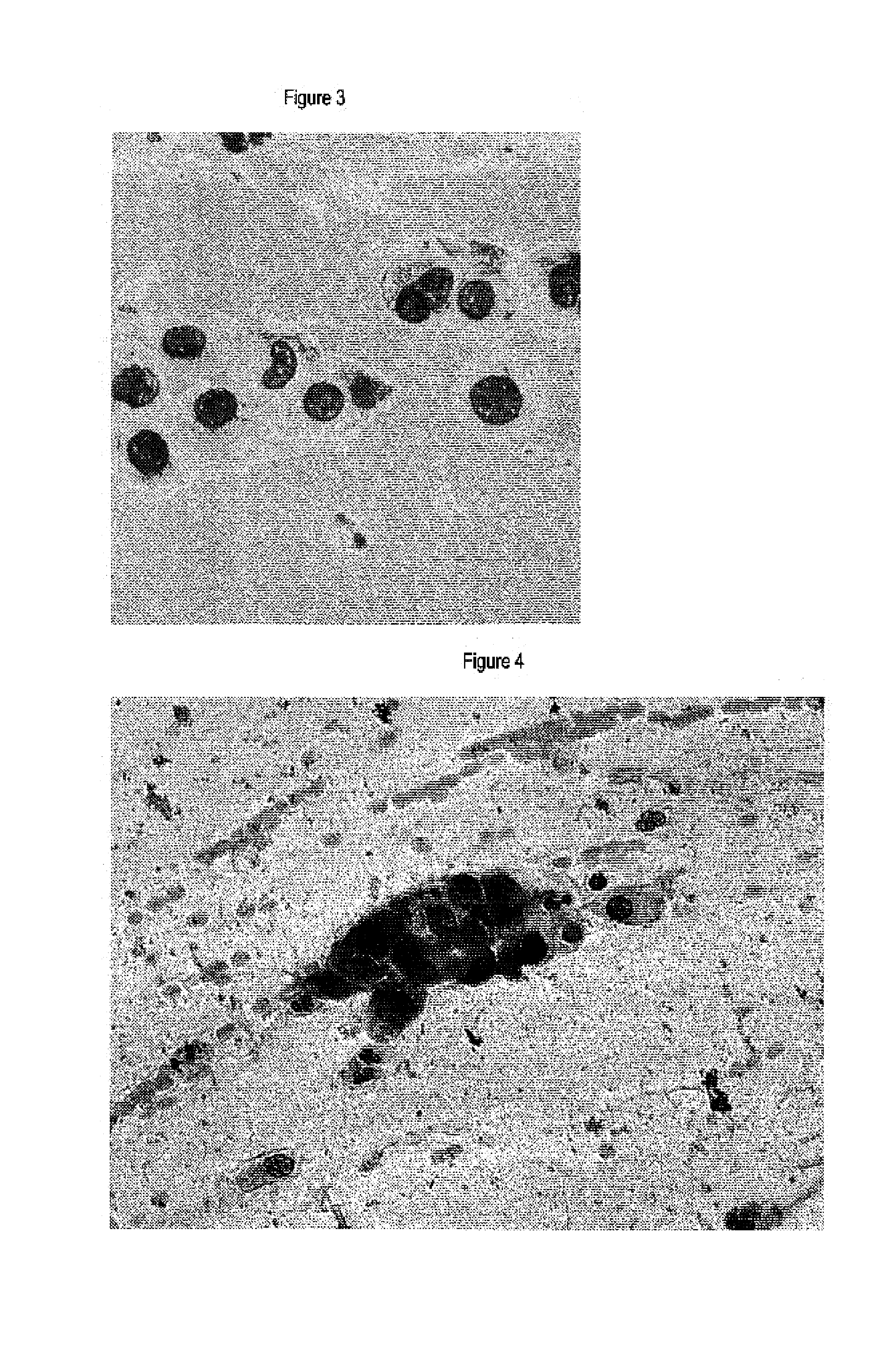
Immunochemical Detection of the Expression of HPV E2, L1 and p16INK4a in Samples of the Uterine Cervix
[0064]Smears of the cervix uteri were immunocytochemically stained using antibodies specific for p16INK4a polyclonal antibodies specific for HPV L1 and polyclonal antibodies specific for HPV E2 protein.
[0065]For rehydration, the spray-fixed smears prepared on slides were incubated in fresh 50% EtOH on a rocking device. The PEG film produced by the fixation procedure was removed by intensive rinsing. The smears were rinsed in aqua bidest. Antigen Retrieval was carried out with 10 mM citrate buffer (pH 6.0). Then the slides were heated in a waterbath for 40 min at 95° C., cooled down to RT for 20 minutes, transferred to washing buffer (PBS / 0.1% Tween20) and finally surrounded with a lipid-pencil.
[0066]For inactivation of endogenous peroxidase, the samples were incubated with 3% H2O2 for 20 min at RT and afterwards washed in PBS / 0.1% Tween20 for 5 min. The proteinblock was carried out ...
example 2
Detection of Cells Expressing HPV E2, HPV L1 or p16INK4a in Samples of the uterine Cervix by In Situ Hybridization
[0071]Smears of the uterine cervix are semi-quantitatively analysed for the mRNA level of p16INK4a and HPV E2 and L2 in an in-situ staining reaction. The staining reaction is performed as follows:
[0072]For rehydration, the spray-fixed smears prepared on slides are incubated in fresh 50% EtOH on a rocking device. The PEG film produced by the fixation procedure is removed by intensive rinsing. Then the smears are rinsed in aqua bidest. The smears are incubated with porteinase K (10 μg / ml in PBS) for 10 min at 37° C. Then the slides are transferred to washing buffer (PBS / 0.1% Tween20) and finally surrounded with a lipid-pencil.
[0073]The hybridization mixture is prepared by mixing 50 μl of ready to use hybridization buffer (DAKO A / S, Glostrup, Danmark) with about 5-10 pmol of the probes. The probes are fluorescein-labelled oligonucleotides of sequences complememtary to the r...
example 3
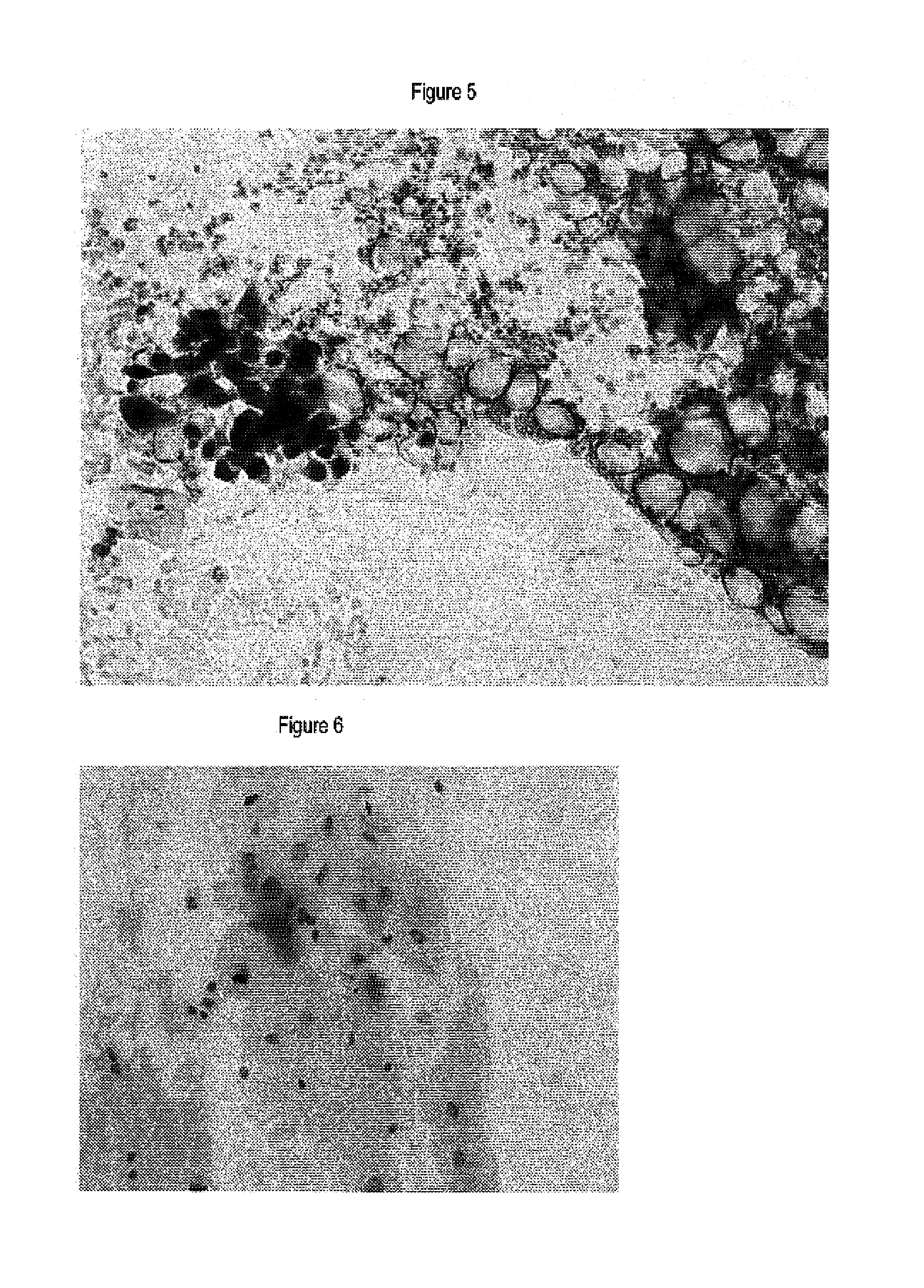
Immunocytochemical Detection of the Expression of HPV E7, and p16INK4a in Samples of the Uterine Cervix
[0077]ThinPrep® thinlayers of smears of the cervix uteri were immunocytochemically stained using antibodies specific for p16INK4a and monoclonal antibodies specific for HPV E7 protein.
[0078]For rehydration, the spray-fixed smears prepared on slides were incubated in fresh 50% EtOH on a rocking device. The PEG film produced by the fixation procedure was removed by intensive rinsing. Then the smears were rinsed in aqua bidest. Antigen Retrieval was carried out with 10 mM citrate buffer (pH 6.0). Thereafter, the slides were heated in a waterbath for 40 min at 95-98° C., cooled down to RT for 20 minutes, transferred to washing buffer (Dakocytomation, S3006) and finally surrounded with a lipid-pencil.
[0079]For inactivation of endogenous peroxidase, the samples were incubated with 3% H2O2 (Dakocytomation; S2023) for 5 min at RT and afterwards washed in wash buffer (Dakocytomation, S3006)...
PUM
| Property | Measurement | Unit |
|---|---|---|
| Fluorescence | aaaaa | aaaaa |
| Chemiluminescence | aaaaa | aaaaa |
| Bioluminescence | aaaaa | aaaaa |
Abstract
Description
Claims
Application Information