
Methods and apparatus for endovascularly replacing a patient's heart valve

a heart valve and endovascular technology, applied in the field of methods and apparatus for endovascular replacement of patients heart valves, can solve the problems of heart failure, stroke, heart attack, adverse reactions to anesthesia medications,

Inactive Publication Date: 2008-11-04
BOSTON SCI SCIMED INC
View PDF367 Cites 500 Cited by
  • Summary
  • Abstract
  • Description
  • Claims
  • Application Information

AI Technical Summary

Benefits of technology

The system enables accurate and stable deployment of the replacement valve with enhanced radial strength, reducing the risk of migration and ensuring proper function by maintaining expansion and radial force, thus improving the success rate of endovascular aortic valve replacement procedures.

Problems solved by technology

Valve replacement surgery is a highly invasive operation with significant concomitant risk.
Risks include bleeding, infection, stroke, heart attack, arrhythmia, renal failure, adverse reactions to the anesthesia medications, as well as sudden death.
Some self-expanding valve anchors have had very poor accuracy in deployment, however.
The stent often jumps to another position once released, making it impossible to know where the ends of the stent will be after release with respect to the native valve, the coronary ostia and the mitral valve.
Due to the jumping action of some self-expanding anchors, and because the replacement valve is often not fully functional before final deployment, visualization of valve function and position prior to final and irreversible deployment is often impossible with these systems.
Another drawback of prior art self-expanding replacement heart valve systems is their relative lack of radial strength.
Expandable stent designs suitable for endovascular delivery for other purposes may not have sufficient radial strength to serve as replacement heart valve anchors.
However when the stent has a valve fastened inside it, and that valve must reside within the heart, as is the case in aortic valve replacement, the anchoring of the stent to vessel walls takes significantly more radial force, especially during diastole.
Moreover, a self-expanding stent without sufficient radial force will end up dilating and contracting with each heartbeat, thereby distorting the valve, affecting its function and possibly causing it to migrate and dislodge completely.
Simply increasing strut thickness of the self-expanding stent is not a good solution as it increases profile and / or a risk of plastic deformation of the self-expanding stent.

Method used


Image

  • Methods and apparatus for endovascularly replacing a patient's heart valve
  • Methods and apparatus for endovascularly replacing a patient's heart valve
  • Methods and apparatus for endovascularly replacing a patient's heart valve

Examples


Embodiment Construction

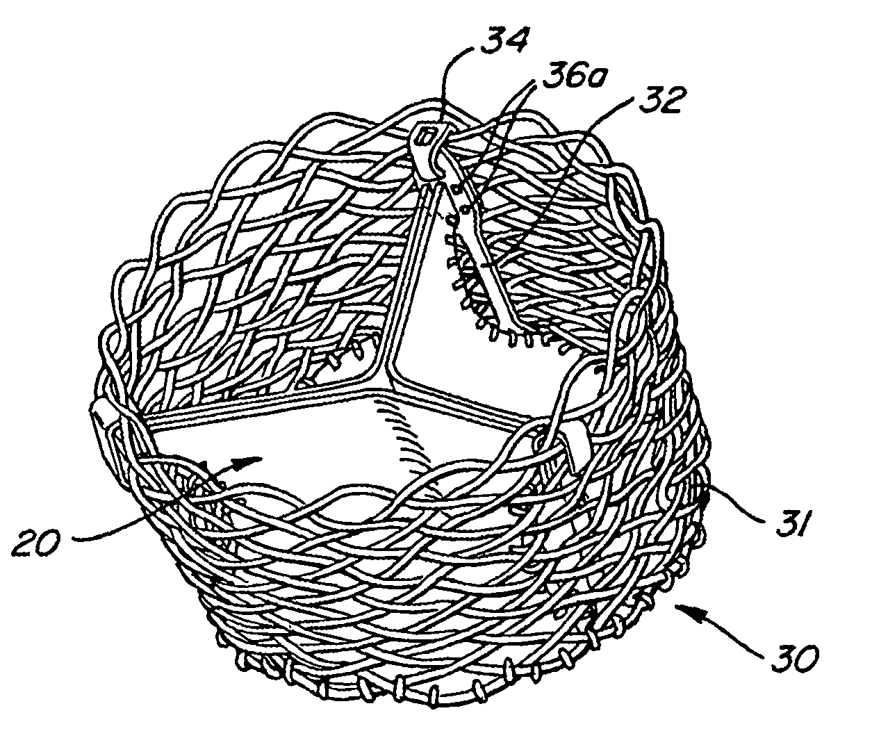
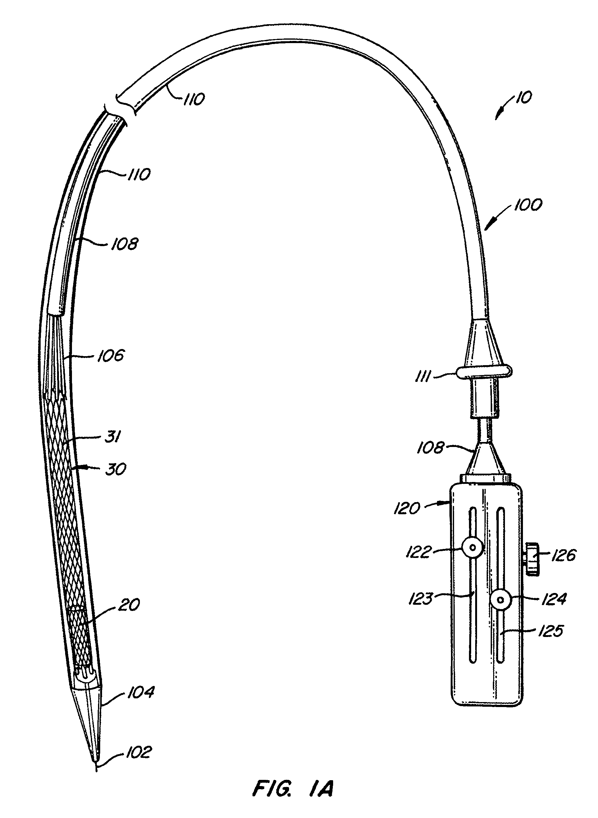
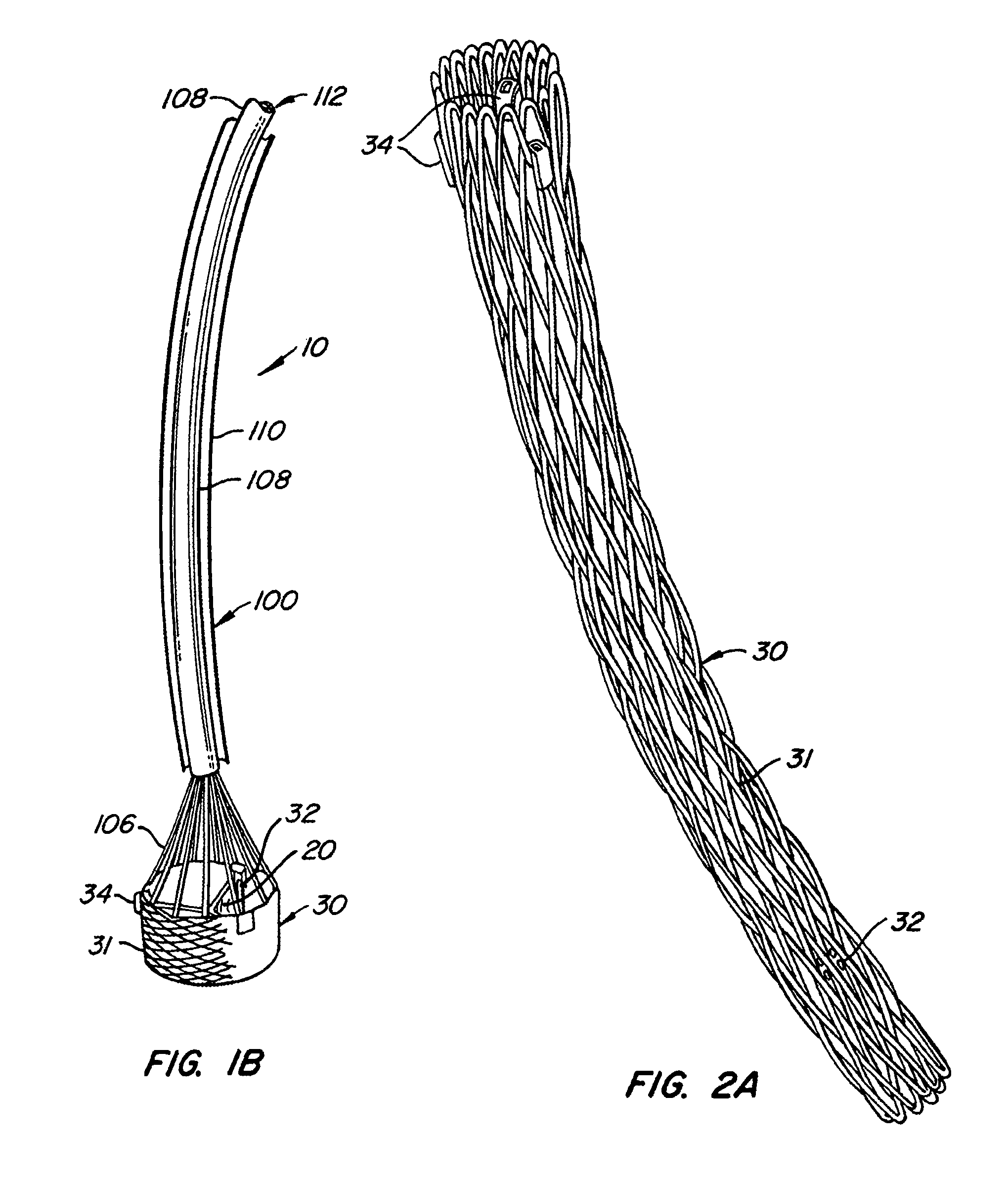
[0033]The present invention relates to a delivery system, apparatus and methods for endovascularly delivering and deploying an aortic prosthesis within a patient's native heart valve, referred to here out as replacing a patients heart valve. The delivery system includes a sheath assembly and a guide wire for placing the apparatus endovascularly within a patient and a user control allowing manipulation of the aortic prosthesis. The apparatus includes an anchor and a replacement valve. The anchor includes an expandable braid. In preferred embodiments, the expandable braid includes closed edges. The replacement valve is adapted to be secured within the anchor, and as such, be delivered endovascularly to patient's heart to replace the patient's native heart valve. More preferably, the apparatus and methods of the present invention contemplate the replacement of a patient's aortic valve.

[0034]FIGS. 1A and 1B illustrate one embodiment of a delivery system and apparatus in accordance with ...


PUM


Abstract

The present invention relates to an apparatus for replacing a native aortic valve, the apparatus includes an expandable anchor adapted to be endovascularly delivered and secured at a site within the native aortic valve. The expandable anchor has a delivery length in a delivery configuration substantially greater than a deployed length in a deployed configuration. The apparatus may also include and a replacement valve configured to be secured within the anchor.

Description

CROSS REFERENCE[0001]This application is a continuation-in-part application of U.S. Ser. No. 10 / 746,280, filed Dec. 23, 2003.BACKGROUND OF THE INVENTION[0002]Heart valve surgery is used to repair or replace diseased heart valves. Valve surgery is an open-heart procedure conducted under general anesthesia. An incision is made through the patient's sternum (sternotomy), and the patient's heart is stopped while blood flow is rerouted through a heart-lung bypass machine.[0003]Valve replacement may be indicated when there is a narrowing of the native heart valve, commonly referred to as stenosis, or when the native valve leaks or regurgitates. When replacing the valve, the native valve is excised and replaced with either a biologic or a mechanical valve. Mechanical valves require lifelong anticoagulant medication to prevent blood clot formation, and clicking of the valve often may be heard through the chest. Biologic tissue valves typically do not require such medication. Tissue valves m...

Claims


Application Information

Patent Timeline
Patent Type & Authority Patents(United States)
IPC IPC(8): A61F2/24A61F2/00A61F2/01A61F2/90
CPCA61F2/013A61F2/2418A61F2/2436A61F2/2427A61F2/2409A61F2/2415A61F2220/0075A61F2230/0054A61F2230/0078A61F2250/0039A61F2250/0036
Inventor SALAHIEH, AMRHAUG, ULRICH R.VALENCIA, HANS F.GESHLIDER, ROBERT A.SAUL, TOMMOREJOHN, DWIGHT P.
Owner BOSTON SCI SCIMED INC
Learn More