Molecular diagnostic method of a cancer tissue or a cancer cell

a cancer cell and molecular diagnostic technology, applied in the field of tissue and cell formation, can solve the problems of significant problems of accompanying cell toxicity and cell turning into a malignant tumor

Active Publication Date: 2009-03-10
SYSMEX CORP
View PDF17 Cites 0 Cited by
  • Summary
  • Abstract
  • Description
  • Claims
  • Application Information

AI Technical Summary

Benefits of technology

[0010]An object of the present invention is to provide a more accurate diagnostic method of a tissue and cell for carrying out definite diagnosis of cancer, drug resistance test and prognosis.

Problems solved by technology

However, such anticancer agents have involved significant problems of accompanying cell toxicity because they also inhibit the proliferation of normal cells.
On the other hand, mutated p53 molecules can not regulate the cell cycle, but lead to proliferation of uncontrollable cells and instability of a genome, resulting in the cell turned into a malignant tumor.
However, on the grounds of unestablished immunohistological techniques depending on specificity for each antibody and the like, ratios of expression of these cyclin E, cyclin D1 and the like in cancer tissues greatly vary with respect to each report.

Method used


Image

  • Molecular diagnostic method of a cancer tissue or a cancer cell
  • Molecular diagnostic method of a cancer tissue or a cancer cell
  • Molecular diagnostic method of a cancer tissue or a cancer cell

Examples


example 1

Method of Cell Examination

1) Preparation of Tissue Sample for Measurement

[0047]Each surgically collected tissue (2 mm3) was ground with a lysis buffer containing Nonidet P-40 (NP-40) (manufactured by Calbiochem), and solubilized with a homogenizer. Insoluble substances were removed by a filter. Thus resulting cell lysate was employed as a tissue sample for the measurement.

2) Measurement of CDK Activity

[0048]A sample containing 100 μg of the total protein was added to 2 μg of an antibody (anti-CDK1, 2, 4 and 6 antibodies, manufactured by Santa Cruz) and 20 μl of protein A beads (manufactured by BioRad) to allow them to react at 4° C. for 1 hour and to precipitate CDK molecules.

[0049]After washing with a buffer (0.1% NP-40, 50 mM Tris-HCl, pH 7.0) three times, 50 μl of a substrate mixture containing proteins was added thereto followed by incubation at 37° C. for 10 minutes while shaking.

[0050]The substrate mixture contains 10 μg of histone H1 corresponding to CDK1 and CDK2 (manufactur...

example 2

CDK Activity

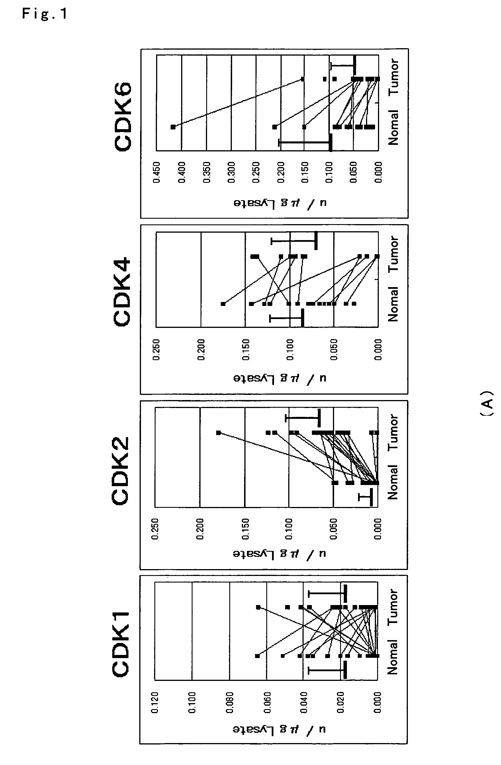
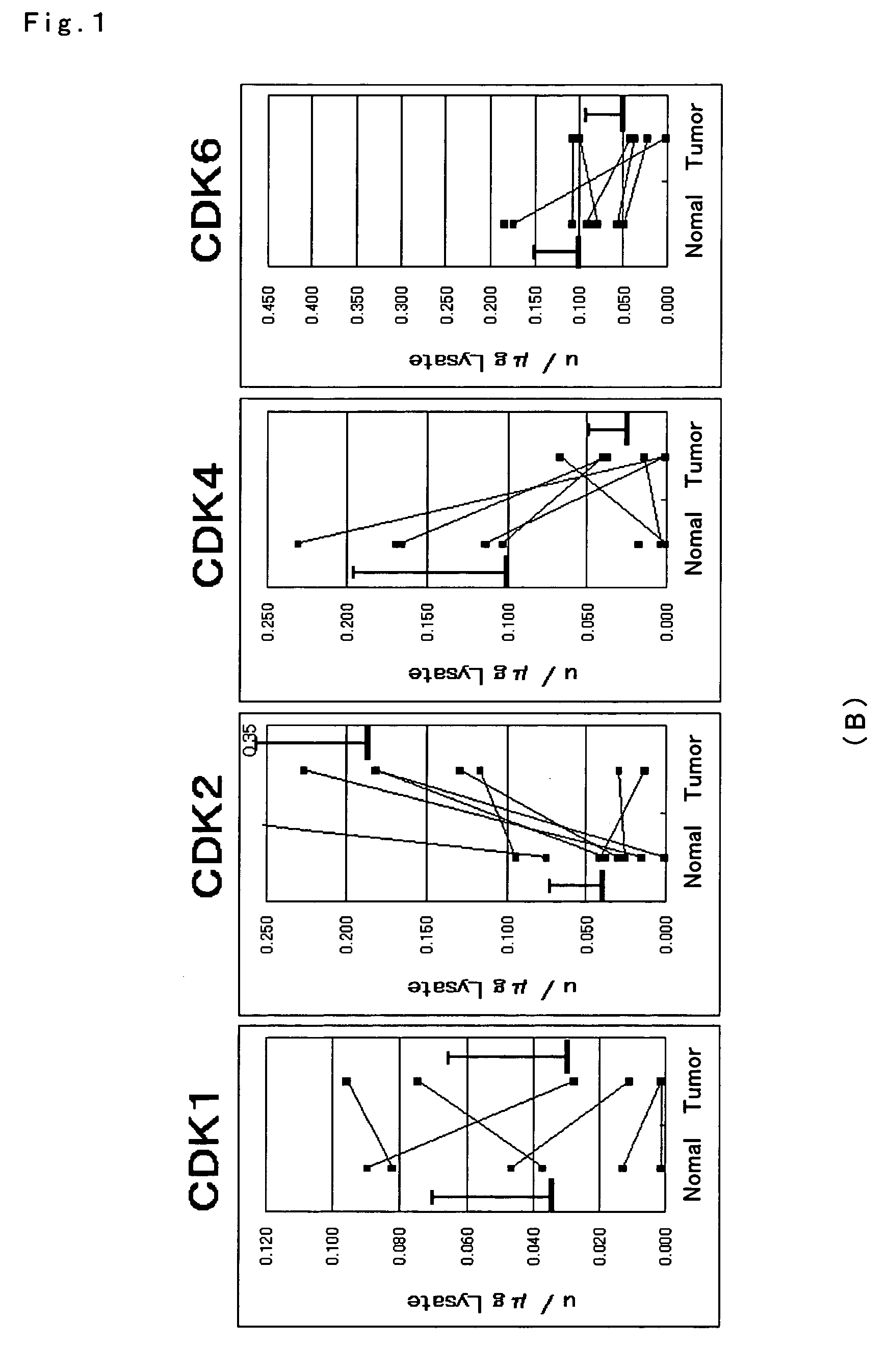
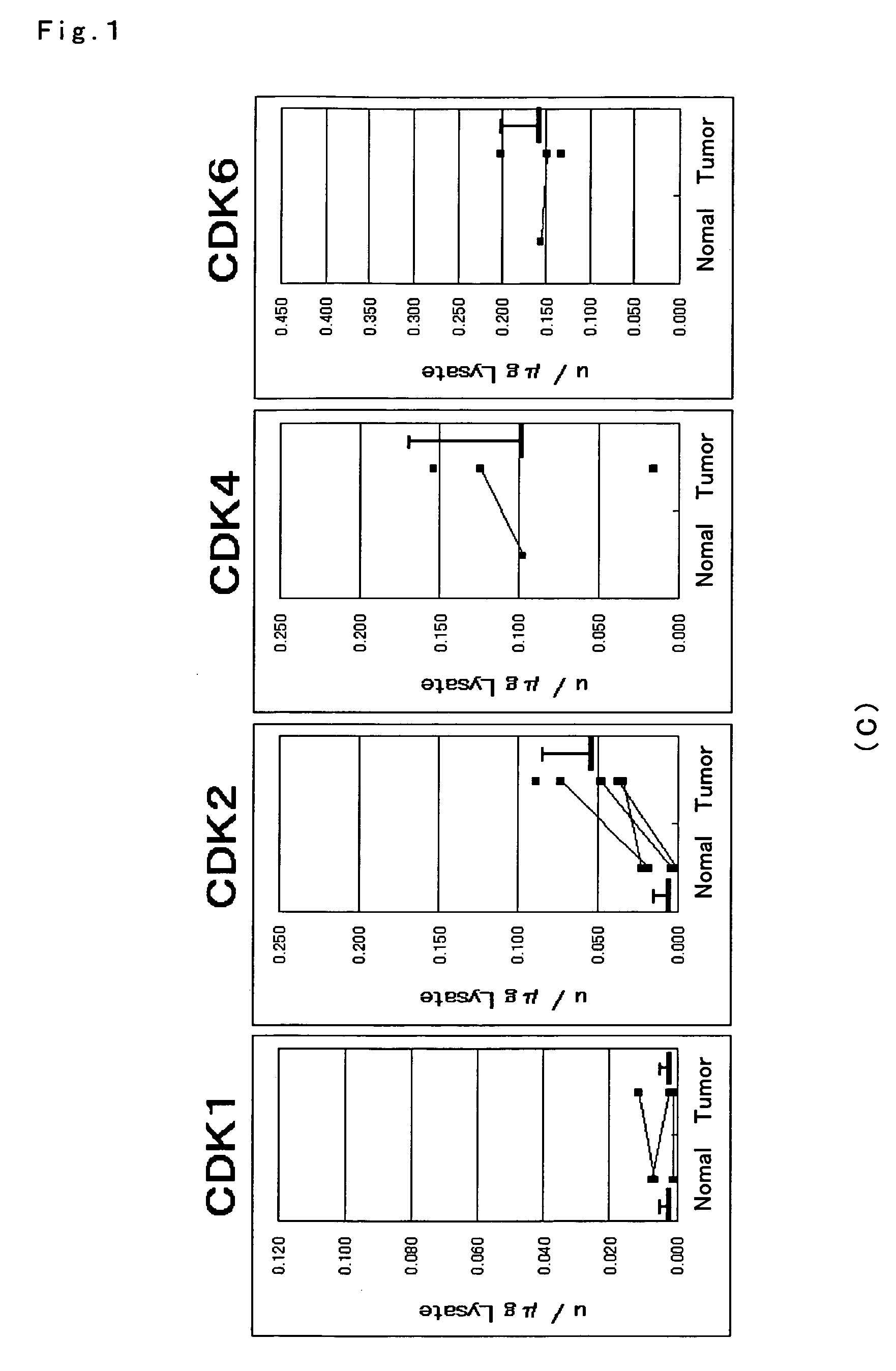
[0058]Each enzyme activity of CDK1, 2, 4 and 6 was measured for gastrointestinal cancer tissues and normal mucosa. Measurement of the activity was conducted with the method described in Example 1, for each of the tissues of (A) large intestine cancer of 22 cases, (B) stomach cancer of 8 cases, and (C) esophagus cancer of 7 cases as well as normal mucosa.

[0059]The results are depicted in FIGS. 1A to C. Consequently, with respect to difference of each enzyme activity between cancer tissues and normal mucosa, statistical significance was found in elevation of the CDK2 activity in the large intestine, stomach and esophagus tissues (P<0.01). On the other hand, CDK4 and CDK6 in the large intestine and stomach cancer tissues exhibited activities that were comparatively lower than those in normal mucosa. Statistical analysis was carried out with a Wilcoxon signed-ranks test. In crease in CDK2 enzyme activity was observed in almost of the large intestine, stomach and esophagus ca...

example 3

Profile of CDK activity

[0060]With respect to CDK (CDK2, 4, 6) in the G1 phase, the activity values of CDK2, 4 and 6 were standardized on the basis of the CDK1 activity value that is G2 / M kinase to determine the profile. Consequently, as shown by a solid line in FIG. 2A, in seven among 8 cases of normal large intestine mucosal tissues, CDK profile in the G1 phase positioned in the fold width of 0.024 to 0.43 for CDK2 / CDK1, 1.2 to 39 for CDK4 / CDK1, and 1.9 to 26 for CDK6 / CDK1, exhibiting a common pattern. This suggests that relative values of the CDK activities are similar among the normal large intestine tissues.

[0061]Next, the profile on the basis of value of CDK1 activity that is G2 / M kinase was similarly determined for CDKs (CDK2, 4, 6) in the G1 phase on randomly selected nine cases of large intestine cancer tissue. Consequently, as shown in FIG. 2B, no profile was observed which was identical to the common profile found in the normal mucosal tissues

[0062]According to the present...


PUM

PropertyMeasurementUnit
pHaaaaaaaaaa
pHaaaaaaaaaa
pHaaaaaaaaaa

Abstract

The present invention relates to a more accurate method of diagnosing cancer in a tissue or a cell by measuring the expression and activity of cell cycle related proteins and analyzing a cell cycle profile. In a cell cycle profile, at least two values of expression or activity of cell cycle related proteins are concurrently measured. The measurement of the cell cycle related protein is not limited as to the measurement method.

Description

TECHNICAL FIELD[0001]The present invention relates to a profiling method of a tissue and a cell by simultaneously measuring cell cycle related proteins on multi parameters, and specifically, relates to a molecular diagnostic method of a cancer tissue or a cancer cell.BACKGROUND ART[0002]Cancer cells are characterized by unlimited proliferation. If means for selectively inhibiting proliferation of cancer cells without impairing the proliferation ability of normal cells can be found, termination of tumor growth is enabled irrespective of extent of differentiation or penetration of the tumor, or degree of metastasis. Development of anticancer agents targeting for proliferating cells has been extensively carried out to provide a therapeutic drug of cancer. However, such anticancer agents have involved significant problems of accompanying cell toxicity because they also inhibit the proliferation of normal cells.[0003]In an attempt to solve such problems, it is interesting to comprehend a...

Claims


Application Information

Patent Timeline
Patent Type & Authority Patents(United States)
IPC IPC(8): C12Q1/48G01N33/50G01N33/574
CPCC12Q1/485G01N33/5011G01N33/574G01N2800/44
Inventor SAKAIISHIHARA, HIDEKI
Owner SYSMEX CORP
Why Eureka
Try Eureka