Systems and methods of identifying biomarkers for subsequent screening and monitoring of diseases

a biomarker and disease technology, applied in the field of imaging systems and methods, can solve the problems of ineffective preventive measures for these diseases, high cost of introducing new drugs to the market, and deepening productivity crisis

Inactive Publication Date: 2009-03-24
VITRIMARK
View PDF6 Cites 2 Cited by
  • Summary
  • Abstract
  • Description
  • Claims
  • Application Information

AI Technical Summary

Benefits of technology

[0008]The present invention provides, in one embodiment, an approach through the use of cryogenic transmission electron microscopy (cryo-TEM), as well as modified freeze fracture direct imaging (M-FFDI) to identify ultra-structural biomarkers, which may subsequently be used for screening and monitoring a range of diseases. The use of cryo-TEM and M-FFDI can generate substantially artifact-free images, unlike images obtained from conventional TEM.
[0010]The present invention also provides a method for generating an image of ultrastructural biomarkers from a biological sample. The method includes, in one embodiment, providing a substantially thin film of a sample to be imaged. The thin film may be generated from blotting or alternatively from sandwiching the sample between two plates. Next, the sample may be immersed in a cryogenic fluid so as to cause the sample to vitrify. This rapid vitrification allows the objects present in the sample to substantially maintain their original morphology. Once vitrified, the sample may be transferred onto a stage for placement in a high contrast imager, such as a transmission electron microscope, under positive dry pressure to minimize the risks of contamination of the sample. The transfer to the high contrast imager also includes keeping the sample at a temperature range of from about −170° C. to about −150° C. in the imager to maintain the integrity of the sample. Thereafter, an image of the thin film sample may be generated for subsequent identification of ultrastructural biomarkers. The generation of the image, in one embodiment, includes producing a substantially artifact-free image in the absence of contrasting agents.

Problems solved by technology

The deepening productivity crisis in the pharmaceutical industry, the high cost to the pharmaceutical industry of introducing new drugs to the market, partly because of expenses related to Phase I, II and III clinical trials, as well as late stage failures for many drug candidates have spurred intense across-the-board activity around biomarker discovery and validation.
As such, preventive measures for these diseases may be ineffective when developed in connection with the management of the disease, or in early evaluation of drug efficacy.
However, the use of conventional TEM has some critical limitations.
For example (i) the high vacuum used in TEM removes solvent, leaving behind structures that are quite different from those present in the original solution, (ii) adequate contrast between the sample features and background is usually not available, necessitating the use of stains (the addition of stains, which usually are heavy metal salts, can cause dramatic changes in aggregate morphology), and (iii) the exposure of the sample to the electron beam often damages the sample.

Method used


Image

  • Systems and methods of identifying biomarkers for subsequent screening and monitoring of diseases
  • Systems and methods of identifying biomarkers for subsequent screening and monitoring of diseases
  • Systems and methods of identifying biomarkers for subsequent screening and monitoring of diseases

Examples


examples

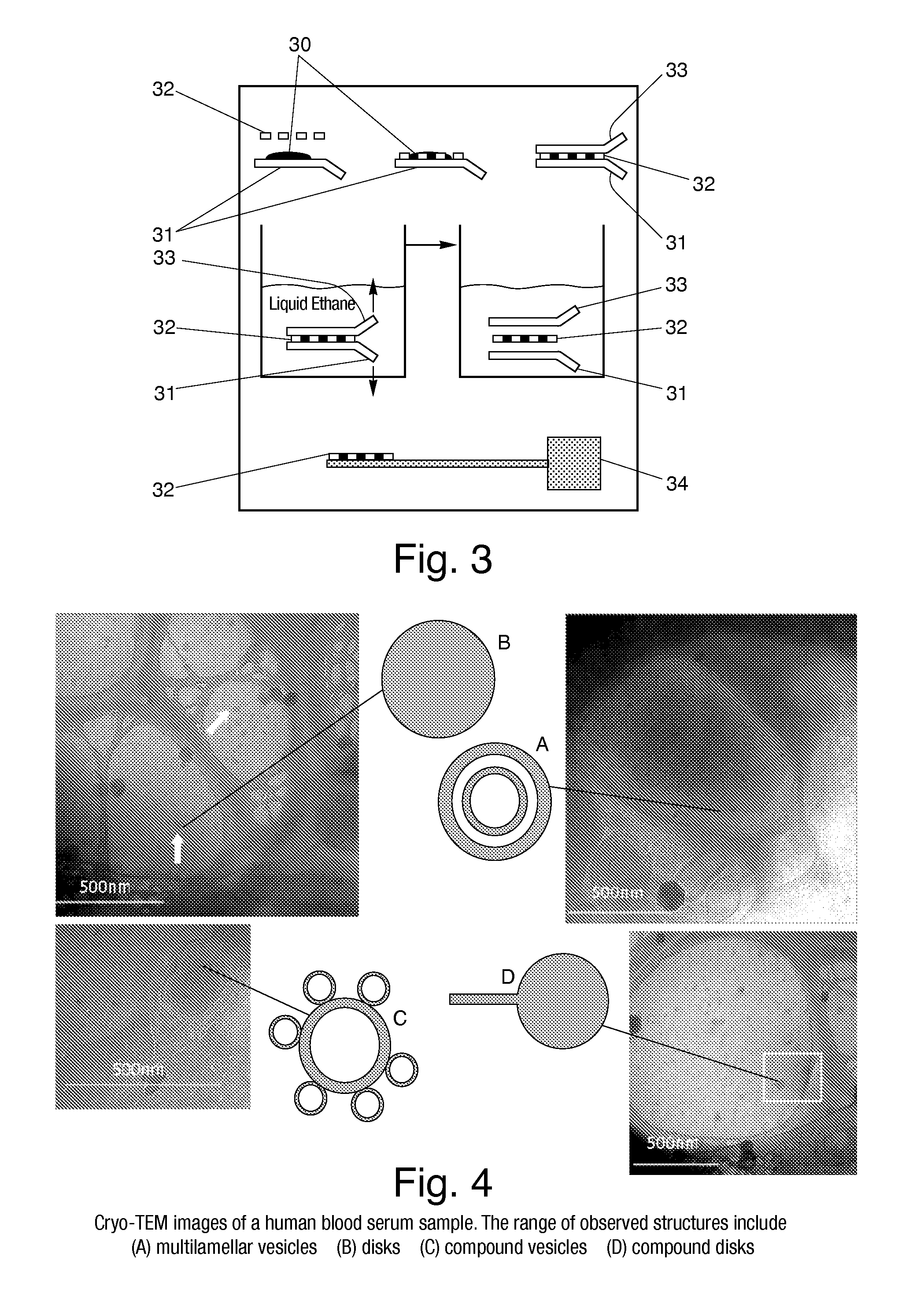
[0033]In an experiment, a serum sample from a test subject was prepared using cryogenic vitrification in the manner set forth above and subsequently imaged through the use of cryo-TEM. The images of the serum sample are illustrated in FIG. 4. In particular, the images are taken from different regions of the same holey carbon grid (i.e. different relatively thin regions of the thin film on the grid). As can be seen, the images show a relatively rich range of structures, including (a) multilamellar vesicles, (b) single and (d) compound discs, and (c) compound vesicles, all at nanoscale resolution of approximately 500 nm or less.

[0034]It should be noted that serum typically contains macromolecules, such as metabolites, lipids, hormones, peptides, and proteins. Certain of these biological macromolecules can also organize into 3-D complexes, which can be biochemically homogeneous or heterogenous in nature. For examples, serum can contain many glycoproteins, glycopeptides, lipoproteins, a...


PUM

PropertyMeasurementUnit
volumeaaaaaaaaaa
thicknessaaaaaaaaaa
temperatureaaaaaaaaaa

Abstract

A system for generating an image of ultrastructural biomarkers from a biological sample is provided. The system includes a grid onto which a sample to be imaged may be placed and a cryogenic reservoir into which the grid and sample may be immersed for vitrification of the sample. The system also includes a stage onto which the grid and sample may be situated for subsequent imaging in a high contrast imager to permit identification of ultrastructural biomarkers therein. A method for generating an image of ultrastructural biomarkers from a biological sample is also provided. The generated image of ultrastructural biomarkers may be used subsequently for screening and monitoring diseases, evaluating drug and therapeutic efficacy, and assessing risks associated with a drug or therapeutic candidate, among other things.

Description

RELATED U.S. APPLICATIONS[0001]The present application claims priority to U.S. Provisional Application Ser. No. 60 / 612,713 filed Sep. 24, 2004, which application is hereby incorporated herein by reference.TECHNICAL FIELD[0002]This invention relates generally to imaging systems and methods, and more particularly to imaging systems and methods for identifying ultrastructural biomarkers for subsequent screening and monitoring of diseases.BACKGROUND ART[0003]The deepening productivity crisis in the pharmaceutical industry, the high cost to the pharmaceutical industry of introducing new drugs to the market, partly because of expenses related to Phase I, II and III clinical trials, as well as late stage failures for many drug candidates have spurred intense across-the-board activity around biomarker discovery and validation. Biomarkers, defined by the FDA as a characteristic that is objectively measured and evaluated as an indicator of normal biologic or pathogenic processes or pharmacolo...

Claims


Application Information

Patent Timeline
Patent Type & Authority Patents(United States)
IPC IPC(8): C12Q1/00
CPCG01N1/42G01N21/6428G01N21/6456G01N33/68B01L3/022G01N2001/2873H01J2237/2001H01J2237/204
Inventor BOSE, ARIJITAZIZ, NAZNEEN
Owner VITRIMARK
Why Eureka
Try Eureka