Diagnostic imaging apparatus

a diagnostic imaging and apparatus technology, applied in the field of diagnostic imaging apparatus, can solve the problems of inaccurate understanding of the inside condition of the surface, inability to and inability to accurately diagnose the dental caries or the attached dental calculus, etc., to achieve accurate diagnostic image information, avoid the adverse effect of disturbance light, and diagnose the primary dental caries

Inactive Publication Date: 2009-08-04
MORITA MFG CO LTD
View PDF34 Cites 34 Cited by
  • Summary
  • Abstract
  • Description
  • Claims
  • Application Information

AI Technical Summary

Benefits of technology

[0015]As mentioned above, there is much room for improvement in any prior art. It has been desired an apparatus which has no fear of X-ray exposure, is easily handled, and timely understands the inside condition of the tooth. The present invention is proposed in consideration with the above-mentioned problems and its object is to provide a diagnostic imaging apparatus in which a diagnostic object in a narrow place such as oral cavity is preferably photographed, and clear image information of not only attached condition of dental calculus and dental plaque on the tooth surface but also dental caries in the area close to the surface of the diagnostic object is recognized. Further, its object is to provide a diagnostic imaging apparatus which is hardly subject to disturbance light.
[0016]The fist object of the present invention is to receive in an imaging means as an X-ray exposure free, compact and handy apparatus the reflected light generated from the diagnostic object base portion on radiation of light with specific wavelength by a luminous means and the fluorescence generated by the exciting light to obtain a predetermined diagnostic image information, thereby easily and advantageously knowing scratch on the tooth surface, dental caries on and in the tooth, and conditions of dental calculus, dental plaque and gingival. Further, the object is to give an accurate diagnosis making the diagnostic imaging apparatus compact. Solid-state image sensing device (CCD and MOS) and an optical means for forming an optical image of the diagnostic object for the solid-state image sensing device are employed for an imaging means so that preferable photography image information can be rapidly obtained at low cost. The apparatus of the present invention is proposed as a compact and handy apparatus. When it is further commercialized as a compact body at low cast and with safety, such an apparatus with limited function is suitable for domestic use. If the imaging means has wide spectroscopic characteristic such that the light with 300-800 nm wavelength can be photographed, photography of fluorescence range can be executed so that it goes without saying that from ultraviolet light to infrared light including visible light can be photographed. Of course, if detection of specific fluorescence is required, an imaging means with a dedicated wide spectroscopic characteristic with wavelength wider than the above-mentioned range is selected.
[0017]In the above-mentioned apparatus, when a luminous means irradiating white light is added, a visible light image (an image obtained by a general intraoral camera with a white light source) can be obtained in addition to the image such as a fluorescence image base portion on radiation of the light with specific wavelength. Namely, the light with specific wavelength and white light can be simultaneously irradiated. When only white light is irradiated and the reflected light is received without passing thought a light receiving filter or is received while passing through a light receiving filter made of glass which passes only visible light, the visible light image of the diagnostic object is obtained. On the other hand, the light with specific wavelength is selectively irradiated, the image (fluorescence image and so on) corresponding to the wavelength characteristic of the diagnostic object can be obtained. Hereby, the obtained visible light image and the obtained image like the fluorescence image are compared, or these images are overlapped and displayed to easily show where the lesion area is on the visible light image and to be easily understood by a patient. Consequently, the image information by which accurate diagnosis is executed while communicating with the patient can be obtained.
[0018]The second object of the present invention is, in addition to the above-mentioned object, the forward portion of the main body is made compact and the thickness thereof is made smaller by employing a optical path changing means as an optical means for forming an optical image on a solid-state image sensing device, thereby improving the qualifications as a dental photography apparatus for use in oral cavity.
[0019]The third object of the present invention is to improve the cleaning performance and maintenance performance of the forward portion by making the forward portion separate into a head portion including the optical path changing means and a base portion including the solid-state image sensing device. The head portion doesn't include a bulky member such as a solid-state image sensing device so that the thickness thereof can be reduced, thereby further improving the aptitude of the apparatus when being used as a dental photography apparatus for use in oral cavity. Expensive solid-state image sensing device is left at the base portion so that the device is commonly used and different kinds of diagnostic image information can be obtained at low cost by changing the head portion on the characteristic of the optical means such as optical path changing means included in the head portion. Further, the light with specific wavelength and the white light can be simultaneously irradiated. When only the white light is irradiated from the luminous means, the reflected light is received without passing through the light receiving filter or is received while passing through the light receiving filter made of glass which only passes through a visible light, thereby obtaining the visible light image of the diagnostic object. On the other hand, when the light with specific wavelength is selectively irradiated, the image (fluorescence image) corresponding to the wavelength characteristic of the diagnostic object can be obtained. Hereby, the obtained visible light image and the image like the obtained fluorescence image are compared, or these images are overlapped and displayed to easily show where the lesion area is on the visible light image and to be easily understood by a patient. Consequently, the image information by which accurate diagnosis is executed while communicating with the patient can be obtained.
[0021]The forth object of the present invention is to provide a light shielding hood for preventing outside light from entering at a light entrance for going out the radiation light and for entering the light from the diagnostic object at the forward portion, thereby avoiding the affect of the disturbance light such as interior illumination and sunbeam and making clear the diagnostic image information taken by the imaging means. More specifically, by providing the light shielding hood so as to be adjacent to the diagnostic object or so as to fix up the diagnostic object, the adverse effect of disturbance light is avoided and a faint fluorescence image from the diagnostic object by radiating exciting light can be distinguished, thereby being useful for diagnosing a primary dental caries. Still further, if the light shielding hood is used so as to be fixed up the diagnostic object, its positioning is achieved and more accurate diagnostic image information can be obtained. “Preventing outside light” described herein means to prevent all the external light from entering and further to prevent the external light to an extent so as not to affect the diagnostic image information base portion on the diagnostic object by radiating from the luminous means.

Problems solved by technology

However, such an intraoral photography apparatus can only photograph under the condition where the surface of the tooth or the oral cavity is irradiated with visible light (wavelength of 380-760 nm) so that inside condition of the surface, dental caries or attached dental calculus haven't been accurately understood.
Therefore, in has been required to execute radiography in the prior art in order to know dental caries, deficit part, crack or attached dental calculus of biopsy such as invisible tooth, thereby causing a fear of X-ray exposure.
However, the apparatus has the following problems.(a) A light guide is required to irradiate light to tooth.(b) Whether a specific part is dental caries or healthy can be determined.
However, detection information when exciting light with specific wavelength is irradiated is only obtained so that dental caries condition can't be understood, therefore such information isn't useful for explaining to a patient.
In other words, dental caries is locally detected but image information to show relative condition of the dental caries for the entire teeth can't be obtained.(c) Image processing using plural images such as a visible light image and an exciting light image can't be executed.
Prior diagnostic imaging apparatus is subject to disturbance light like interior illumination and sunbeam so that clear diagnostic image information has been hardly obtained.
Moreover, according to the apparatus disclosed in the above-mentioned prior arts, a standard photography by which the same area is photographed from the same position and from the same angle has been impossible.
Still further, a standard photography with radiation light with different wavelength has been also impossible in the prior arts.

Method used


Image

  • Diagnostic imaging apparatus
  • Diagnostic imaging apparatus
  • Diagnostic imaging apparatus

Examples


embodiment 1

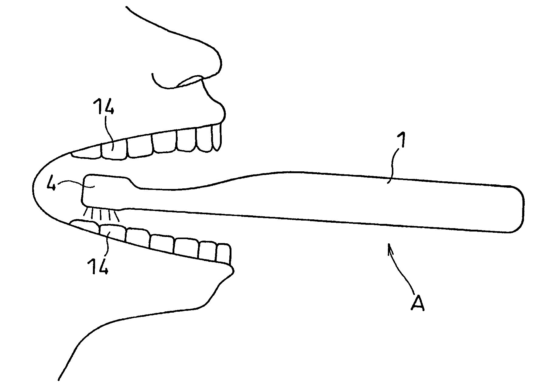
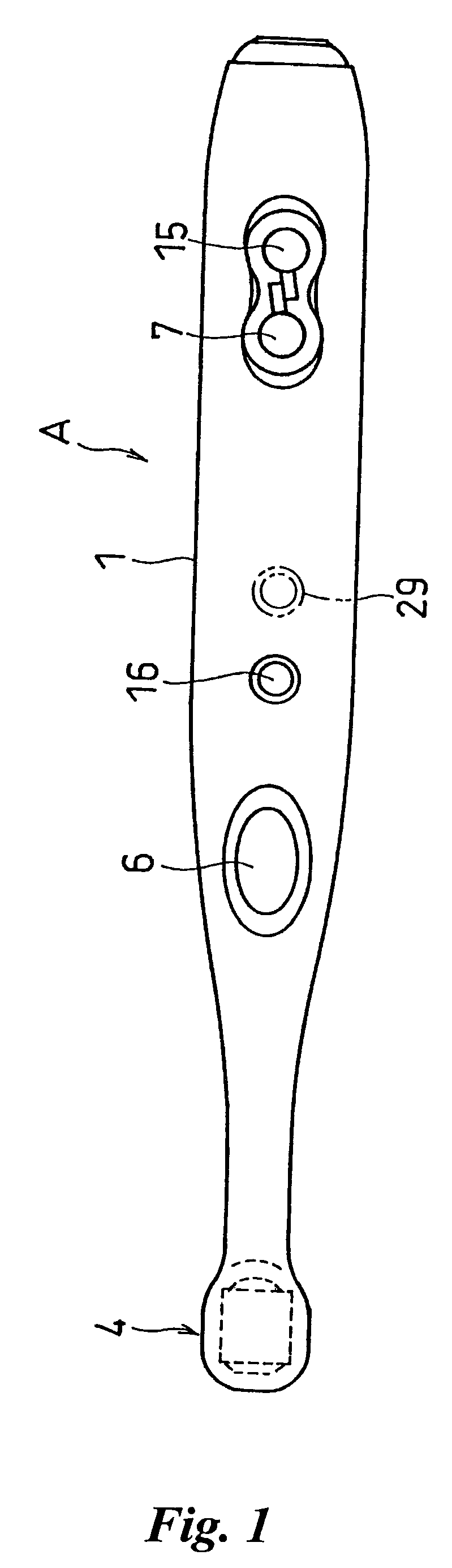
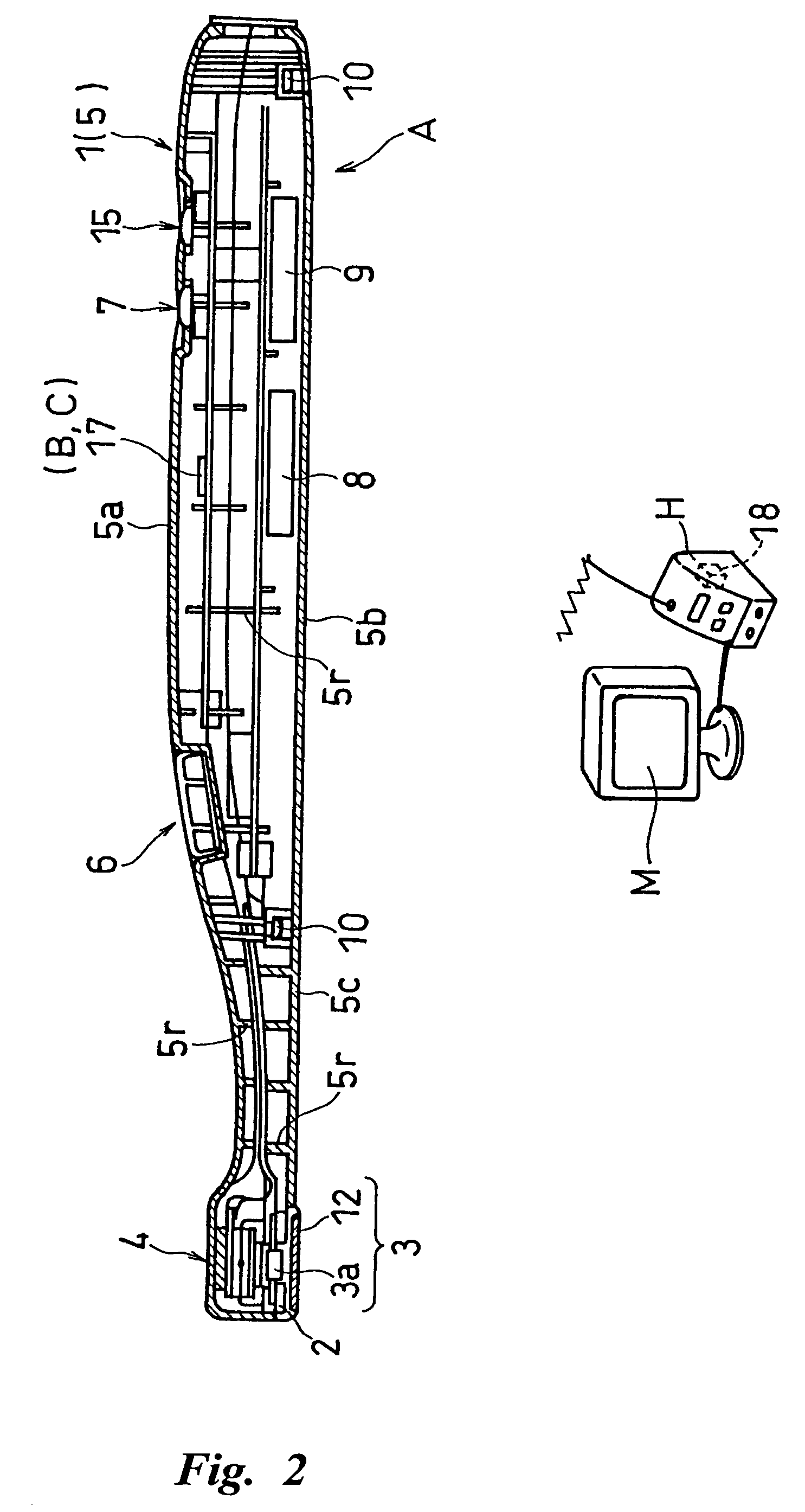
[0088]The diagnostic imaging apparatus A as shown in FIG. 1-FIG. 5 is comprised of a main body 1 like a dental handpiece freely supported with hands and fingers and a forward portion 4 provided with a luminous means 2 for irradiating at least one of lights among exciting light, infrared light, ultraviolet light, and white light (light emitting member 2a, 2b, 2c) and with an imaging means 3 comprised of CCD (charge coupled device) 3a. The diagnostic imaging apparatus A is preferable for diagnosing mainly dental caries, deficit part, crack, lesioned part, and attachment of dental calculus, dental plaque and bio-film on tooth in oral cavity. The apparatus A can photograph not only the surface of tooth but also the inside of the tooth surface (about 1 mm inside of the surface) to recognize a lesioned part in the surface of the tooth. Moreover, if it is constructed as a cordless type, signals are transmitted to a control box H (see FIG. 2) via cordless manner and the obtained image can b...

embodiment 2

[0122]FIG. 13 shows one embodiment of a radiation source selection means D (corresponding to the light source selection switch 7 in FIG. 1). The radiation source selection means D constitutes a luminous means 2 comprised of four kinds of light emitting members 2a-2d (plural light emitting members radiating the light with different wavelength), that is, infrared ray, white light, ultraviolet ray 1 and ultraviolet ray 2, all of them are LEDs. Any one (or plural ones) of the plural light emitting means 2a-2d is (are) selectively driven. The radiation source selection means D is provided with four analog switches sw1-sw4 connected between a power source 8 and each light emitting member 2a-2d, four light source selection switches hs1-hs4, and a switch controller 19.

[0123]On operation of the first light source selection switch hs1 enables the first analog switch sw1 to be operated and to drive the infrared LED 2b. In the same manner, on operation of the second light source selection switc...

embodiment 3

[0124]FIG. 14 is one embodiment of the light receiving filter changing means. The filter changing means F in the figure is designed such that a support frame (filter unit) 24 provided with two light receiving filters 12, 12 with different cut-off wavelength range at both ends respectively is arranged so as to be freely rotated by hands around the center P of the axis parallel to the optical axis of the imaging means 3 (or luminous means 2). When the support frame 24 is rotated 180 angles around the axis center P, the light receiving filter 12 as shown in FIG. 14 is switched to another light receiving filter 12 (having different permeability).

[0125]The support frame 24 is constructed in a manner that a penetrating window 24a is formed between the axis center P and the light receiving filters 12, 12 respectively so as not to prevent the light from the luminous means 2 from transmitting. The support frame 24 may be rotated in the reverse direction as mentioned above or more than three ...


PUM


Abstract

A diagnostic imaging apparatus of hand piece type suitable for medical or dental use, comprising a main body held by operator's fingers, a luminous means for irradiating at least one of lights selected from excitation light, infrared light and ultraviolet light, and an imaging means provided in a forward portion of the main body. The imaging means comprises a solid-state image sensing device and an optical means for forming an optical image of a diagnosis object to be examined on the solid-state image sensing device and is so constructed as to output a predetermined diagnostic image information by receiving the light reflected from the diagnosis object and / or the fluorescence generated from the diagnosis object when irradiation of light from the luminous means to the diagnosis object is performed.

Description

DETAILED DESCRIPTION OF THE INVENTION[0001]1. Field of the Invention[0002]The present invention relates to a diagnostic imaging apparatus for diagnosing dental caries, deficit tooth, crack of tooth, lesioned tooth, attachment of dental calculus or dental plaque, lesioned part of root canal, gum, bucca or tang in oral cavity, for diagnosing otolaryngology area, and for diagnosing rectum tumor. More specifically, the present invention relates to technology which can diagnose in an inner area of a diagnostic object, such as tooth, near the surface as well as on the surface. Further, the apparatus of the present invention isn't limited to an apparatus for doctors and it is preferably used as a diagnostic imaging apparatus for domestic use for confirming the appearance of tooth, dental caries, attached dental calculus or dental plaque.[0003]2. Prior Art[0004]For example, a diagnostic imaging apparatus for diagnosing oral cavity is required to have compactly designed imaging means because...

Claims


Application Information

Patent Timeline
Patent Type & Authority Patents(United States)
IPC IPC(8): A61B5/05A61B1/247A61B1/24A61B5/00
CPCA61B1/00089A61B1/00096A61B1/00101A61B1/0684A61B1/24A61B5/0088A61B1/0615A61B1/0676A61B1/00016A61C19/004A61B1/0605G06F13/382G06F2213/0042
Inventor KATSUDA, NAOKIKINO, KENJIMATOBA, KAZUNARIIMAZATO, MINORU
Owner MORITA MFG CO LTD
Why Eureka
Try Eureka