
Methods and system for treating subcutaneous tissues

a subcutaneous tissue and subcutaneous tissue technology, applied in the field of methods and systems for treating subcutaneous tissues, can solve the problems of affecting the treatment effect,

Active Publication Date: 2009-09-15
ULTHERA INC
View PDF172 Cites 126 Cited by
  • Summary
  • Abstract
  • Description
  • Claims
  • Application Information

AI Technical Summary

Benefits of technology

This method provides a minimally invasive, safer treatment for cellulite and other skin irregularities, achieving sustained aesthetic results with fewer side effects and potentially permanent reduction of fat deposits and septae, allowing for localized tissue disruption without heat-induced damage.

Problems solved by technology

As the ultrasound driving pressure increases, more complex phenomenon occurs, for example, with bubble expansion larger than contraction.
Furthermore, there may be relatively slow expansion followed by rapid collapse.
However, HIFU is disadvantageous in that it produces a significant amount of heat and thermal damage to tissues.
Although low acoustic pressure ultrasound may somewhat heat the tissue, the tissue is not heated sufficiently to cause direct tissue disruption or to enhance the ablation.

Method used


Image

  • Methods and system for treating subcutaneous tissues
  • Methods and system for treating subcutaneous tissues
  • Methods and system for treating subcutaneous tissues

Examples


example 1

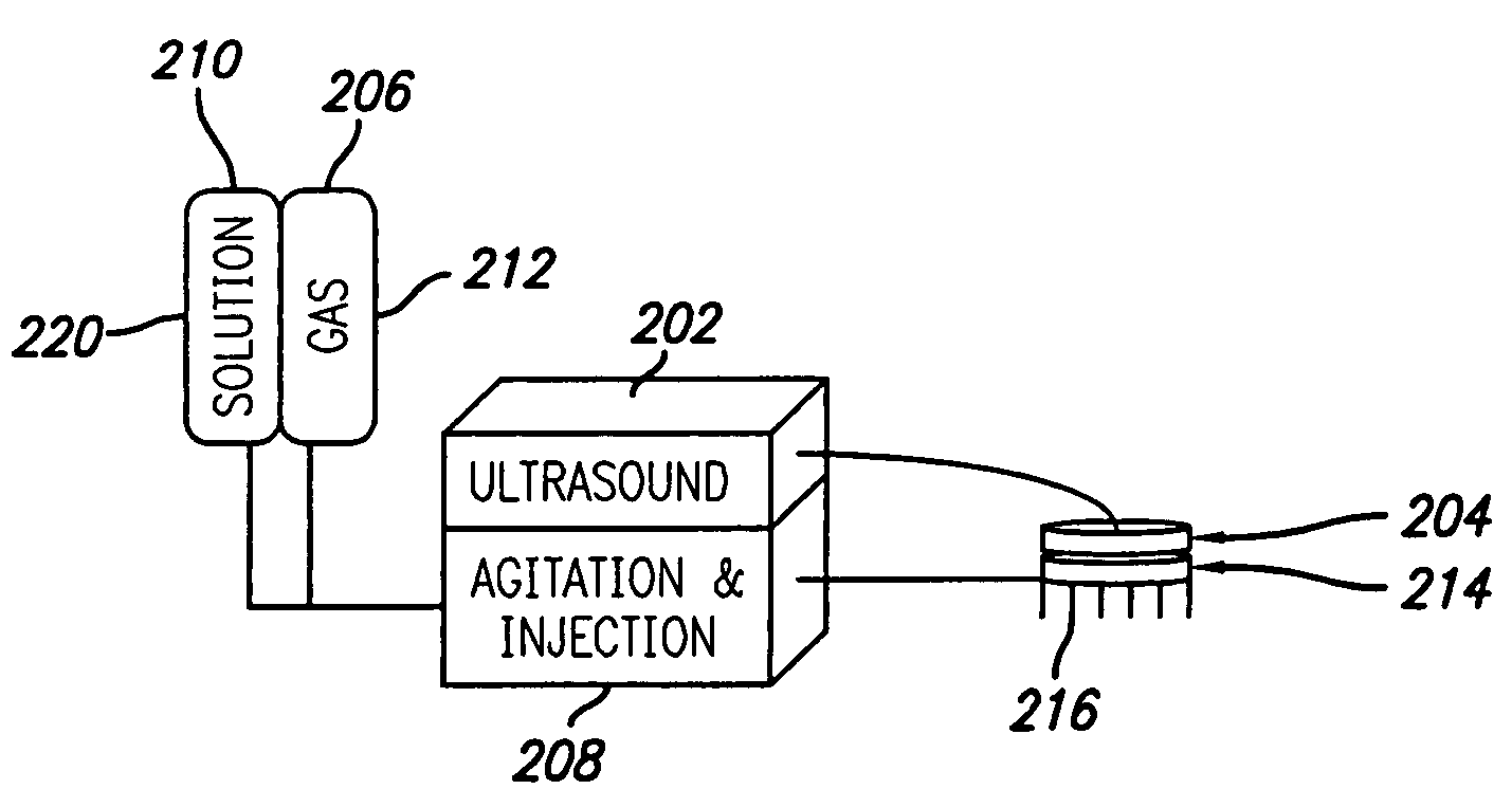
[0156]Referring specifically now to FIG. 12, in one embodiment of the present invention all components are built into a single unit or apparatus 200. Disposable canisters provide the supply of gas 206 and a container 220 providing solution 210 are included in the system. An electrical control unit 228 including input, output, memory, and processor is provided for the assembly. The control unit provides for flow of solution and gas into the agitator 208 that then agitates the solution-gas mixture. The control unit then further directs the injector 214 to injects the solution including gaseous bodies into the tissue. The control unit then further controls the ultrasound generator 202 and transducer 204 to insonate the tissue to be treated according to a pre-programmed or user defined algorithm.

example 2

[0157]Referring specifically now to FIG. 13, in one embodiment of the invention the control unit 228 drives the ultrasound generator 202 and transducer 204 only. The gas supply 206, the solution 210, the agitator, 208, and the injector 214 are all contained in a single use, disposable, pre-filled unitary disposable module 230. In one embodiment the transducer is reusable. In yet another embodiment, the transducer is disposable and built into the unitary disposable module. In at least one embodiment, the hypodermic needles 216 may pass through openings in the transducer. In at least one embodiment, the unitary disposable module includes a solution agitator and a solution injection member that are self contained and self powered. In yet at least one embodiment, the control unit powers and / or controls the injection and insonation algorithm for the unitary disposable module.

example 3

[0158]Referring specifically now to FIG. 14, in yet one other embodiment of the invention a dedicated ultrasound generator 202 and a separate unit including a solution agitator 208 and a solution injection member 214 are provided. The ultrasound unit may be, for example, substantially similar to one presently used by those skilled in the art for physical therapy applications. Such ultrasound units are commercially available. A tumescent solution 210 injection system substantially similar to one known in the art and commercially available may also be provided. The transducer 204 and the injection member 214 may be connected together, wherein the injection of solution and the insonation may occur generally simultaneously. As with other embodiments described herein, the transducer and / or injection member may either by reusable or disposable. Furthermore, a source of gas 206 and a container 220 for the solution is provided.


PUM


Abstract

An apparatus and method for treating subcutaneous tissues using acoustic waves in the range of low acoustic pressure ultrasound waves is disclosed. The method includes injections of enhancing agents, wherein disruption of subcutaneous tissues and subcutaneous cavitational bioeffects are produced by ultrasound waves having a power that will not produce tissue cavitation in the absence of the enhancing agents. The apparatus and method of use is useful for treatment of subcutaneous abnormalities including cellulite, lipomas, and tumors.

Description

CROSS-REFERENCES TO RELATED APPLICATIONS[0001]The present application is a continuation of U.S. Ser. No. 11 / 292,950 filed on Dec. 2,2005 entitled “METHOD FOR TREATING SUBCUTANEOUS TISSUES” which claims priority to U.S. Provisional Ser. No. 60 / 715,398 filed Sep. 7, 2005 entitled “SYSTEM AND METHOD FOR DISRUPTING SUBCUTANEOUS STRUCTURES”, the entirety of which are incorporated herein by reference.FIELD OF THE INVENTION[0002]The present invention relates generally to the use of externally applied energy to biologic tissues wherein destruction or disruption of the biologic tissue is provided. More specifically, the present invention relates to the use of acoustic waves to produce the destruction or disruption of biologic tissues and cells. Even more specifically, the present invention relates to methods and apparatus that include ultrasound wave application to a tissue infiltrated with a solution for the treatment of subcutaneous structures in a mammalian body, for example, for the trea...

Claims


Application Information

Patent Timeline
Patent Type & Authority Patents(United States)
IPC IPC(8): A61H1/00
CPCA61K41/0028A61M37/0092A61N1/0412A61N1/0492A61N7/00A61B2017/00747A61B2017/22008A61K9/0009A61N2007/0039A61N1/327A61N2007/0008A61M2037/003
Inventor DEEM, MARK E.GIFFORD, III, HANSON S.SUTTON, DOUGLAS
Owner ULTHERA INC
Learn More