Biodegradable glaucoma implant

a biodegradable, glaucoma technology, applied in the field of medical devices and methods for reducing intraocular pressure, can solve the problems of significant side effects, untreated blindness, and patients' significant blindness, and achieve the effects of reducing surgical morbidity, eliminating the risk of hypotony, and avoiding hypotony

Inactive Publication Date: 2005-12-29
GHARIB MORTEZA +2
View PDF2 Cites 170 Cited by
  • Summary
  • Abstract
  • Description
  • Claims
  • Application Information

AI Technical Summary

Benefits of technology

[0027] Glaucoma surgical morbidity would greatly decrease if one were to bypass the focal resistance to outflow of aqueous only at the point of resistance, and to utilize remaining, healthy aqueous outflow mechanisms. This is in part because episcleral aqueous humor exerts a backpressure that prevents intraocular pressure from going too low, and one could thereby avoid hypotony. Thus, such a surgery would virtually eliminate the risk of hypotony-related maculopathy and choroidal hemorrhage. Furthermore, visual recovery would be very rapid, and the risk of infection would be very small, reflecting a reduction in incidence from 2-5% to about 0.05%.
[0028] Techniques performed in accordance with the aspects of the invention may be referred to generally as “trabecular bypass surgery.” Advantages of this surgery include lowering intraocular pressure in a manner which is simple, effective, disease site-specific, and can potentially be performed on an outpatient basis.
[0035] In one embodiment, at least one of the two bifurcatable elements has a tapered distal end, adapted for insertion ease. The trabecular shunt may have its surface coated with a coating material selected from one or more of the following: polytetrafluoroethylene (e.g., Teflon™), polyimide, hydrogel, heparin, hydrophilic compound, anti-angiogenic factor, anti-proliferative factor, therapeutic drugs, and the like. The surface coating material may also provide a mechanism for site-specific therapies.
[0036] In one embodiment, the device may include a flow-restricting member for restricting at least one component in fluid. The flow-restricting member may be a filter comprising one or more filtration materials selected from the following: expanded polytetrafluoroethylene, cellulose, ceramic, glass, Nylon, plastic, fluorinated material, or the like. The flow-restricting member may advantageously be a filter selected from the following group of filter types: hydrophobic, hydrophilic, membrane, microporous, and non-woven. The flow-restricting member acts to limit or prevent the reflux of any undesired component or contaminant of blood, such as red blood cells or serum protein, from the aqueous veins into the anterior chamber. It is useful to restrict one or more of the following components or contaminants: platelets, red blood cells, white blood cells, viruses, bacteria, antigens, and toxins.
[0038] Among the advantages of trabecular bypass surgery is its simplicity. The microsurgery may potentially be performed on an outpatient basis with rapid visual recovery and greatly decreased morbidity. There is a lower risk of infection and choroidal hemorrhage, and there is a faster recovery, than with previous techniques.

Problems solved by technology

Glaucoma causes pathological changes in the optic nerve, visible on the optic disk, and it causes corresponding visual field loss, resulting in blindness if untreated.
Patients may suffer substantial, irreversible vision loss prior to diagnosis and treatment.
However, there are secondary open-angle glaucomas which may include edema or swelling of the trabecular spaces (e.g., from corticosteroid use), abnormal pigment dispersion, or diseases such as hyperthyroidism that produce vascular congestion.
However, these drug therapies for glaucoma are sometimes associated with significant side effects, such as headache, blurred vision, allergic reactions, death from cardiopulmonary complications, and potential interactions with other drugs.
However, long-term review of surgical results showed only limited success in adults.
In retrospect, these procedures probably failed due to cellular repair and fibrosis mechanisms and a process of “filling in.” Filling in is a detrimental effect of collapsing and closing in of the created opening in the trabecular meshwork.
Once the created openings close, the pressure builds back up and the surgery fails.
However, the relatively small hole created by this trabeculopuncture technique exhibits a filling-in effect and fails.
Although morbidity was zero in both trials, success rates did not warrant further human trials.
Failure was again from filling in of surgically created defects in the trabecular meshwork by repair mechanisms.
This is an ab interno (from the inside), mechanically disruptive technique that uses an instrument similar to a cyclodialysis spatula with a microcurrette at the tip.
The risk of placing a glaucoma drainage device also includes hemorrhage, infection, and diplopia (double vision).
All of the above embodiments and variations thereof have numerous disadvantages and moderate success rates.
They involve substantial trauma to the eye and require great surgical skill in creating a hole through the full thickness of the sclera into the subconjunctival space.
However, modifying existing filtering surgery techniques in any profound way to increase their effectiveness appears to have reached a dead end.

Method used


Image

  • Biodegradable glaucoma implant
  • Biodegradable glaucoma implant
  • Biodegradable glaucoma implant

Examples


Embodiment Construction

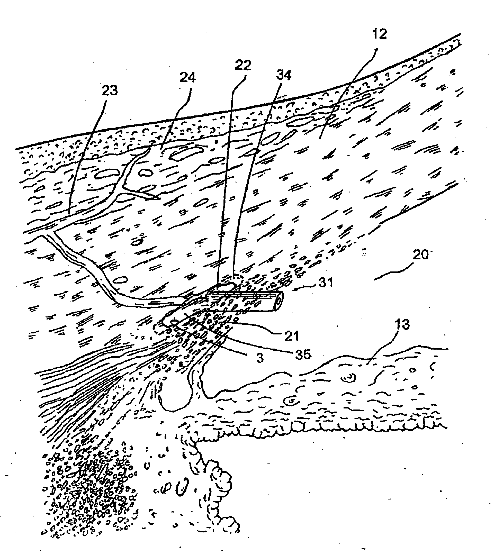
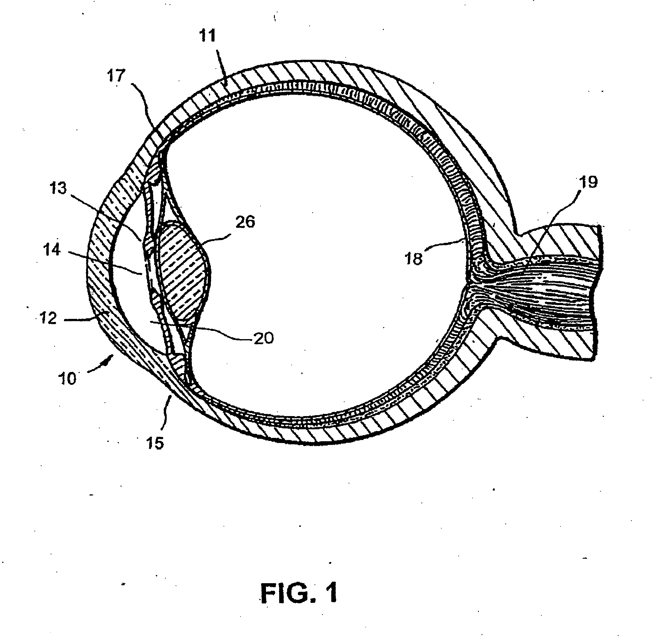
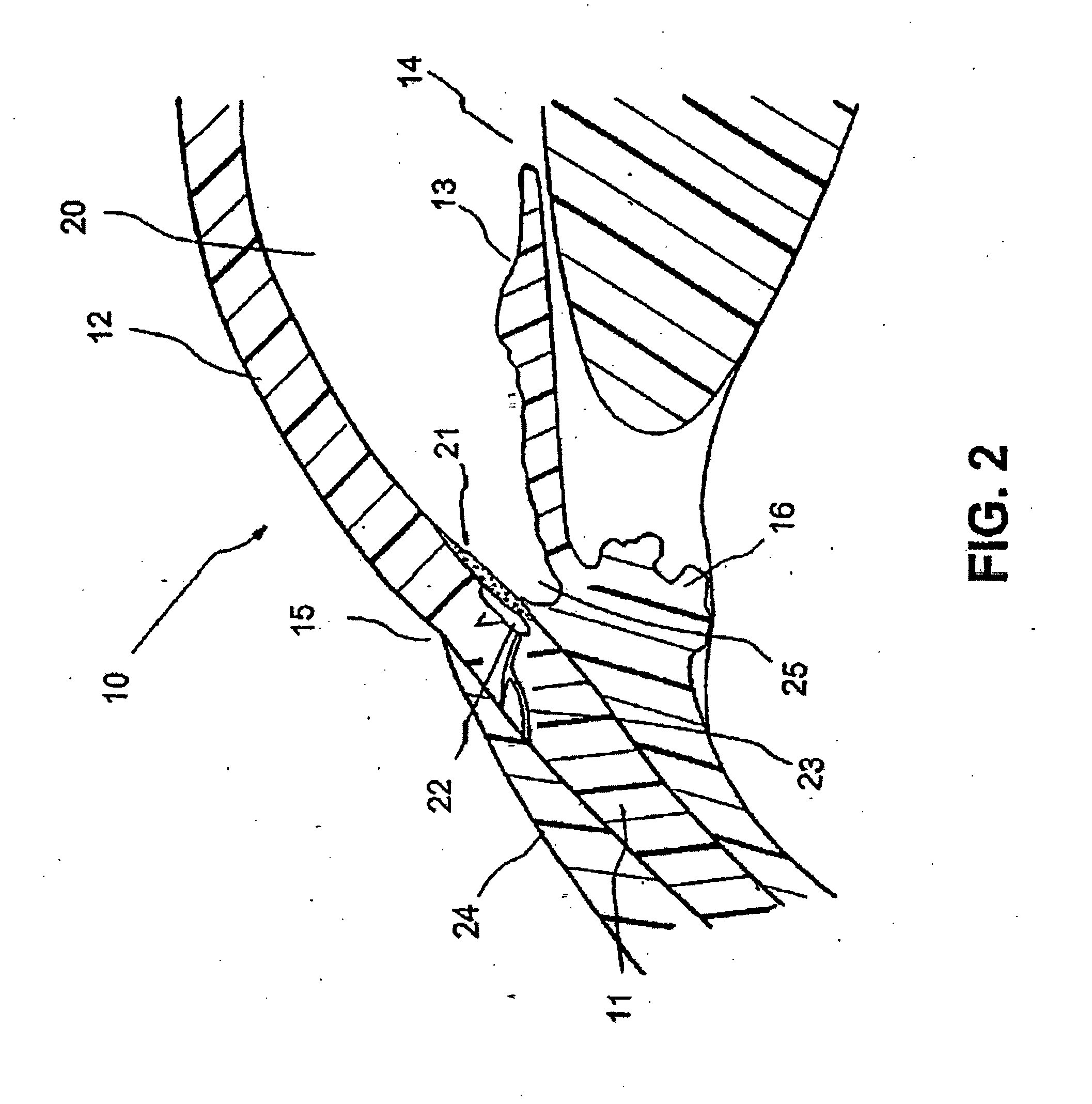
[0053] FIGS. 1 to 8 illustrate an apparatus for the treatment of glaucoma by trabecular bypass surgery.

[0054]FIG. 1 is a sagittal sectional view of an eye 10, while FIG. 2 is a close-up view, showing the relative anatomical locations of trabecular meshwork 21, the anterior chamber 20, and Schlemm's canal 22. Thick collagenous tissue known as sclera 11 covers the entire eye 10 except that portion covered by the cornea 12. The cornea 12 is a thin transparent tissue that focuses and transmits light into the eye and through the pupil 14, which is the circular hole in the center of the iris 13 (colored portion of the eye). The cornea 12 merges into the sclera 11 at a juncture referred to as the limbus 15. The ciliary body 16 extends along the interior of the sclera 11 and is coextensive with the choroid 17. The choroid 17 is a vascular layer of the eye 10, located between the sclera 11 and retina 18. The optic nerve 19 transmits visual information to the brain and is the anatomic struct...


PUM


Abstract

A trabecular shunt and methods for treating glaucoma are disclosed. One of the methods comprises transporting fluid from the anterior chamber of an eye to Schlemm's canal through an implant, the implant extending between the anterior chamber and Schlemm's canal; sensing an intraocular pressure using a sensor incorporated into the implant; and transmitting a signal indicative of the sensed pressure to an external receiver.

Description

CROSS-REFERENCE TO RELATED APPLICATION [0001] This application is a continuation application of U.S. patent application Ser. No. 10 / 626,181, filed Jul. 24, 2003, entitled “Implant with Pressure Sensor for Glaucoma Treatment,” which is a continuation application of U.S. patent application Ser. No. 09 / 847,523, filed May 2, 2001, and entitled “Bifurcatable Trabecular Shunt for Glaucoma Treatment,” now U.S. Pat. No. 6,666,841, the entirety of each of which is hereby incorporated by reference.BACKGROUND OF THE INVENTION [0002] The present invention generally relates to medical devices and methods for reducing intraocular pressure in the animal eye. More particularly, the present invention relates to the treatment of glaucoma by permitting aqueous humor to flow out of the anterior chamber through a surgically implanted pathway. [0003] The human eye is a specialized sensory organ capable of light reception and able to receive visual images. The trabecular meshwork serves as a drainage chan...

Claims


Application Information

Patent Timeline
Patent Type & Authority Applications(United States)
IPC IPC(8): A61F9/007
CPCA61B3/16A61F9/00781
Inventor GHARIB, MORTEZATU, HOSHENGBERGHEIM, OLAV
Owner GHARIB MORTEZA
Why Eureka
Try Eureka