
Galactosidase-treated prosthetic devices

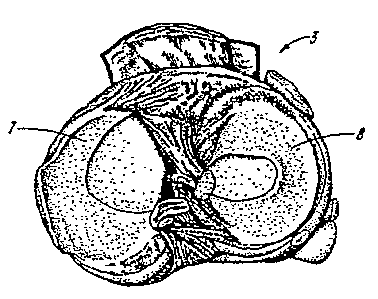
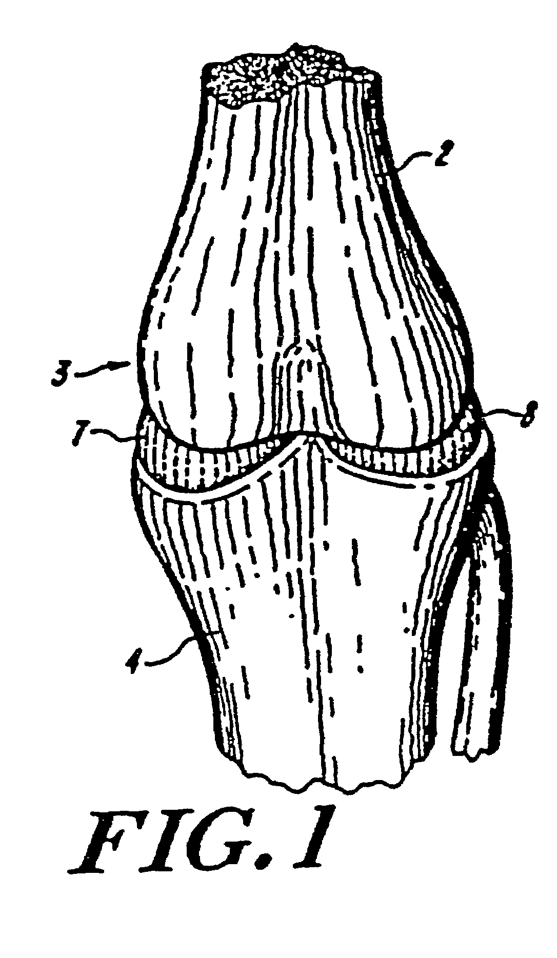
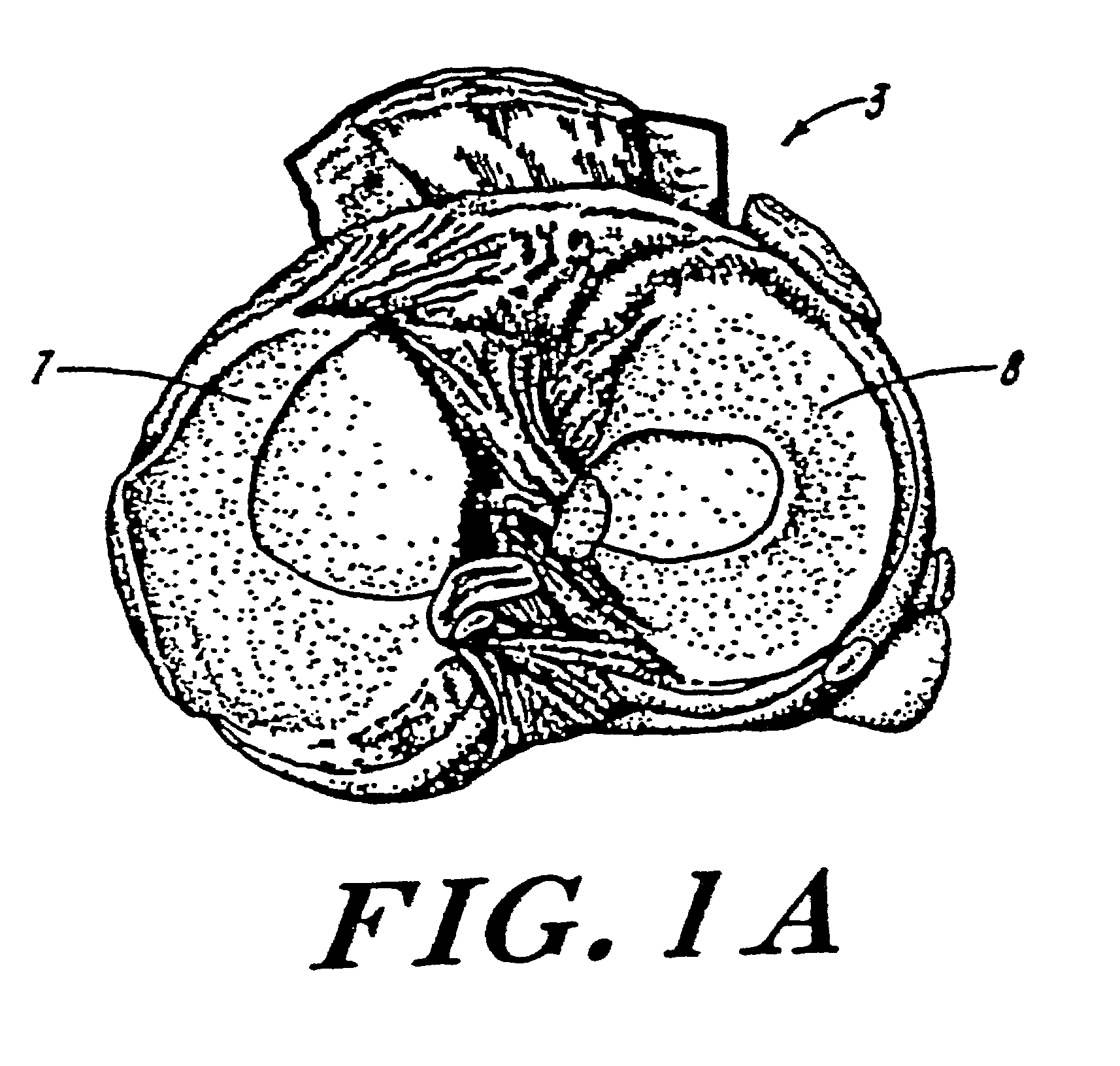
a technology of galactosidase and prosthetic devices, applied in the field of implantable prosthetic devices, can solve the problems of reducing the immunogenicity affecting the healing effect of the knee joint, and the inability to replace the meniscus with permanent artificial materials, etc., and achieve the effect of reducing the immunogenicity

Inactive Publication Date: 2009-09-29
APERION BIOLOGICS
View PDF7 Cites 9 Cited by
  • Summary
  • Abstract
  • Description
  • Claims
  • Application Information

AI Technical Summary

Benefits of technology

The device reduces immunogenicity, supports natural meniscal load forces, and promotes tissue regeneration by creating a biocompatible scaffold that integrates with the body's tissue, reducing the risk of immunological rejection and improving joint mechanics.

Problems solved by technology

These phenomena can result in premature development of arthritis.
The replacement of meniscal tissue with structures consisting of permanent artificial materials, however, is often unsuccessful.
This lack of success is due principally to the fact that opposing articular cartilage of human and animal knee joints is fragile.
The articular cartilage in the knee joint will not withstand abrasive interfaces, or compliance variances from normal, which eventually result from implanted artificial prosthetic menisci.
Thus far, permanent artificial menisci have not been composed of materials having natural meniscal properties, nor have they been able to be positioned securely enough to withstand such routine forces.
However, the reconstitution process can leave antigenic components, causing an immunological reaction when the prosthetic meniscus is implanted into a subject.

Method used


Image

  • Galactosidase-treated prosthetic devices
  • Galactosidase-treated prosthetic devices
  • Galactosidase-treated prosthetic devices

Examples


example 1

Device I Fabrication

[0111](A) Approximately 700 g of a Type I collagen dispersion (treated with ∝-galactosidase to eliminate ∝-Gal epitopes) is weighed into a 2 liter vacuum flask. Approximately 120 ml 0.66 ammonium hydroxide is added to the dispersion to coacervate the collagen. About 80 ml 20% NaCl is then added to the coacervated fibers to further reduce the solution imbibition between the fibers.

[0112](B) The fully coacervated fibers are dehydrated to about 70 g to 80 g in a perforated mesh basket to remove the excess solution from the fibers.

[0113](C) The partially dehydrated collagen fibers are inserted into a mold of specified dimension related to the dimensions of the defect to be remedied. Further dehydration is ongoing in the mold using a constant (between 300 grams to 700 grams) weight to slowly remove the water from the fibers, yet maintaining the same density throughout. This slow dehydration process lasts for about 24 hours until the desired dimension (about 8 mm in th...

example 2

Device II Fabrication

[0121]The procedure for fabricating Device II is identical to that described in Example 2, except that approximately 700 g of a Type II collagen dispersion (treated with ∝-galactosidase to eliminate ∝-Gal epitopes) are used.

example 3

Device III Fabrication

[0122](A) The collagen content of the highly purified type I collagen fibrils (treated with ∝-galactosidase to eliminate ∝-Gal epitopes) is determined either by gravimetric methods or by determining the hydroxyproline content assuming a 13.5% by weight of hydroxyproline in Type I collagen. The amount of purified material needed to fabricate a given density of a meniscal augmentation device is then determined and weighed.

[0123](B) A solution of fibrillar collagen is fit into a mold of specified dimensions, e.g., according to the exemplary meniscal augmentation devices described above. Collagen fibers are laid down in random manner or in an oriented manner. In the oriented manner, circumferential orientation of the fibers is produced by rotation of the piston about its principal axis as the material is compressed in the mold; radial orientation is produced by manual painting of the collagen fibers in a linear, radially directed fashion.

[0124](C) The fibers are fr...


PUM

PropertyMeasurementUnit
heightaaaaaaaaaa
heightaaaaaaaaaa
heightaaaaaaaaaa

Abstract

Matrix material for a substantially immunologically-compatible meniscal augmentation device of biocompatible and partially bioresorbable fibers is prepared by treatment with oc-galactosidase to eliminate oc-Gal epitopes. Upon implantation into a segmental defect of a meniscus, the composite of the meniscal augmentation device and the meniscus establishes a scaffold adapted for ingrowth of meniscal fibrochondrocytes.

Description

FIELD OF THE INVENTION[0001]The invention relates to implantable prosthetic devices, and more particularly to the regeneration of tissue using substantially immunologically-compatible devices as in vivo scaffolds.BACKGROUND OF THE INVENTION[0002]The medial and lateral menisci are biconcave, generally C-shaped wedges of fibrocartilage interposed between the condyles of the femur and the tibia. Together, the menisci act as crucial stabilizers, providing a mechanism for force distribution, and a lubricant between the tibia and the femur. Without functional menisci, stress concentration occurs in the knee in conjunction with abnormal joint mechanics. These phenomena can result in premature development of arthritis.[0003]The replacement of meniscal tissue with structures consisting of permanent artificial materials, however, is often unsuccessful. This lack of success is due principally to the fact that opposing articular cartilage of human and animal knee joints is fragile. The articula...

Claims


Application Information

Patent Timeline
Patent Type & Authority Patents(United States)
IPC IPC(8): A61F2/36A61F2/38A61F2/00A61L27/00A61F2/02A61F2/08A61F2/28A61F2/30A61F2/44A61L27/24A61L27/26A61L27/36A61L27/46A61L27/48A61L27/56A61L27/58
CPCA61F2/30756A61L27/58A61L27/56A61L27/48A61L27/46A61L27/3687A61F2/3872A61L27/24A61L27/26A61L27/3604A61L27/3645C08L89/06C08L5/08A61F2/30965A61F2/442A61F2002/2817A61F2002/30062A61F2002/30131A61F2002/3023A61F2002/30535A61F2002/30736A61F2002/30766A61F2002/30957A61F2210/0004A61F2230/0013A61F2230/0069A61F2250/0058A61F2310/00293A61F2310/00365Y10T442/2525
Inventor STONE, KEVIN R.
Owner APERION BIOLOGICS
Learn More