Intracellular antibodies

a technology of antibodies and intracellular, applied in the field of intracellular antibodies, can solve the problems of limited half-life of antibody domains, low expression levels, and design of intracellular expression formats

Inactive Publication Date: 2009-10-27
SISSA SCUOLA INT SUPERIORE DE STUDI AVANZATI +1
View PDF9 Cites 44 Cited by
  • Summary
  • Abstract
  • Description
  • Claims
  • Application Information

AI Technical Summary

Benefits of technology

[0019]By ‘aligning the (amino acid) sequences’, it is meant that the (amino acid) sequences are arranged or lined up such that those amino acid residues which are the same or similar between the sequences are apparent. Thus ‘aligning the sequences’ as herein defined permits the simple and efficient comparison of the residue similarities and differences between two or more amino acid sequences. Sequences are advantageously aligned as set forth in Kabat, “Sequences of Proteins of Immunological Interest”, US Department of Health and Human Services, using the Kabat numbering system which is known to those skilled in the art.
[0024]Analysis of the antibody sequences contained in VIDA makes it possible to identify a subset of the amino acid residues that are conserved in human and murine intracellular antibodies. This subset of residues is designated ICS (intrabody consensus sequence), and enables us to define an ICS for the VL chain and one for the VH chain for each species (human ICS-VH, human ICS-VL, mouse ICS-VH, mouse ICS-VL). Comparative analysis of the ICSs of different species for the same chain made it possible to identify the amino acids in common and therefore an ICS-VH hxm (man mouse) and an ICS-VL hxm, i.e. (minimum) general ICSs. Obviously the ICSs will be different depending on the threshold of homology between all the antibodies present in the VIDA database (absolute consensus, 90% consensus, etc.).
[0028]Analysis of the antibody sequences contained in VIDA makes it possible to identify a subset of the amino acid residues that are conserved in human and murine intracellular antibodies. This subset of residues is designated ICS (intrabody consensus sequence), and enables us to define an ICS for the VL chain and one for the VH chain for each species (human ICS-VH, human ICS-VL, mouse ICS-VH, mouse ICS-VL). Comparative analysis of the ICSs of different species for the same chain made it possible to identify the amino acids in common and therefore an ICS-VH hxm (man mouse) and an ICS-VL hxm, i.e. (minimum) general ICSs. Obviously the ICSs will be different depending on the threshold of homology between all the antibodies present in the VIDA database (absolute consensus, 90% consensus, etc.).
[0083]Furthermore, according to the present invention, the term ‘randomising’ (one or more CDR regions) means to ‘shuffle’, rearrange or otherwise change by amino acid insertion, deletion or substitution some or all the amino acid residues comprising those one or more CDR regions so as to create a repertoire of immunoglobulin molecules sharing a common framework region and comprising CDR regions which comprise the same amino acid constituents as the other immunoglobulin molecules within the library but wherein the identity of at least some of those residues differs between the individual immunoglobulin molecules. Those skilled in the art will appreciate that the randomisation of CDR residues enables the generation of libraries comprising immunoglobulin molecules having a number of different antigen binding specificities.

Problems solved by technology

However, when antibodies are expressed in the cell cytoplasm (where the redox conditions are unlike those found in the ER) folding and stability problems occur resulting in low expression levels and the limited half-life of antibody domains.
A further problem is the design of expression formats for intracellular antibodies and much effort has be expended on using scFv in which the VH and VL segments (i.e. the antibody combining site) are linked by a polypeptide linker at the C-terminus of VH and the N-terminus of VL (Bird, R. E., et al.
While this is the most successful form for intracellular expression, it has a drawback in the lowering of affinity when converting from complete antibody (e.g. from a monoclonal antibody) to a scFv.

Method used


Image

  • Intracellular antibodies
  • Intracellular antibodies
  • Intracellular antibodies

Examples


example 1

Bacterial Protein Expression and Purification of Antigens

[0219]BCR antigen: A plasmid for expression of histidine tagged SH2 binding domain of BCR in bacteria, pRSET-BCRSH2BD, was made by amplifying the sequences encoding amino acids 185-417 of BCR and cloning the PCR product into mini pRSET vector (a gift from O. Perisic) as BamHI-EcoRI fragment.

[0220]ABL antigen: A plasmid for bacterial expression of histidine tagged ABL protein SH2 domain (amino acid 26-348) (pRSET-ABL) was constructed by PCR of the corresponding sequences and subcloning the PCR product as BamHI-EcoRI fragment into mini pRSET vector. PCR conditions were 30 cycles of 1 minute each at 95° C., 50° C. and 72° C. using specific primers (5′cagggatccgagcgcggcctggtgaag3′ / 5′ caggaattcatcgttgggccagatctg3′ for pRSET-BCRSH2BD and 5′cagggatccgaagcccttcagcggcca3′ / 5′caggaattccgagatctgagtggccat3′ for pRSET-ABL) and pE1A2 (BCR-ABL p190, a gift from Dr. G. Grosveld) as template.

[0221]The plasmids were transformed into E. coli C41 ...

example 2

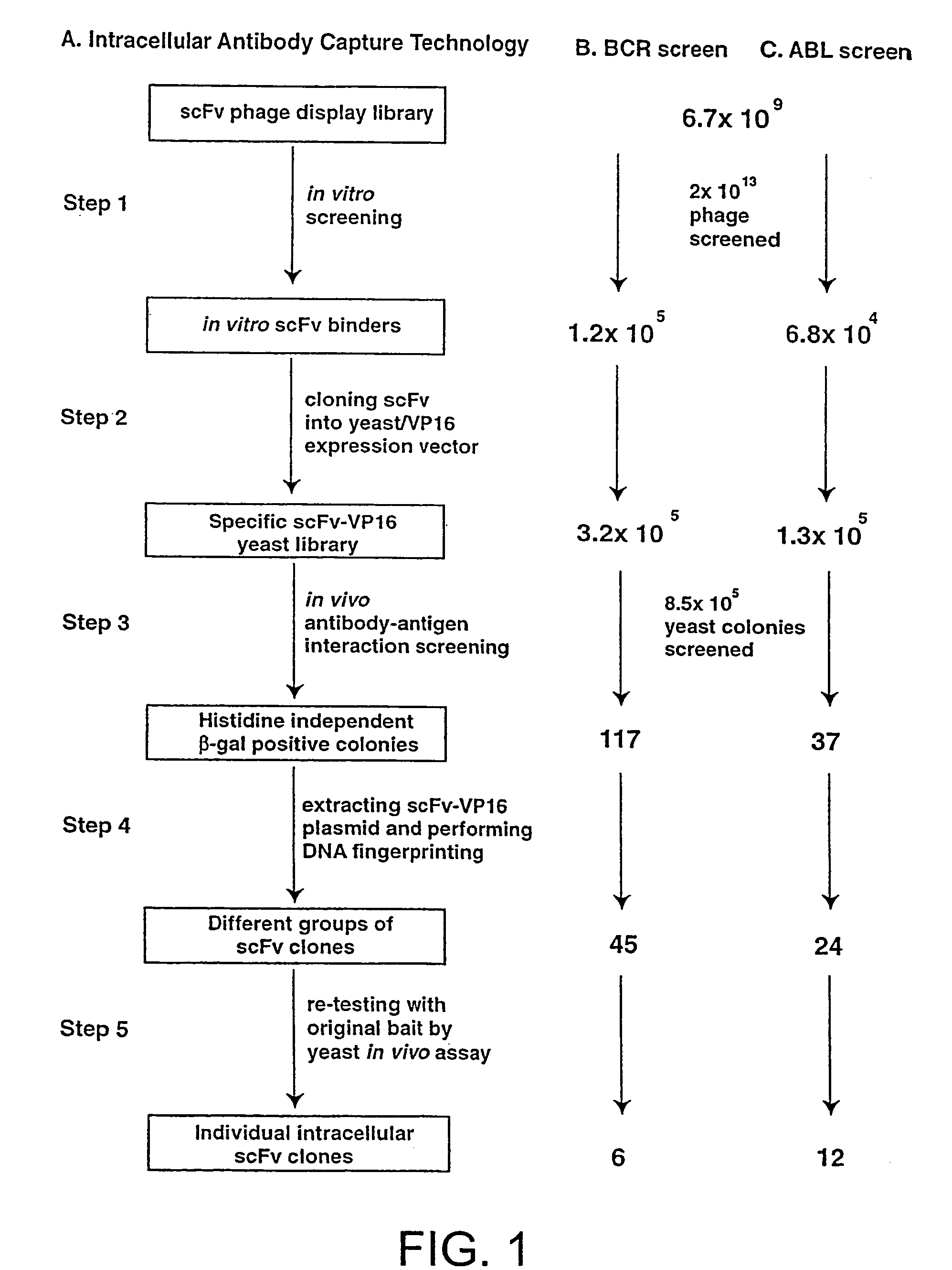
In Vitro scFv Phage Display Library Screening and Preparation of SpecificscFv-VPI6 Yeast Library

[0222]A detailed protocol for the IAC methodology is described elsewhere (Tse, E., Chung., G & Rabbitts, T., H K, Turksen, Editor, 2000, Humana Press: Totawa). Purified His-tagged SH2 binding domain of BCR protein or the SH2 domain of ABL protein were coated onto immunotubes (Nunc) at a concentration of 50 μ / ml in PBS for overnight at 4° C. 2×10¹³ phage displaying scFv (Sheets, M. D., et al (1998) Proc. Nat. Acad Sci, USA 95, 6157-6162) were incubated with the antigen-coated tubes and the bound phage were eluted with 100 mM triethylamine, neutralised by 1M Tris, pH 7.4 and were used to infect E. coli TG1 bacteria. The transduced bacteria were amplified by plating onto ampicillin supplemented agar plate from which phagemid DNA was extracted. The collected phagemid DNA was digested with SfiI and NotI restriction enzyme and the 700-800 bp scFv DNA fragment was gel purified. The purified scFv...

example 3

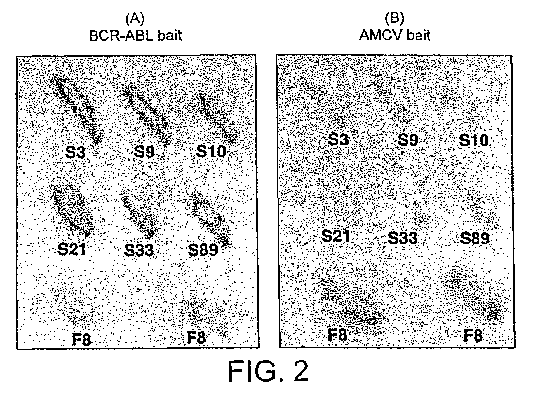
In Vivo Antibody—Antigen Interaction Screening in Yeast

[0223]L40 Yeast (Statagene) was grown at 30° C. for 3-4 days, in YAPD medium (1% yeast extract, 2% Bacto-Peptone, 2% glucose, and 0.1% mg / ml adenine buffered at pH5.8) or in synthetic minimal YC medium (0.12% yeast nitrogen base, without amino acids and 0.5% ammonium sulphate, 0.1% succinic acid, 0.6% NaOH, 2% glucose and, as required, 2% agar) containing 0.075% amino acid supplements (lacking Trp, Leu, Ura, Lys, and His; 0.1% each of adenine sulphate, Arg, Cys, Thr; 0.05% each of Asp, Ile, Met, Phe, Pro, Ser, and Tyr) buffered at pH5.8. When necessary, 0.01% each of Trp, Ura, Lys, Leu and 0.005% His were supplemented to the media.

[0224]The bait antigen expressing plasmid comprised LexA linked to BCR-ABL (pBTM / E1A2) was made by subcloning the 4 kb blunted EagI fragment of pE1A2 into BamHI blunted pBTM / 116 vector. For yeast in vivo scFv library screening, 1 mg of pBTM / E1A2 and 500 μg of the yeast scFv-VP16AD (where AD is activati...


PUM

PropertyMeasurementUnit
frequency thresholdaaaaaaaaaa
concentrationaaaaaaaaaa
body weightaaaaaaaaaa

Abstract

A method of identifying at least one consensus sequence for an intracellular antibody (ICS) comprising the steps of: creating a database comprising sequences of validated intracellular antibodies (VIDA database) and aligning the sequences of validated intracellular antibodies according to Kabat; determining the frequency with which a particular amino acid occurs in each of the positions of the aligned antibodies; selecting a frequency threshold value (LP or consensus threshold) in the range from 70% to 100%; identifying the positions of the alignment at which the frequency of a particular amino acid is greater than or equal to the LP value; and identifying the most frequent amino acid, in the position of said alignment.

Description

[0001]This application is a continuation-in-part of International Application No. PCT / GB02 / 03512, filed Aug. 1, 2002, which claims the priority of Great Britain Application No. GB 0119004.0, filed Aug. 3, 2001, Great Britain Application No. GB 0121577.1, filed Sep. 6, 2001, Italian Application No. IT RM2001A000633, filed Oct. 25, 2001, Great Britain Application No. GB 0200928.0, filed Jan. 16, 2002, and Great Britain Application No. GB 0203569.9, filed Feb. 14, 2002. Each of these applications is incorporated herein in its entirety by reference, including figures, tables and sequence listings.[0002]The present invention relates to molecules which can function in an intracellular environment. In particular, the invention relates to the characteristics of immunoglobulin molecules which can bind selectively to a ligand within an intracellular environment. Uses of these molecules are also described.BACKGROUND TO THE INVENTION[0003]Intracellular antibodies or intrabodies have been demons...

Claims


Application Information

Patent Timeline
Patent Type & Authority Patents(United States)
IPC IPC(8): A61K51/00A61M36/14C12N5/06C12N5/16C07K16/18G16B30/10C07K16/00G01N33/48G01N33/68G06F17/00
CPCG06F19/22G16B30/00G16B30/10
Inventor CATTANEO, ANTONINOMARITAN, AMOSVISINTIN, MICHELARABBITTS, TERRENCESETTANNI, GIOVANNI
Owner SISSA SCUOLA INT SUPERIORE DE STUDI AVANZATI
Why Eureka
Try Eureka