

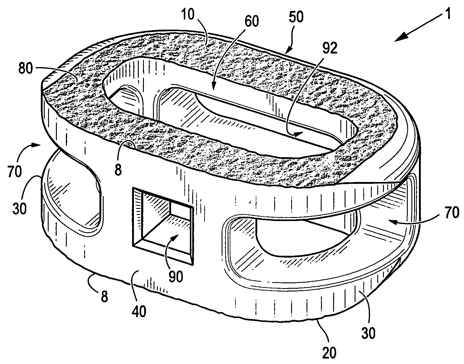
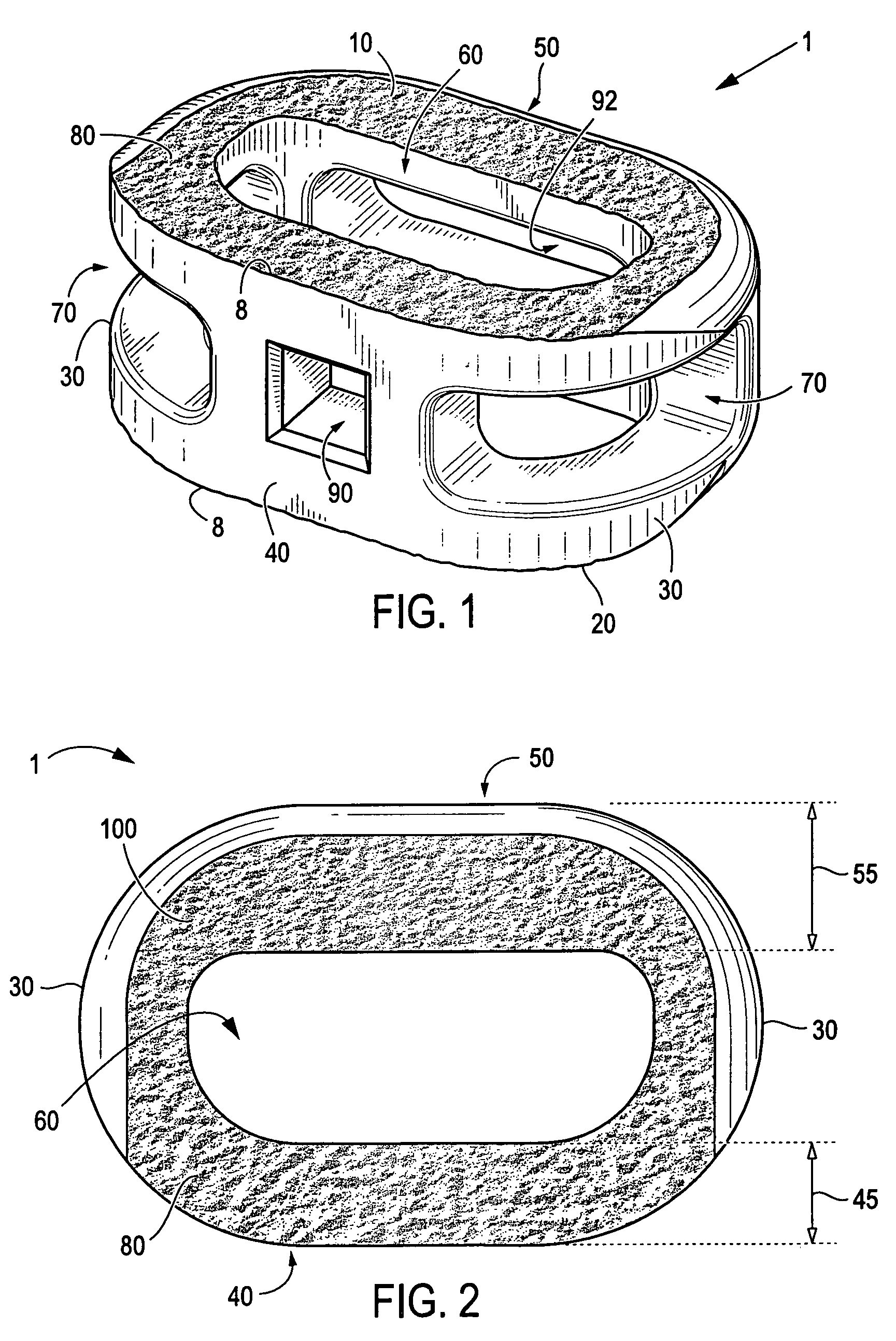
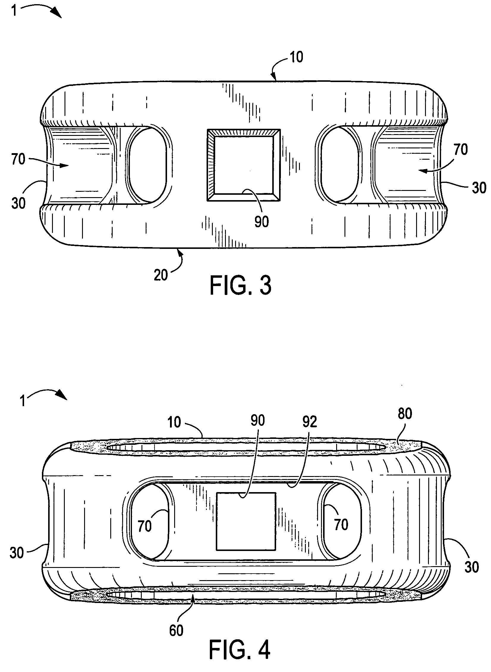
Anterior interbody spinal implant
a spinal implant and interbody technology, applied in the field of spinal implants, can solve the problems of discs affecting the anatomical functions of vertebrae, discs may become diseased or infected, discs may develop deformities, tear/cracks, etc., and achieve the effects of improving the utilization of the vertebral apophyseal rim, improving the seating position of the disc, and facilitating implantation
- Summary
- Abstract
- Description
- Claims
- Application Information
AI Technical Summary
Benefits of technology
Problems solved by technology
Method used
Image

Examples
example 1
[0031]Certain embodiments of the present invention are particularly suited for use during interbody spinal implant procedures currently known in the art. For example, the disc space may be accessed using a standard mini open retroperitoneal laparotomy approach. The center of the disc space is located by anterior-posterior (AP) fluoroscopy taking care to make sure the pedicles are equidistant from the spinous process. The disc space is then incised by making a window in the annulus for insertion of certain embodiments of the present spinal implant. The endplates are cleaned of all cartilage with a curette and a size-specific rasp may then be used.
[0032]A lateral c-arm fluoroscopy can be used to follow insertion of the rasp in the posterior disc space. The smallest height rasp that touches both endplates (e.g., the superior and inferior endplates) in first chosen. After the disc space is cleared of all soft tissue and cartilage, distraction is then accomplished by using implant trials...
PUM
 Abstract
Description
Claims
Application Information